Description
The exogenous promoter and human PD-L1 CDS were inserted into the mouse Pd-l1 exon 3. The exogenous promoter and human TPBG CDS were inserted into mouse Tpbg locus site in B-hPD-L1 plus/hTPBG MC38. Human PD-L1 and TPBG are detected on the surface of B-hPD-L1 plus/hTPBG MC38 cells.
Targeting strategy
Gene targeting strategy for B-hPD-L1 plus/hTPBG MC38. The exogenous promoter and human PD-L1 CDS were inserted into the mouse Pd-l1 exon 3. The exogenous promoter and human TPBG CDS were inserted into mouse Tpbg locus site in B-hPD-L1 plus/hTPBG MC38.
Protein expression analysis
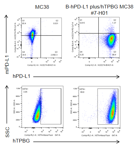
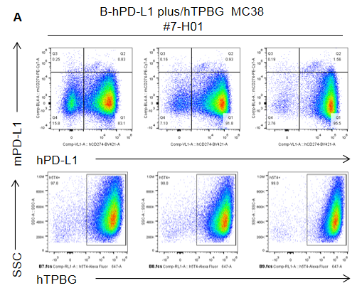
PD-L1 and TPBG expression analysis in B-hPD-L1 plus/hTPBG MC38 cells by flow cytometry. Single cell suspensions from B-hPD-L1 plus/hTPBG MC38 cultures were stained with anti-PD-L1 and anti-TPBG antibodies. Human PD-L1 and TPBG were detected on the surface of B-hPD-L1 plus/hTPBG MC38 cells.
Tumor growth curve & Body weight changes
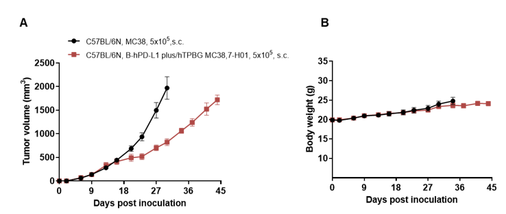
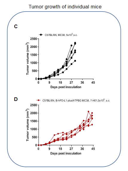
Subcutaneous homograft tumor growth of B-hPD-L1 plus/hTPBG MC38 cells. B-hPD-L1 plus/hTPBG MC38 cells and wild-type MC38 cells were subcutaneously implanted into C57BL/6N mice (female, 9-week-old, n=6). Tumor volume and body weight were measured twice a week. (A) Average tumor volume ± SEM. (B) Body weight (Mean± SEM). (C, D) Tumor volume of individual mice in each group. Volume was expressed in mm3 using the formula: V=0.5 X long diameter X short diameter2. As shown in panel, B-hPD-L1 plus/hTPBG MC38 cells were able to establish tumors in vivo.
PD-L1 and TPBG expression analysis in tumor cells
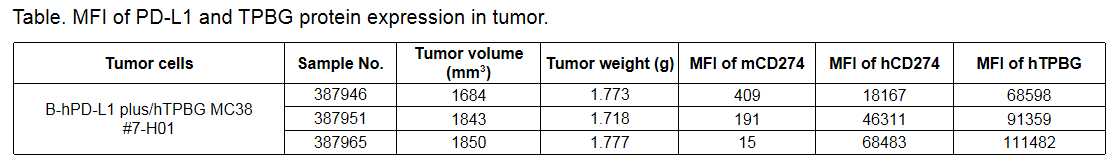
PD-L1 and TPBG expression analysis in tumor cells. Tumor cells were harvested at the end point of the experiment and assessed for m/hPD-L1 and hTPBG expression by flow cytometry. As shown in the panel A, human PD-L1 and TPBG were expressed on the surface of tumor cells. The MFI value was shown in the table.