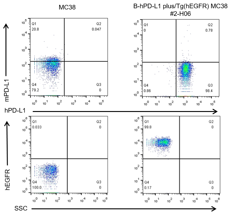
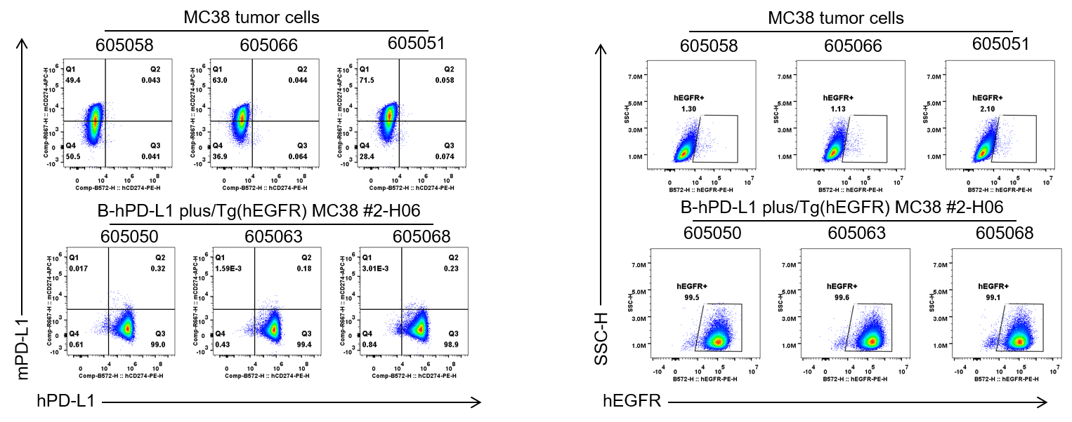
B-hPD-L1 plus/Tg(hEGFR) MC38
Catalog Number: 322357
Strain Name: NA
Strain Background: C57BL/6
NCBI gene ID: 60533,13649 (Human)
Aliases: B7h1; Pdl1; Pdcd1l1; Pdcd1lg1; A530045L16Rik; Wa5; wa2; Erbb; Errp; wa-2; Errb1; 9030024J15Rik
---
Licensing option available