
Basic Information
-
Description

-
Homozygous mice for the targeted mutation are viable, fertile, normal in size and do not display any gross physical or behavioral abnormalities.
-
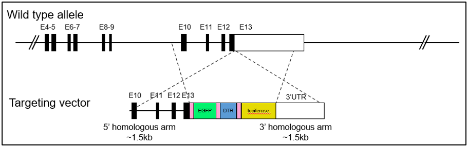
Gene editing strategy
-
-
Analysis of spleen leukocyte subpopulations by FACS
-
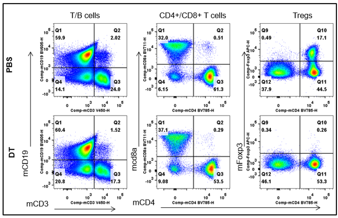
Splenocytes were isolated from male B-Foxp3-EGFP-DTR-Luc mice (n=3, 7-month-old) injected with PBS or DT (30ng/ per body weight) for two consecutive days. Flow cytometry analysis of the splenocytes was performed to assess leukocyte subpopulations.
-
Analysis of lymph node leukocyte subpopulations by FACS
-
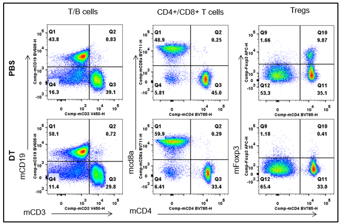
Lymph node were isolated from male B-Foxp3-EGFP-DTR-Luc mice (n=3, 7-month-old) injected with PBS or DT (30ng/ per body weight) for two consecutive days. Flow cytometry analysis was performed to assess leukocyte subpopulations.
-
Analysis of EGFP expression in spleen of B-Fxop3-EGFP-DTR-Luc mice by FACS
-
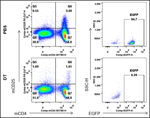
Splenocytes were isolated from B-Foxp3-EGFP-DTR-Luc mice mice(n=3, 7-month-old) injected with PBS or DT (30ng/per body weight) for two consecutive days. Flow cytometry analysis was performed to assess EGFP expression.
-
BLI analysis of homozygous B-Foxp3-EGFP-DTR-Luc mice after DT injection
-
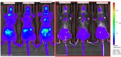
Homozygous B-Foxp3-EGFP-DTR-Luc mice i.p. injected with PBS (n=3) or DT (n=3) for two consecutive days were anaesthetized for the bioluminescence imaging. Mice were imaged 10min after i.p. injection of 150mg/kg D-Lucifenrin potassium salt using IVIS Lumina LT Inst Series III imaging system.
The ratio of Treg cells in homozygous B-Foxp3-EGFP-DTR-Luc mice is comparable with that in wild type mice; EGFP was exclusively detectable in CD4+; CD25+ populations from Foxp3-EGFP-DTR-Luc mice and could be as a marker for Treg cells in vivo; besides Bioluminescence imaging could also be used for tracing Tregs cells; After DT injection, Treg cells showed dramatically decreased at EGFP, Foxp3 and bioluminescence imaging levels.
