
Basic Information
-
Targeting strategy

-
Gene targeting strategy for B-h4-1BB/hTNFR2 mice plus. The exon 2-7 of mouse 4-1BB gene that encode the extracellular domain was replaced by human 4-1BB counterpart gene in B-h4-1BB/hTNFR2 mice plus. The exons 2~6 of mouse Tnfr2 gene that encode the extracellular domain was replaced by human TNFR2 counterpart gene in B-h4-1BB/hTNFR2 mice plus.
-
Protein expression analysis
-
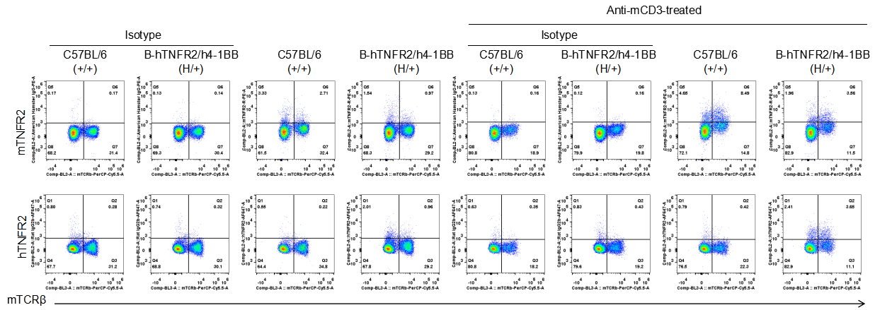
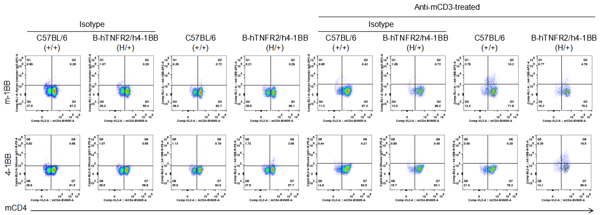
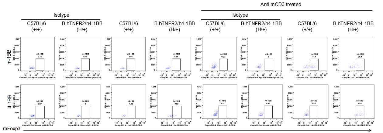
Strain specific TNFR2 expression analysis in heterozygous B-h4-1BB/hTNFR2 mice plus by flow cytometry. Splenocytes were collected from wild-type mice (+/+) and heterozygous B-h4-1BB/hTNFR2 mice plus (H/+) stimulated with anti-CD3ε in vivo (7.5 μg/mice, 24 hours, i.p.), and analyzed by flow cytometry with species-specific anti-TNFR2 antibody. Mouse TNFR2 was detectable in wild-type mice and heterozygous B-h4-1BB/hTNFR2 mice plus, but human TNFR2 was only detectable in heterozygous B-h4-1BB/hTNFR2 mice plus.
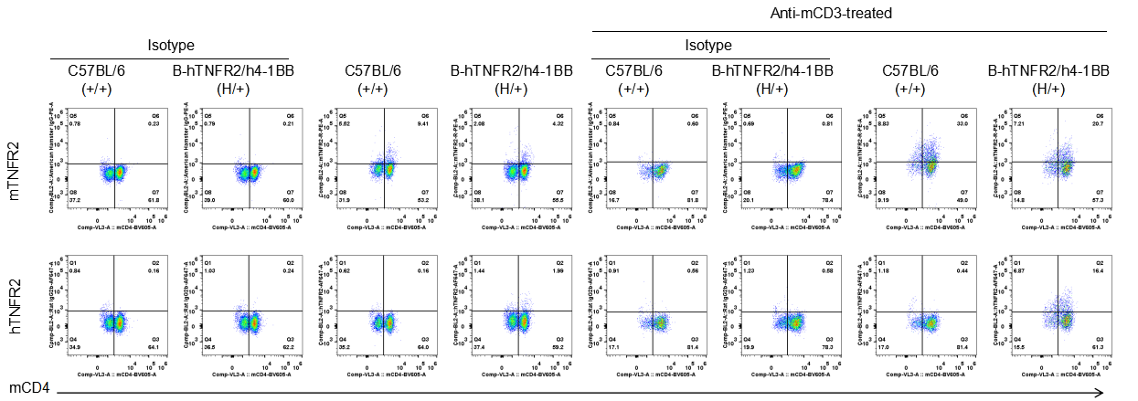
Strain specific TNFR2 expression analysis in heterozygous B-h4-1BB/hTNFR2 mice plus by flow cytometry. Splenocytes were collected from wild-type mice (+/+) and heterozygous B-h4-1BB/hTNFR2 mice plus (H/+) stimulated with anti-CD3ε in vivo (7.5 μg/mice, 24 hours, i.p.), and analyzed by flow cytometry with species-specific anti-TNFR2 antibody. Mouse TNFR2 was detectable in wild-type mice and heterozygous B-h4-1BB/hTNFR2 mice plus, but human TNFR2 was only detectable in heterozygous B-h4-1BB/hTNFR2 mice plus.
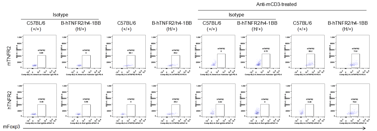
Strain specific TNFR2 expression analysis in heterozygous B-h4-1BB/hTNFR2 mice plus by flow cytometry. Splenocytes were collected from wild-type mice (+/+) and heterozygous B-h4-1BB/hTNFR2 mice plus (H/+) stimulated with anti-CD3ε in vivo (7.5 μg/mice, 24 hours, i.p.), and analyzed by flow cytometry with species-specific anti-TNFR2 antibody. Mouse TNFR2 was detectable in wild-type mice and heterozygous B-h4-1BB/hTNFR2 mice plus, but human TNFR2 was only detectable in heterozygous B-h4-1BB/hTNFR2 mice plus.
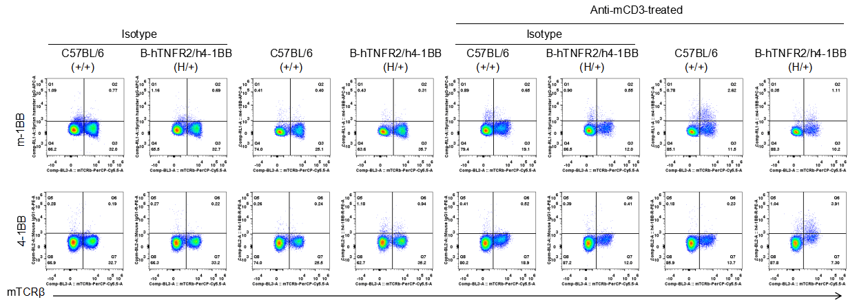
Strain specific 4-1BB expression analysis in heterozygous B-h4-1BB/hTNFR2 mice plus by flow cytometry. Splenocytes were collected from wild-type mice (+/+) and heterozygous B-h4-1BB/hTNFR2 mice plus (H/+) stimulated with anti-CD3ε in vivo (7.5 μg/mice, 24 hours, i.p.), and analyzed by flow cytometry with species-specific anti-4-1BB antibody. Mouse 4-1BB was detectable in wild-type mice and heterozygous B-h4-1BB/hTNFR2 mice plus, but human 4-1BB was only detectable in heterozygous B-h4-1BB/hTNFR2 mice plus.
Strain specific 4-1BB expression analysis in heterozygous B-h4-1BB/hTNFR2 mice plus by flow cytometry. Splenocytes were collected from wild-type mice (+/+) and heterozygous B-h4-1BB/hTNFR2 mice plus (H/+) stimulated with anti-CD3ε in vivo (7.5 μg/mice, 24 hours, i.p.), and analyzed by flow cytometry with species-specific anti-4-1BB antibody. Mouse 4-1BB was detectable in wild-type mice and heterozygous B-h4-1BB/hTNFR2 mice plus, but human 4-1BB was only detectable in heterozygous B-h4-1BB/hTNFR2 mice plus.
Strain specific 4-1BB expression analysis in heterozygous B-h4-1BB/hTNFR2 mice plus by flow cytometry. Splenocytes were collected from wild-type mice (+/+) and heterozygous B-h4-1BB/hTNFR2 mice plus (H/+) stimulated with anti-CD3ε in vivo (7.5 μg/mice, 24 hours, i.p.), and analyzed by flow cytometry with species-specific anti-4-1BB antibody. Mouse 4-1BB was detectable in wild-type mice and heterozygous B-h4-1BB/hTNFR2 mice plus, but human 4-1BB was only detectable in heterozygous B-h4-1BB/hTNFR2 mice plus.
-
Tumor growth curve
-
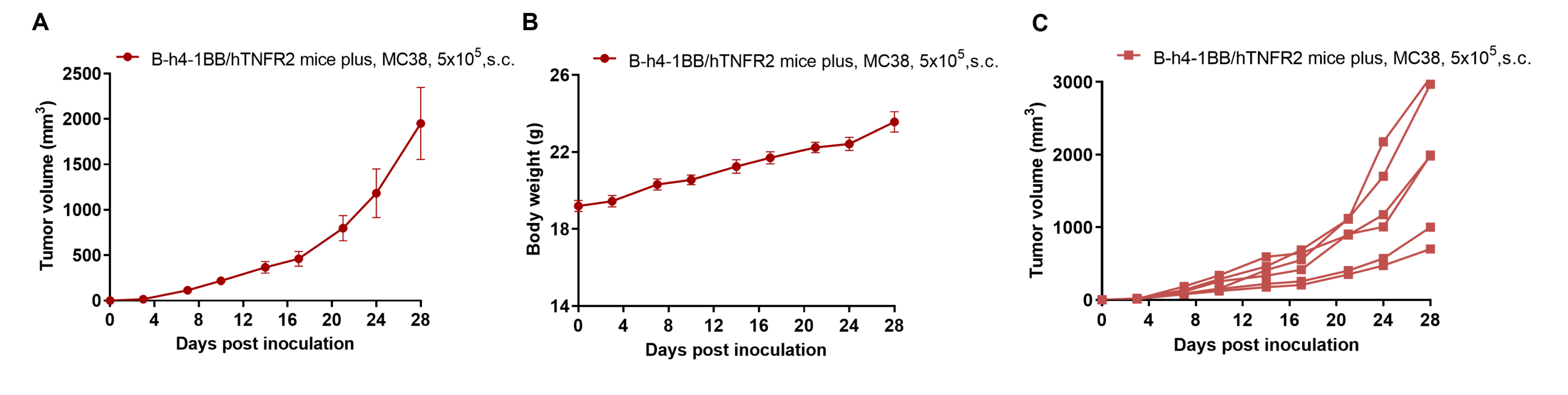
Subcutaneous homograft tumor growth of MC38 cells in homozygous B-h4-1BB/hTNFR2 mice plus. Wild-type MC38 cells (5×10⁵) were subcutaneously implanted into B-h4-1BB/hTNFR2 mice plus (female, 8-week-old, n=6). Tumor volume and body weight were measured twice a week. (A) Average tumor volume ± SEM. (B) Body weight (Mean± SEM). (C) Tumor growth curve of individual mice. Volume was expressed in mm3 using the formula: V=0.5 X long diameter X short diameter2. As shown in panel A, MC38 cells were able to establish tumors in vivo and can be used for efficacy studies.
