
Basic Information
-
Targeting strategy

-
The human CD89 CDS was inserted into into the ROSA26 locus in B-hCD89 mice.
-
mRNA expression analysis
-
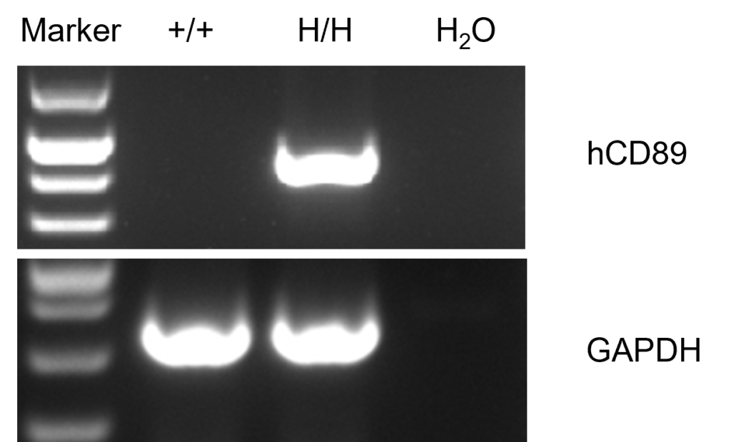
Strain specific analysis of CD89 gene expression in wild type (WT) mice and B-hCD89 mice by RT-PCR. Human CD89 mRNA was detectable only in splenocytes of homozygous B-hCD89 mice (H/H) but not WT mice (+/+).
-
Protein expression analysis
-
Protein expression analysis in NK cells
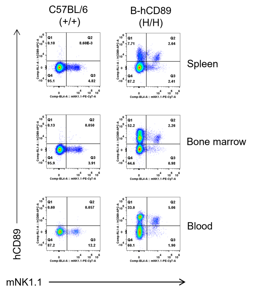
Strain specific CD89 expression analysis in homozygous B-hCD89 mice by flow cytometry. Spleen, bone marrow and blood were collected from wild type (WT) mice (+/+) and homozygous B-hCD89 mice (H/H), and analyzed by flow cytometry with species-specific anti-CD89 antibody. Human CD89 was exclusively detectable in NK cells of homozygous B-hCD89 mice (H/H).
Protein expression analysis in CD11b+ cells
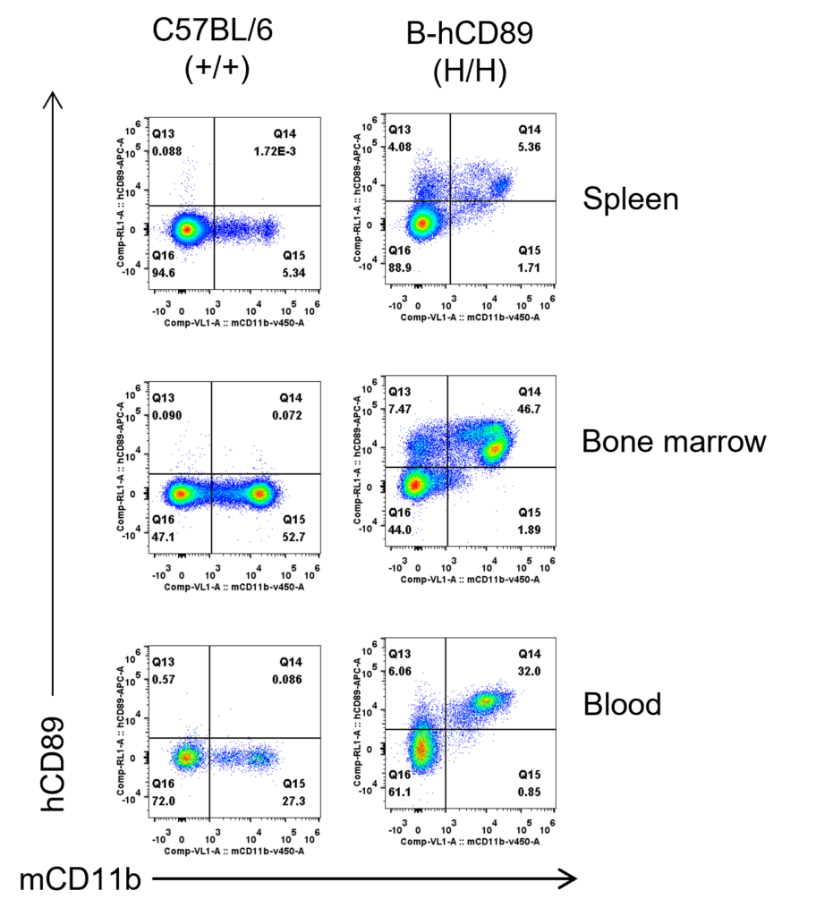
Strain specific CD89 expression analysis in homozygous B-hCD89 mice by flow cytometry. Spleen, bone marrow and blood were collected from wild type (WT) mice (+/+) and homozygous B-hCD89 mice (H/H), and analyzed by flow cytometry with species-specific anti-CD89 antibody. Human CD89 was exclusively detectable in CD11b+ cells of homozygous B-hCD89 mice (H/H).
-
References
-
1.Mkaddem S B, Rossato E, Heming N, et al. Anti-inflammatory role of the IgA Fc receptor (CD89): from autoimmunity to therapeutic perspectives[J]. Autoimmunity reviews, 2013, 12(6): 666-669.,
