
Basic Information
Description
The CDS of human C5 was inserted to replace part of murine exon 2 in B-hC5 MC38 cells. Human C5 is highly expressed on the surface of B-hC5 MC38 cells.
-
Targeting Strategy

-
Gene targeting strategy for B-hC5 MC38 cells. The CDS of human C5 was inserted to replace part of murine exon 2. The insertion disrupts the endogenous murine C5 gene, resulting in a non-functional transcript.
-
Protein Expression Analysis
-
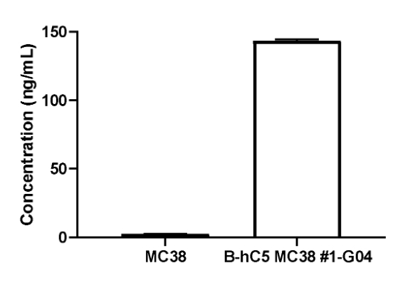
C5 expression analysis in B-hC5 MC38 cells by ELISA. Supernatant from wild-type MC38 and B-hC5 MC38 cultures were stained with species-specific anti-C5 antibody. Human C5 was detectable in the supernatant of B-hC5 MC38 cells but not in that of wild-type MC38 cells. The 1-G04 clone of B-hC5 MC38 cells was used for in vivo experiments.
-
Tumor growth curve & Body weight changes
-
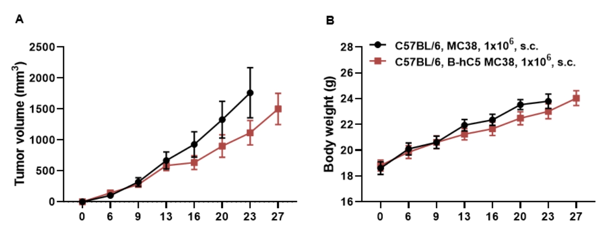
Subcutaneous homograft tumor growth of B-hC5 MC38 cells. B-hC5 MC38 cells (1×10⁶) and wild-type MC38 cells (1×10⁶) were subcutaneously implanted into C57BL/6N mice (female, 7-week-old, n=5). Tumor volume and body weight were measured twice a week. Three mice from each group were sacrificed on day 14 and the other mice were sacrificed at the endpoint. (A) Average tumor volume. (B) Body weight. Volume was expressed in mm3 using the formula: V=0.5 X long diameter X short diameter2. As shown in panel A, B-hC5 MC38 cells were able to establish tumors in vivo and can be used for efficacy studies. Values are expressed as mean ± SEM.
-
Protein expression analysis of C5 and C5a in serum and tumor tissues
-
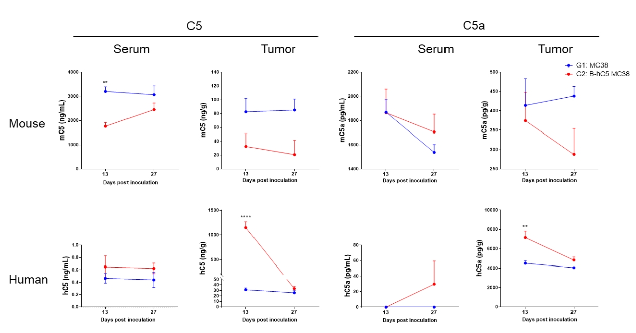
Strain specific C5 and C5a expression analysis in serum and tumor tissues by ELISA. Wild-type MC38 cells and B-hC5 MC38 cells were subcutaneously inoculated into C57BL/6 mice. Serum and tumors were collected at two time point. One time point is when tumor volume reached 300-500 mm3 (Groups G1-1, G2-1). The other is when part of tumor volume reached 3000 mm3 (Groups G1-2, G2-2). mouse and human C5 and C5a were analyzed by ELISA with species specific anti-C5 antibody and anti-C5a antibody. Mouse C5 and C5a were detectable in serum and tumor tissues collected from mice inoculated with wild-type MC38 cells and B-hC5 MC38 cells. Human C5 and C5a were detectable in tumor tissues collected when tumor volume reached 300-500 mm3 from mice inoculated with B-hC5 MC38 cells. Human C5 and C5a were not detectable in tumor tissues when tumor volume reached about 3000 mm3.
-
Protein expression analysis of C5AR1 in cell line and tumor tissues
-
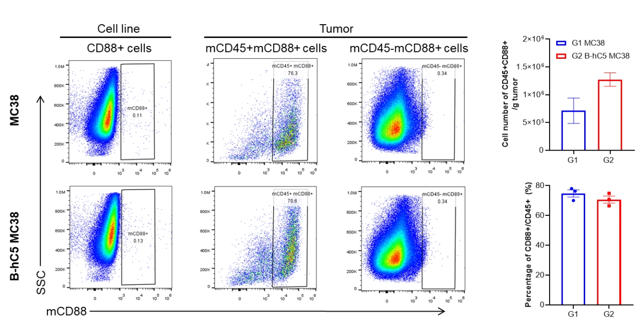
Strain specific CD88 (C5AR1) expression analysis in MC38 cell line and tumor by flow cytometry. Wild-type MC38 cells and B-hC5 MC38 cells were subcutaneously inoculated into C57BL/6 mice, and tumors were removed and prepare tumor cells when part of tumor volume reached 3000 mm3. Cultured wild-type MC38 cell line and tumor cells were collected and analyzed by flow cytometry with anti-mCD88 antibody. Mouse CD88 was not detectable on cultured MC38 cell line. But in tumor tissue, mCD88 was expressed in most of the CD45+ cells and it was expressed in a small number of CD45− cells.
