
Basic Information
Description
The mouse H2-t23 and Pdl1 gene were replaced by human HLA-E and PD-L1 coding sequence in B-HLA-E plus/hPD-L1 MC38 cells. Human HLA-E and PD-L1 are highly expressed by B-HLA-E plus/hPD-L1 MC38.
-
Gene targeting strategy

-
Gene targeting strategy for B-HLA-E plus/hPD-L1 MC38 cells . The exogenous promoter and human HLA-E coding sequence was inserted to replace part of murine exon 3 and all of exons 4-7. The exogenous promoter and human PD-L1 coding sequence was inserted to replace part of murine exon 3. The insertion disrupts the endogenous murine H2-t23 and Pdl1 gene, resulting in non-functional transcripts.
-
Protein expression analysis
-
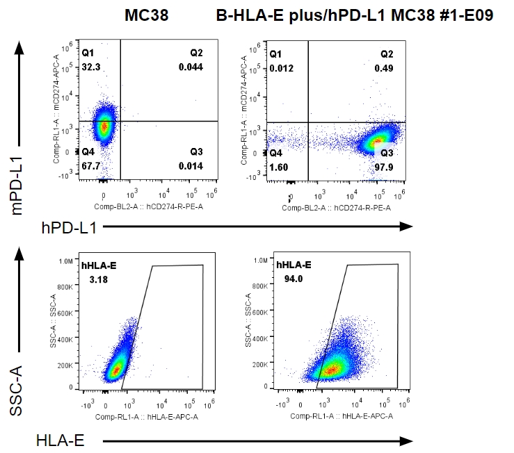
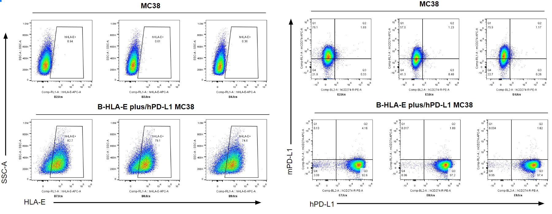
PD-L1 and HLA-E expression analysis in B-HLA-E plus/hPD-L1 MC38 cells by FACS. Supernatant from wild-type MC38 and B-HLA-E plus/hPD-L1 MC38 cultures were stained with species-specific anti-PD-L1 and anti-HLA-E antibody. Human PD-L1 and HLA-E were detectable in the supernatant of B-HLA-E plus/hPD-L1 MC38 cells but not in that of wild-type MC38 cells. The 1-E09 clone of B-HLA-E plus/hPD-L1 MC38 cells was used for in vivo experiments.
-
Tumor growth curve & Body weight changes
-
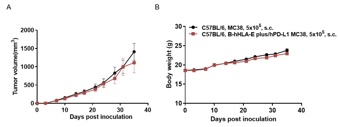
Subcutaneous homograft tumor growth of B-HLA-E plus/hPD-L1 MC38 cells. B-HLA-E plus/hPD-L1 MC38 cells (5×10⁵) and wild-type MC38 cells (5×10⁵) were subcutaneously implanted into C57BL/6 mice (female, 8-week-old). Tumor volume and body weight were measured twice a week. (A) Average tumor volume ± SEM. (B) Body weight (Mean± SEM). Volume was expressed in mm3 using the formula: V=0.5 X long diameter X short diameter2. As shown in panel A, B-HLA-E plus/hPD-L1 MC38 cells were able to establish tumors in vivo and can be used for efficacy studies.
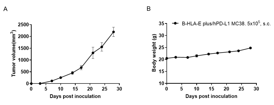
Subcutaneous homograft tumor growth of B-HLA-E plus/hPD-L1 MC38 cells. B-HLA-E plus/hPD-L1 MC38 cells (5×10⁵) were subcutaneously implanted into B-hPD-1/hPD-L1/hCD94/hNKG2A mice (female, 8-week-old, n=5). Tumor volume and body weight were measured twice a week. (A) Average tumor volume ± SEM. (B) Body weight (Mean± SEM). Volume was expressed in mm3 using the formula: V=0.5 X long diameter X short diameter2. As shown in panel A, B-HLA-E plus/hPD-L1 MC38 cells were able to establish tumors in vivo and can be used for efficacy studies.
-
Protein expression analysis of tumor cells
-
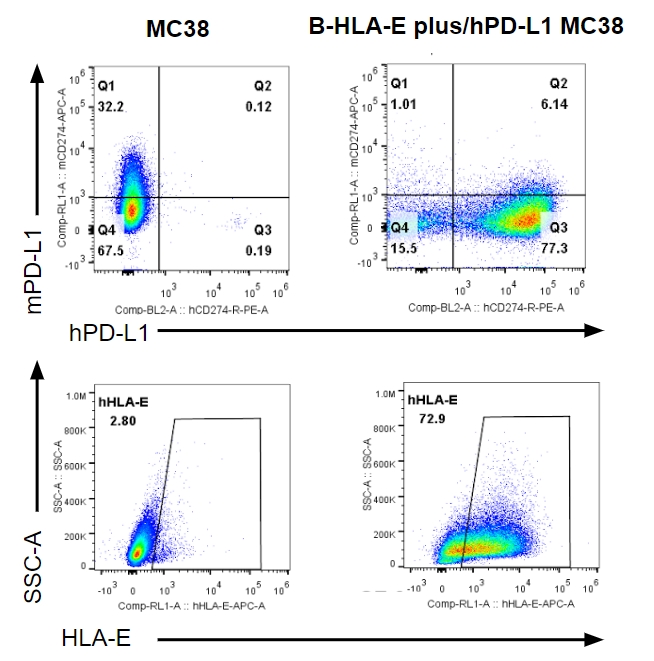
B-HLA-E plus/hPD-L1 MC38 cells were subcutaneously transplanted into C57BL/6 mice (n=6), and on 35 days post inoculation, tumor cells were harvested and assessed for human PD-L1 and HLA-E expression by FACS. As shown, human PD-L1 and HLA-E were highly expressed by tumor cells. Therefore, B-HLA-E plus/hPD-L1 MC38 cells can be used for in vivo efficacy studies.
B-HLA-E plus/hPD-L1 MC38 cells were subcutaneously transplanted into B-hPD-1/hPD-L1/hCD94/hNKG2A mice (n=6), and on 31 days post inoculation, tumor cells were harvested and assessed for human PD-L1 and HLA-E expression by FACS. As shown, HLA-E and human PD-L1 were highly expressed by tumor cells. Therefore, B-HLA-E plus/hPD-L1 MC38 cells can be used for in vivo efficacy studies.
