
Basic Information
Description
Biocytogen developed the immunodeficient B-NDG hSIRPA mouse model. The targeting strategy used was that exon 2 of the mouse Sirpα gene, which encodes the extracellular domain, was replaced by human SIRPα in B-NDG mice. This mouse combines a highly immunodeficient B-NDG mouse background (completely lacking mature T, B and NK cells and is deficient in cytokine signaling) with the extracellular domain of the human SIRPα protein.
-
Details

-
Generation of gene editing mouse model and expression analysis
Protein expression analysis (heterozygous mice)
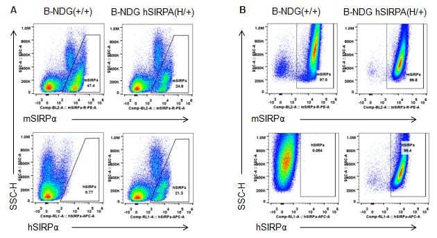
Species specific SIRPα expression analysis in B-NDG hSIRPA mice by flow cytometry. Splenocytes (A) and peritoneal lymphocyte (B) from B-NDG and heterozygous B-NDG hSIRPA (H/+) mice were analyzed by flow cytometry with anti-SIRPα antibodies. Mouse SIRPα was detectable in B-NDG and heterozygous B-NDG hSIRPA mice. Human SIRPα were exclusively detectable in heterozygous B-NDG hSIRPA but not B-NDG mice.
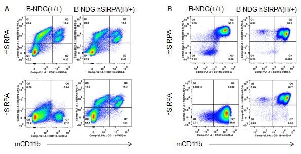
Species specific SIRPα expression analysis in B-NDG hSIRPA mice by flow cytometry. Splenocytes (A) and peritoneal lymphocyte (B) from B-NDG and heterozygous B-NDG hSIRPA (H/+) mice were analyzed by flow cytometry with anti-SIRPα and anti-CD11b antibodies. Mouse SIRPα were detectable in macrophages of B-NDG and heterozygous B-NDG hSIRPA mice. Human SIRPα were exclusively detectable in macrophages of heterozygous B-NDG hSIRPA but not B-NDG mice.
Protein expression analysis (homozygous mice)
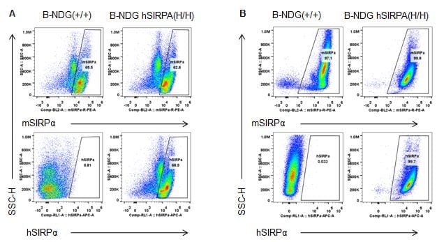
Species specific SIRPα expression analysis in B-NDG hSIRPA mice by flow cytometry. Splenocytes (A) and peritoneal lymphocyte (B) from B-NDG and homozygous B-NDG hSIRPA (H/H) mice were analyzed by flow cytometry with anti-SIRPα antibodies. Mouse SIRPα was detectable in B-NDG and homozygous B-NDG hSIRPA mice. This anti-mouse SIRPα antibody also cross reacts with human SIRPα. Human SIRPα were exclusively detectable in homozygous B-NDG hSIRPA but not B-NDG mice.
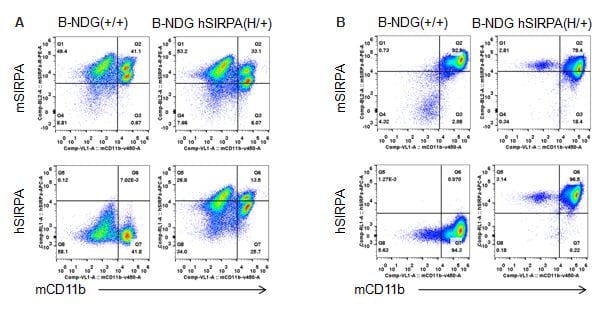
Species specific SIRPα expression analysis in B-NDG hSIRPA mice by flow cytometry. Splenocytes (A) and peritoneal lymphocyte (B) from B-NDG and homozygous B-NDG hSIRPA (H/H) mice were analyzed by flow cytometry with anti-SIRPα antibodies. Mouse SIRPα was detectable in macrophages of B-NDG and homozygous B-NDG hSIRPA mice. This anti-mouse SIRPα antibody also cross reacts with human SIRPα. Human SIRPα were exclusively detectable in macrophages of homozygous B-NDG hSIRPA but not B-NDG mice.
Analysis of spleen leukocytes cell subpopulations in B-NDG hSIRPA mice
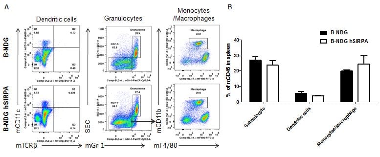
Analysis of spleen leukocyte subpopulations by FACS
Splenocytes were isolated from male B-NDG and B-NDG hSIRPA mice (n=3, 7-week-old). Flow cytometry analysis of the splenocytes was performed to assess leukocyte subpopulations. A. Representative FACS plots. Single live cells were gated for CD45 population and used for further analysis as indicated here. B. Results of FACS analysis. Percent of Monocyte, DC and macrophage cells in homozygous B-NDG hSIRPA mice were similar to those in the B-NDG mice, demonstrating that introduction of hSIRPα in place of its mouse counterpart does not change the overall development, differentiation or distribution of these cell types in spleen.
In vivo efficacy of anti human CD47 antibodies
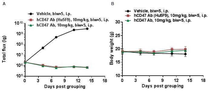
Antitumor activity of anti-human CD47 antibodies in B-NDG hSIRPA mice. (A) Human B-luciferase-GFP Raji cells (B lymphocytes) (5.0E+05) were inoculated into homozygous B-NDG hSIRPA mice (fmale, 5-week-old, n=5). Mice were grouped when the fluorescence intensity reached 1.0E+06 , at which time they were treated with anti-human CD47 antibody with doses and schedules indicated in panel. (B) Body weight changes during treatment. As shown in panel A, anti-human CD47 antibody was efficacious in controlling tumor growth in this model. Values are expressed as mean ± SEM.
-
Summary
-
1.Species specific SIRPα expression analysis in B-NDG hSIRPA mice by flow cytometry. Human SIRPα were exclusively detectable in macrophages of homozygous B-NDG hSIRPA
2.Analysis of spleen leukocyte subpopulations by FACS. Percent of Monocyte, DC and macrophage cells in homozygous B-NDG hSIRPA mice were similar to those in the B-NDG mice. demonstrating that introduction of hSIRPα in place of its mouse counterpart does not change the overall development, differentiation or distribution of these cell types in spleen.
