
Basic Information
-
Gene targeting strategy

-
Gene targeting strategy for B-hEGFR mice. The exons 2-17 of mouse Egfr gene that encode the extracellular domain were replaced by human EGFR exons 2-17 in B-hEGFR mice.
-
mRNA expression analysis
-
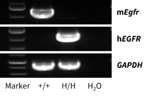
Strain specific analysis of EGFR gene expression in wild type (WT) mice and B-hEGFR mice by RT-PCR. Mouse Egfr mRNA was detectable only in liver of WT mice (+/+). Human EGFR mRNA was detectable only in homozygous B-hEGFR mice (H/H) but not in WT mice (+/+).
-
IHC analysis
-
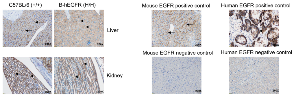
Immunohistochemical (IHC) analysis of EGFR expression in homozygous B-hEGFR mice. Liver and kidney tissues were collected from wild-type C57BL/6 (+/+) and homozygous B-hEGFR (H/H) mice and analyzed by IHC using anti-EGFR antibodies. Mouse EGFR was detected in wild-type mice, while human EGFR was detected in B-hEGFR mice. The arrow indicates tissue cells with positive EGFR staining (brown).
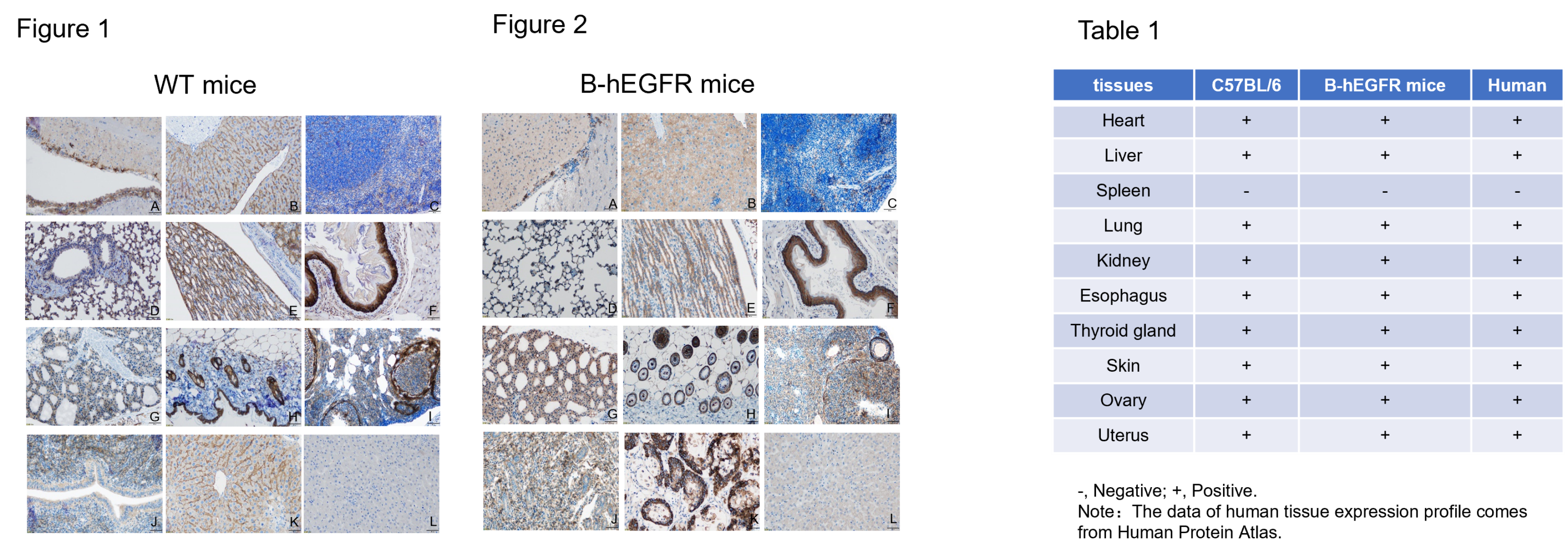
Immunohistochemical (IHC) analysis of EGFR protein expression in wild-type (WT) mice and B-hEGFR mice. Ten major tissues were collected from WT mice and homozygous B-hEGFR mice (2 females, 8 week-old), and analyzed by IHC with anti-EGFR antibodies. Humanized EGFR was detected in the heart (A), liver (B), lung (D), kidney (E), esophagus (F), thyroid (G), skin (H), ovary (I) and uterus (J) of homozygous B-hEGFR mice (Figure 2) and human EGFR positive control (K), but not in spleen (C) and human EGFR negative control (L). This is similar to the mouse EGFR expression profile of WT mice (Figure 1, Table 1). The results showed that humanized EGFR did not change the expression site of EGFR protein in B-hEGFR mice, and the expression profile of EGFR in B-hEGFR mice was similar to that in normal human tissues (Table 1). B-hEGFR mice can be used to evaluate the toxicity of EGFR drugs.
-
Blood routine test
-
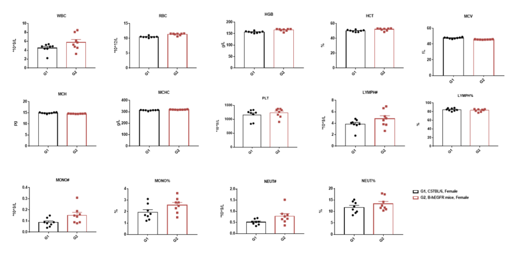
Complete blood count (CBC). Blood from C57BL/6 and B-hEGFR mice (female, 9 week-old,n=8) was collected and analyzed for CBC. There was no differences among any measurement between C57BL/6 and B-hEGFR mice, indicating that introduction of hEGFR in place of its mouse counterpart does not change blood cell composition and morphology. Values are expressed as mean ± SEM.
-
Blood chemistry test
-

Blood chemistry tests of B-hEGFR mice. Serum from the C57BL/6 and B-hEGFR mice (female, 9 week-old, n=8) was collected and analyzed for levels of ALT and AST. There was no differences on either measurement between C57BL/6 and B-hEGFR mice, indicating that introduction of hEGFR in place of its mouse counterpart does not change ALT and AST levels or health of liver. Values are expressed as mean ± SEM.
-
Tumor growth curve & Body weight changes
-
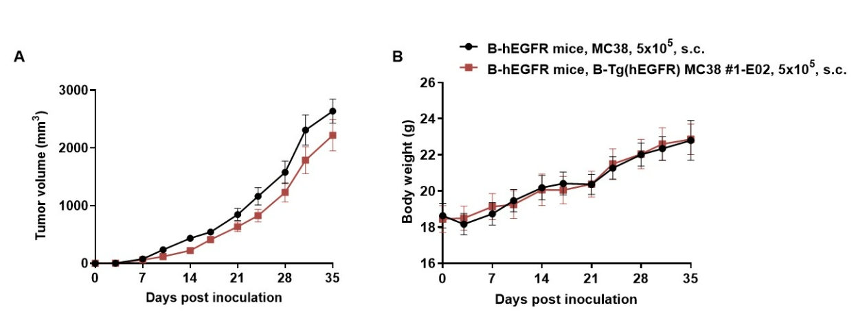
Subcutaneous homograft tumor growth of B-Tg(hEGFR) MC38 cells.
B-Tg(hEGFR) MC38 cells (5×10⁵) and wild-type MC38 cells (5×10⁵) were subcutaneously implanted into homozygous B-hEGFR mice (female, 6-9-week-old, n=6). Tumor volume and body weight were measured twice a week. (A) Average tumor volume ± SEM. (B) Body weight (Mean ± SEM). Volume was expressed in mm3 using the formula: V=0.5 X long diameter X short diameter2. As shown in panel A, B-Tg(hEGFR) MC38 cells were able to form tumors in vivo and can be used for efficacy studies.
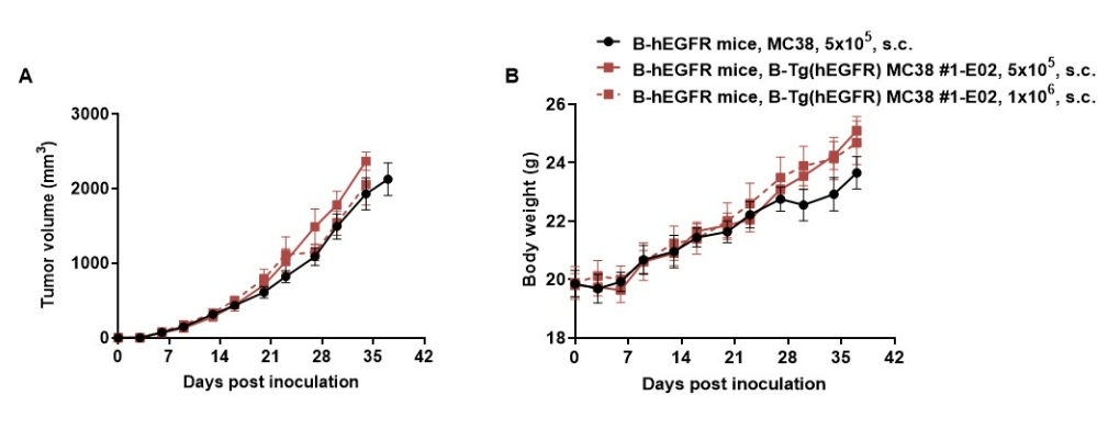
Subcutaneous homograft tumor growth of B-Tg(hEGFR) MC38 cells.
B-Tg(hEGFR) MC38 cells (5×10⁵, 1×10⁶) and wild-type MC38 cells (5×10⁵) were subcutaneously implanted into homozygous B-hEGFR mice (female, 5-8-week-old, n=6). Tumor volume and body weight were measured twice a week. (A) Average tumor volume ± SEM. (B) Body weight (Mean ± SEM). Volume was expressed in mm3 using the formula: V=0.5 X long diameter X short diameter2. As shown in panel A, B-Tg(hEGFR) MC38 cells were able to form tumors in vivo and can be used for efficacy studies.
-
Protein expression analysis of tumor cells
-
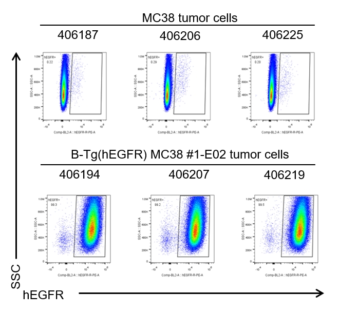
B-Tg(hEGFR) MC38 cells were subcutaneously transplanted into homozygous B-hEGFR mice (n=6), and on 35 days post inoculation, tumor cells were harvested and assessed for human EGFR expression by flow cytometry. As shown, human EGFR was highly expressed on the surface of tumor cells. Therefore, B-Tg(hEGFR) MC38 cells can be used for in vivo efficacy studies of EGFR therapeutics.
