Description
APOE: A major genetic risk factor for late-onset Alzheimer’s disease
- Gene Information: Human APOE is a 34 kDa glycoprotein, and there was three APOE polymorphic alleles in human, ε2, ε3 and ε4, in which ε4 increases the risk, whereas ε2 reduces the risk compared with ε3. The APOE isoforms encoded by the three corresponding gene alleles differ from one another only at position 112 and 158.
- Protein Expression: In the CNS, APOE is primarily expressed in astrocytes and microglia, and in the periphery, APOE is mainly expressed in liver and macrophages.
- Signaling Pathway: APOE4 is associated with an increased risk and lower age of onset through multiple pathways, such as the inhibition of amyloid-β (Aβ) clearance, promotion of Aβ aggregation, influencing on tau pathology, impairing microglial responsiveness and lipid transport.
- Therapeutic Inhibition: APOE represents a promising therapeutic target for Alzheimer’s disease. By inhibition the expression of APOE4, correcting APOE4 conformation or AAV-mediated APOE2 gene therapies/gene transition were being evaluated.
Targeting Strategy
APOE4
- Part of exon 2 to exon 4 of the mouse Apoe gene, which encode the entire protein (from ATG to stop codon) and the 3’UTR, are replaced with the corresponding human sequences.
- The endogenous mouse promoter and 5’UTR are retained, allowing human APOE expression to be driven by the native mouse Apoe promoter, while endogenous mouse Apoe transcription and translation are abolished.
mRNA Expression by RT-PCR
- Human APOE4 mRNA was specifically and correctly expressed in B-hAPOE4 mice.
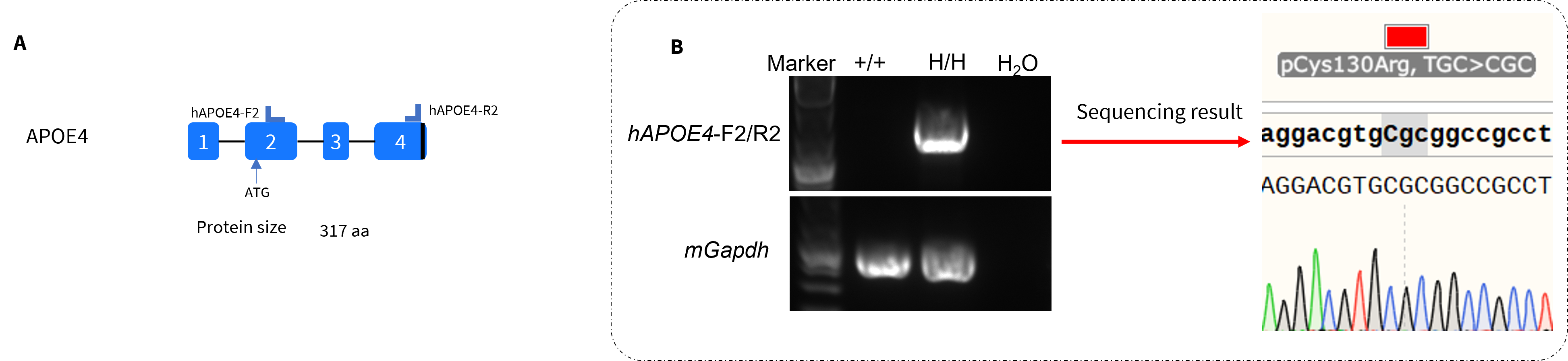
Strain specific analysis of APOE4 mRNA expression in wild-type mice and homozygous B-hAPOE4 mice by RT-PCR and sequencing. Liver RNA was isolated from wild-type C57BL/6JNifdc mice (+/+) and homozygous B-hAPOE4 mice (H/H). (A) Primers were designed to detect human APOE4 expression in the liver. (B) Human APOE4 mRNA was detectable in B-hAPOE4 mice, but not in wild-type mice. Sequencing of the PCR products confirmed that Arg at 130 position was successfully mutated in B-hAPOE4 mice.
APOE4 Protein Expression Analysis in Serum
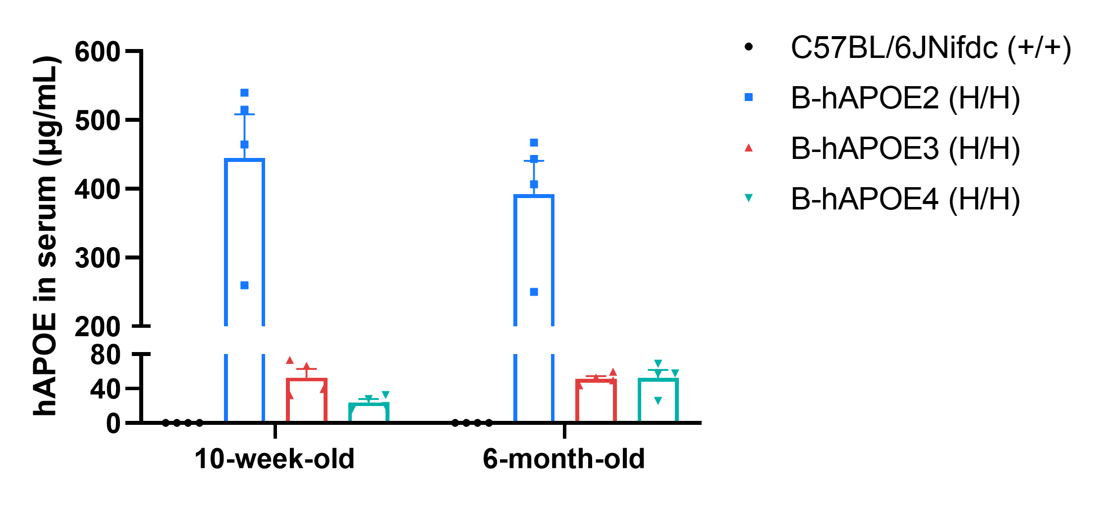
- Human APOE4 was detected in serum from homozygous B-hAPOE4 mice, but not from wild-type mice.
Strain specific analysis of human APOE expression in homozygous B-hAPOE2, B-hAPOE3, and B-hAPOE4 mice by ELISA. Serum was collected from wild-type C57BL/6JNifdc mice (+/+), homozygous B-hAPOE2 mice (H/H), B-hAPOE3 mice (H/H) and B-hAPOE4 mice (H/H) at 10-week-old and 6-month-old (n=4 each group, Female), and serum were then analyzed by ELISA with human-specific APOE ELISA kit (Abcam, ab233623). No human APOE was detected in wild-type mice. Among the knock-in models, B-hAPOE2 mice exhibit the highest serum APOE levels at both time points, whereas APOE expression in B-hAPOE3 and B-hAPOE4 mice was comparable at 6 months of age.
APOE4 Protein Expression Analysis in Brain
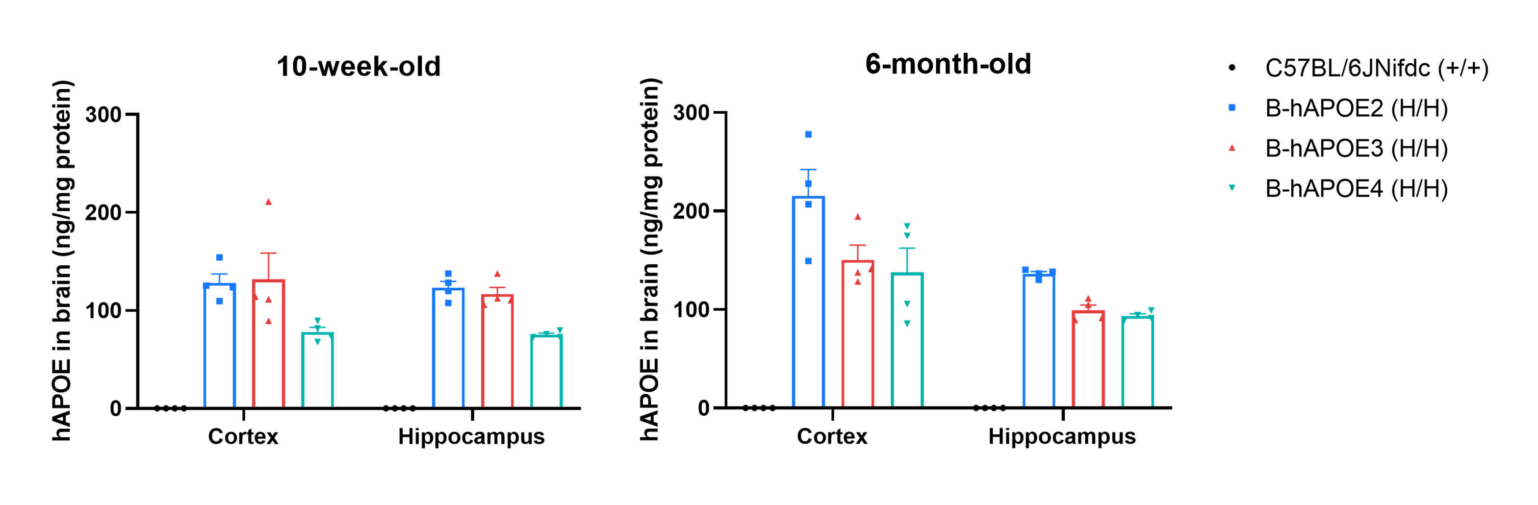
- Human APOE4 was detected in brain from homozygous B-hAPOE4 mice, but not from wild-type mice.
Strain specific analysis of human APOE expression in homozygous B-hAPOE2, B-hAPOE3, and B-hAPOE4 mice by ELISA. Cortex and hippocampus were collected from wild-type C57BL/6JNifdc mice (+/+), homozygous B-hAPOE2 mice (H/H), B-hAPOE3 mice (H/H) and B-hAPOE4 mice (H/H) at 10-week-old and 6-month-old (n=4 each group, Female), and samples were then analyzed by ELISA with human-specific APOE ELISA kit (Abcam, ab233623). No human APOE was detected in wild-type mice.
The Inhibitory Efficiency of the Nucleic Acid Drugs Against Human APOE4
- The human APOE4 levels in the treatment group were reduced compared to the control group, demonstrating that B-hAPOE4 mice provide a powerful preclinical model for in vivo evaluation of human APOE4-targeted nucleic acid drugs.
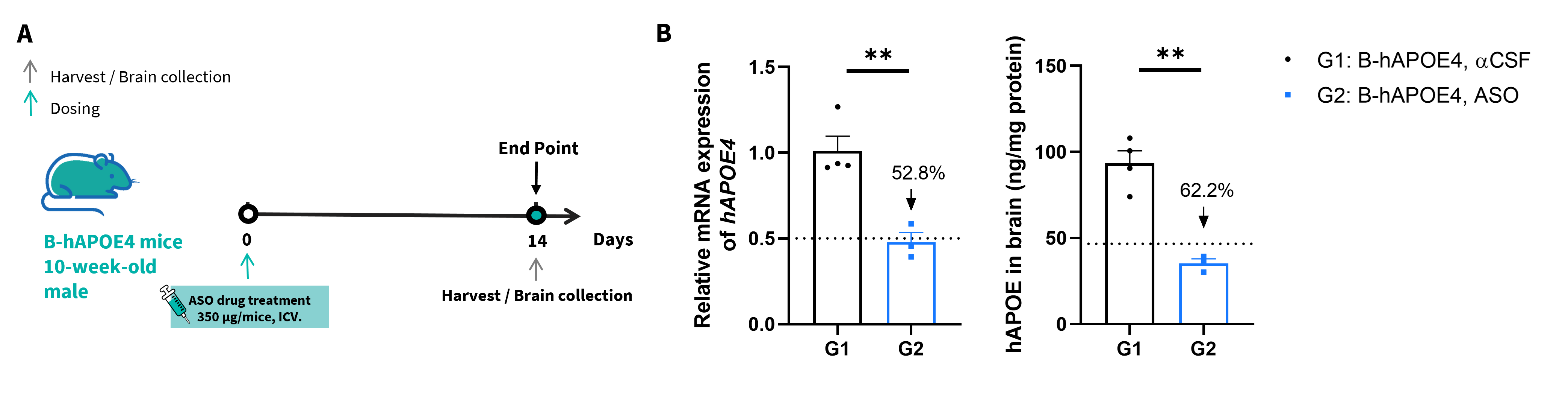
The inhibitory efficiency of the APOE4-targeted small nucleic acid drug in homozygous B-hAPOE4 mice. B-hAPOE4 mice were randomly divided into 2 groups (n=3, 10-week-old, male). The human APOE4-targeted nucleic acid drug (from literature, doi: 10.1016/j.neuron.2017.11.014) and αCSF (artificial cerebrospinal fluid) were administered to the mice individually. The mice were sacrificed on day 14, and the brains (cortex) were collected to detect the human APOE4 expression by qRT-PCR and ELISA. (A) The schematic diagram of experimental processing. (B) The expression of human APOE4 mRNA and APOE4 protein in cortex. The human APOE4 mRNA and APOE protein in the treatment group were significantly reduced compared to the control group. Values are expressed as mean ± SEM. Significance was determined by unpaired t-test. *P < 0.05, **P < 0.01, ***P < 0.001.
* When publishing results obtained using this animal model, please acknowledge the source as follows: The animal model [B-hAPOE4 mice] (Cat# 112564) was purchased from Biocytogen.