Premature Aging & Progeria Mouse Models Introduction
The global population is aging at an unprecedented pace, increasing the burden of chronic diseases, including cancer, cardiovascular disorders, neurodegenerative diseases, autoimmune diseases, and metabolic disorders, all of which are closely associated with the aging process.
Aging is no longer viewed simply as an irreversible natural process, but is increasingly recognized as a decipherable and therapeutically actionable biological process. Key mechanisms, including cellular senescence, DNA damage response, inflammaging, mTOR signaling, and immunosenescence, have been systematically elucidated, positioning aging biology as a new frontier for innovative drug discovery.
Biocytogen has developed three genetically engineered premature aging and progeria mouse models: B-Klotho KO, B-Zmpste24 KO, and B-hLMNA*G608G. These models rapidly recapitulate hallmark aging-associated phenotypes, including osteoporosis, skin and adipose tissue atrophy, skeletal abnormalities, bone microarchitecture deterioration, and progeria-related pathological features, providing efficient in vivo tools for target validation, pathway investigation, and therapeutic efficacy evaluation.
Results
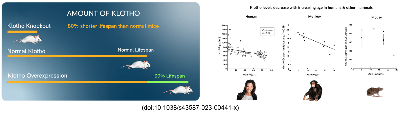
B-Klotho KO Mouse
Gene Targeting Strategy & Validation
- Gene Targeting Strategy of B-Klotho KO Mice
Gene targeting strategy for Klotho knockout mouse generation. The exons 2~5 of mouse Klotho gene were knocked out in B-Klotho KO mice.
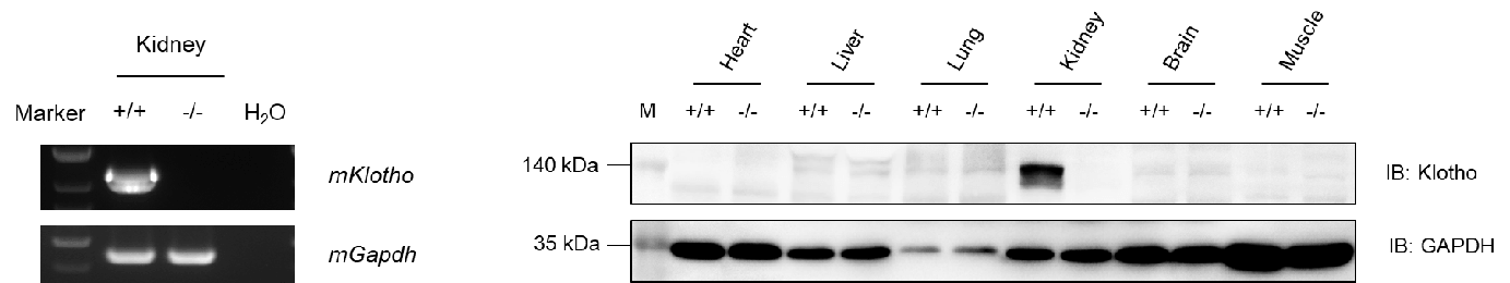
- Validation of mKlotho Expression in B-Klotho KO Mice
mKlotho was only detectable in kidney in wild-type C57BL/6 mice but not in homozygous B-Klotho mice.
Micro-CT Bone Microstructure Analysis
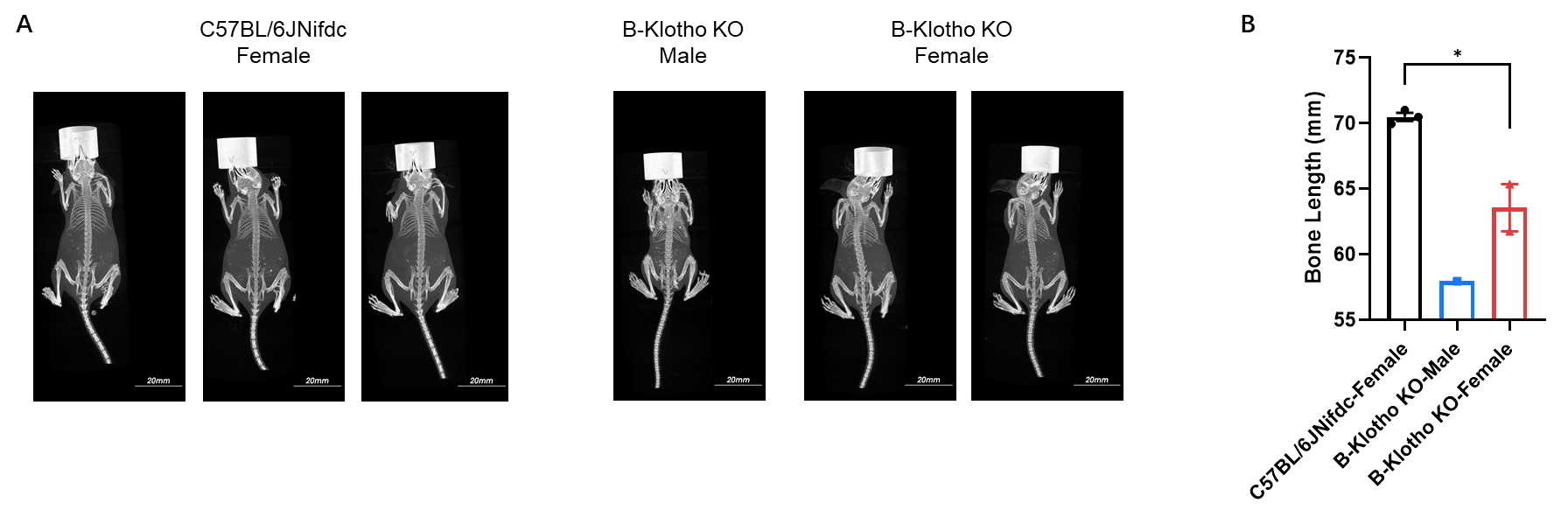
- Whole-body and bone length Micro-CT imaging
Whole-body micro-CT imaging of homozygous B-Klotho KO mice. Body size of homozygous B-Klotho KO mice was smaller than wild-type C57BL/6JNifdc mice. Data are presented as mean ± SEM. *P < 0.05, **P < 0.01, ***P < 0.001. Scale bar = 20 mm.
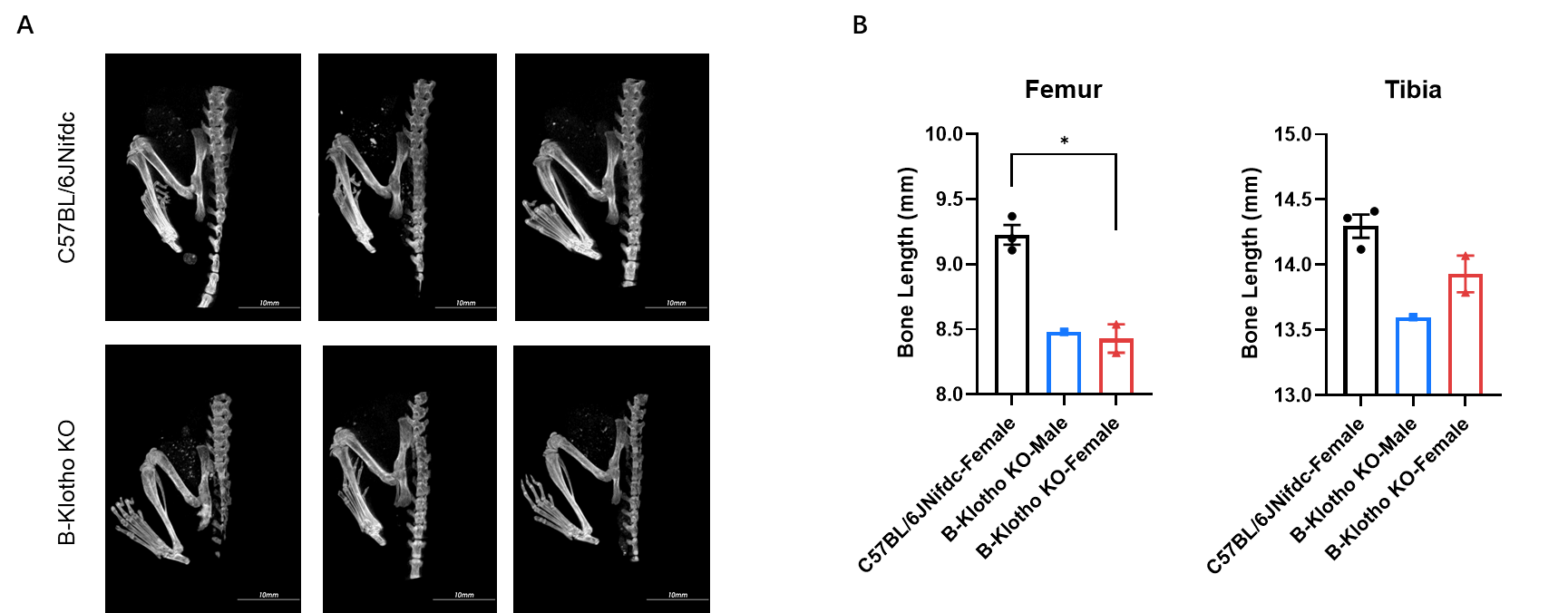
Micro-CT analysis of femur and tibia length in homozygous B-Klotho KO mice. In the homozygous B-Klotho KO mice, the lengths of the femur and tibia were shortened. Data are presented as mean ± SEM. *P < 0.05, **P < 0.01, ***P < 0.001. Scale bar = 10 mm.
Histopathological Analysis
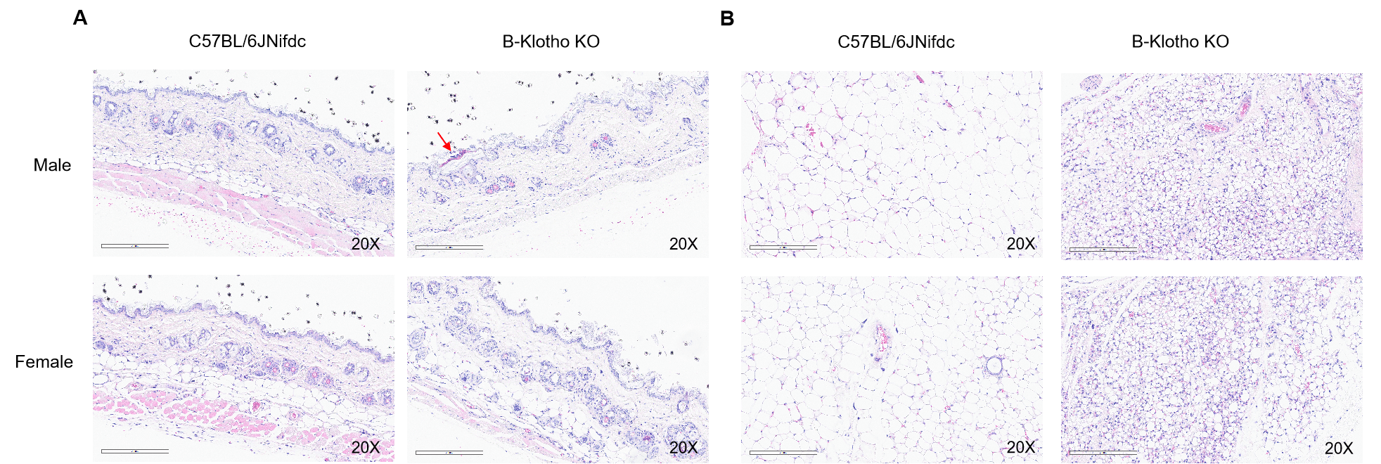
- Histopathological analysis of adipocyte atrophy in B-Klotho KO mice
Histopathological analysis of dorsal skin and subcutaneous adipose tissue in homozygous B-Klotho KO mice. (A) H&E-stained sections showed skin pathological changes with punctate crusts, hyperkeratosis and subcutaneous fat layer was not visible in homozygous B-Klotho KO mice (-/-, 7-week-old, 1 male and 1 female) compared with wild-type C57BL/6JNifdc mice (+/+, 7-week-old, 1 male and 1 female). (B) H&E-stained subcutaneous adipose tissue showed adipocyte atrophy in homozygous B-Klotho KO mice compared with wild-type C57BL/6JNifdc. Scale bars: 200 µm.
B-Zmpste24 KO mice
Gene Targeting Strategy & Validation
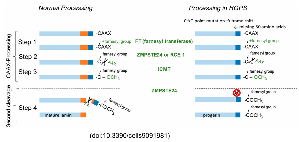
- Gene targeting strategy of B-Zmpste24 KO mice
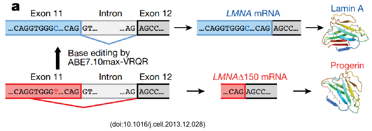
Zmpste24 is a zinc metalloproteinase and Lamin A is processed by Zmpste24. Failure to cleave a truncated form of prelamin A—also called progerin—causes Hutchinson–Gilford progeria syndrome a well-known premature aging disease.
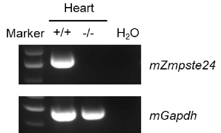
Strain specific analysis of Zmpste24 mRNA expression in wild-type C57BL/6JNifdc mice and B-Zmpste24 KO mice by RT-PCR. Heart RNA was isolated from wild-type C57BL/6JNifdc mice (+/+) and homozygous B-Zmpste24 KO mice (-/-). Mouse Zmpste24 mRNA was only detectable in wild-type C57BL/6JNifdc mice, but not in B-Zmpste24 KO mice.
Micro-CT Bone Microstructure Analysis
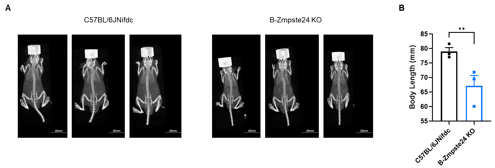
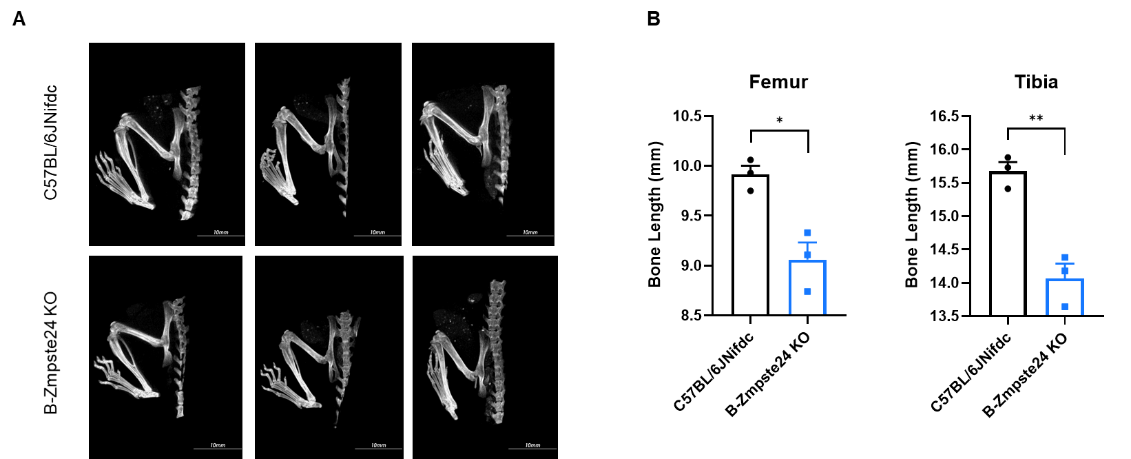
- Whole-body and bone Micro-CT imaging
Whole-body micro-CT imaging of homozygous B-Zmpste24 KO mice. Body size of homozygous B-Zmpste24 KO mice was smaller than wild-type C57BL/6JNifdc mice. Data are presented as mean ± SEM. *P < 0.05, **P < 0.01, ***P < 0.001. Scale bar = 20 mm.
Micro-CT analysis of femur and tibia length in homozygous B-Zmpste24 KO mice. In the homozygous B-Zmpste24 KO mice, the lengths of the femur and tibia were shortened. Data are presented as mean ± SEM. *P < 0.05, **P < 0.01, ***P < 0.001. Scale bar = 10 mm.
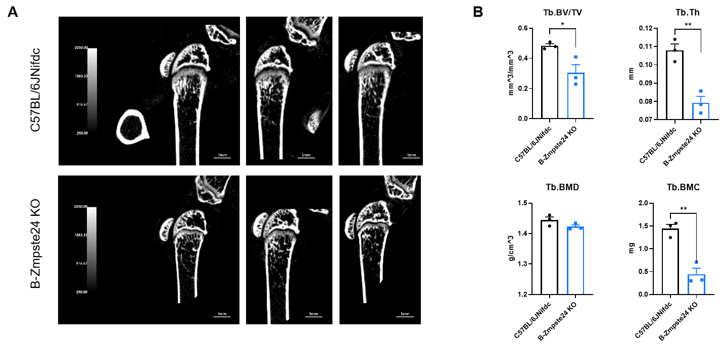
Bone Microarchitecture Analysis — Osteoporosis
- Micro-CT analysis of bone microarchitecture in B-Zmpste24 KO mice
Micro-CT analysis of bone microarchitecture in homozygous B-Zmpste24 KO mice. (A) Representative micro-CT images of trabecular bone from wild-type C57BL/6JNifdc mice (n=3, male, 6-week-old) and homozygous B-Zmpste24 KO mice (n=3, male, 6-week-old). (B) Quantitative analysis of bone volume fraction (Tb.BV/TV), trabecular thickness (Tb.Th), trabecular bone mineral density (Tb.BMD) and trabecular bone mineral content (Tb.BMC). Tb.BV/TV, Tb.Th, Tb.BMC decreased in homozygous B-Zmpste24 KO mice compared with wild-type mice, indicating a deterioration or loss of trabecular bone structure, which is consistent with characteristics of osteoporosis. Data are presented as mean ± SEM. *P < 0.05, **P < 0.01, ***P < 0.001.
B-hLMNA*G608G mice
Gene Targeting Strategy & Validation
- Gene Targeting Strategy of B-hLMNA*G608G Mouse
The exons 1-12 of mouse Lmna gene that encode whole protein domains are replaced by human counterparts containing p.G608G, c.C1824T mutation in B-hLMNA*G608G mice. The promoter, 5’UTR and 3’UTR region of the mouse gene are retained.
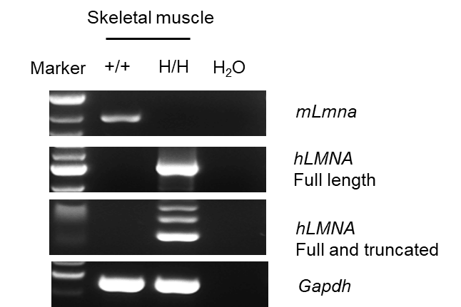
- Validation of LMNA*G608G Expression in B-hLMNA*G608G KO Mice
Strain specific analysis of LMNA mRNA expression in wild-type C57BL/6JNifdc mice and homozygous B-hLMNA*G608G mice by RT-PCR. Skeletalmuscle RNA were isolated from wild-type C57BL/6JNifdc mice (+/+) and homozygous B-hLMNA*G608G mice (H/H) (n=1, 7-week-old, male), then cDNAlibraries were synthesized by reverse transcription, followed by PCR with LMNA primers. Mouse Lmna was only detected in wild-type C57BL/6JNifdc mice,and human LMNA was exclusively detectable in homozygous B-hLMNA*G608G mice. Truncated length of hLMNA (150 bp deletion near the C-terminusbecause of the p.G608G, c.C1824T mutation) was detected in homozygous B-hLMNA*G608G mice, and sequence was confirmed by Sanger sequencing.
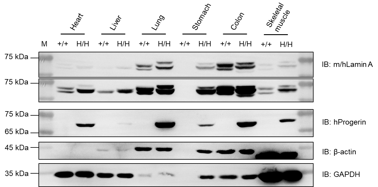
Western blot analysis of Lamin A and progerin protein expression in wild-type C57BL/6JNifdc mice and homozygous B-hLMNAG608G mice. Various tissues, including heart, liver, lung, colon, stomach and skeletal muscle, were collected from wild-type C57BL/6JNifdc mice (+/+) and homozygous B-hLMNAG608G mice (H/H). A total of 30 μg protein was loaded for western blot analysis, with GAPDH used as an internal control. Lamin A was detected using an anti-Lamin antibody (CST, 2032T), which cross-reacts with both mouse and human Lamin A; the upper band represents full-length Lamin A and the lower band represents progerin. Human progerin was further detected using a species-specific anti-human progerin antibody (Abcam, ab66587), and was exclusively observed in tissues from homozygous B-hLMNA*G608G mice. M, marker.
Bone Microarchitecture Analysis
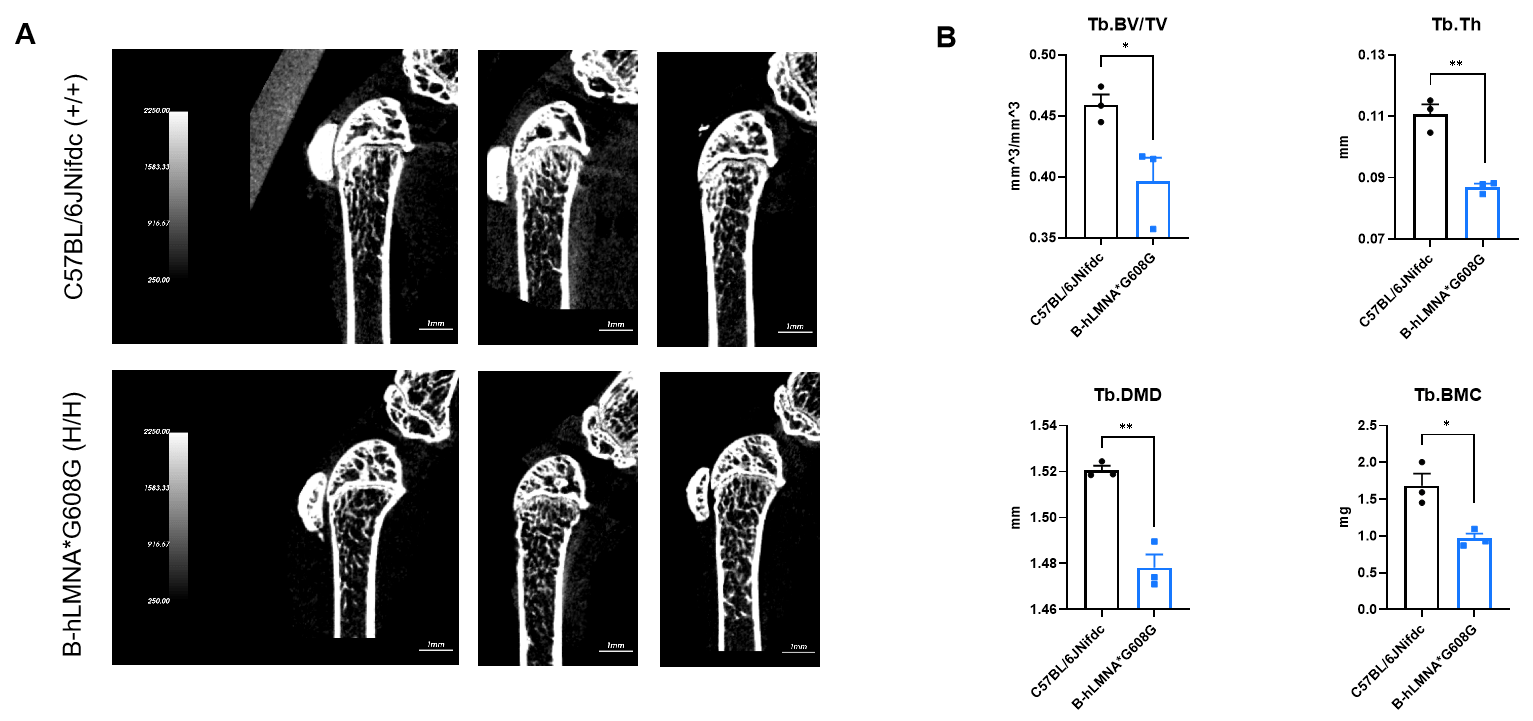
- Micro-CT analysis of bone microarchitecture in B-hLMNA*G608G mice
Micro-CT analysis of bone microarchitecture in homozygous B-hLMNA*G608G mice. (A) Representative micro-CT images of trabecular bone from wild-type C57BL/6JNifdc mice (n=3, male, 11-week-old) and homozygous B-hLMNA*G608G mice (n=3, male, 11-week-old). (B) Quantitative analysis of bone volume fraction (Tb.BV/TV), trabecular thickness (Tb.Th), trabecular bone mineral density (Tb.BMD) and trabecular bone mineral content (Tb.BMC). Tb.BV/TV, Tb.Th, Tb.BMC decreased in homozygous B-hLMNA*G608G mice compared with wild-type mice, indicating a deterioration or loss of trabecular bone structure, which is consistent with characteristics of osteoporosis. Data are presented as mean ± SEM. *P < 0.05, **P < 0.01, ***P < 0.001.