FR2-Camelized RenNano® Mice
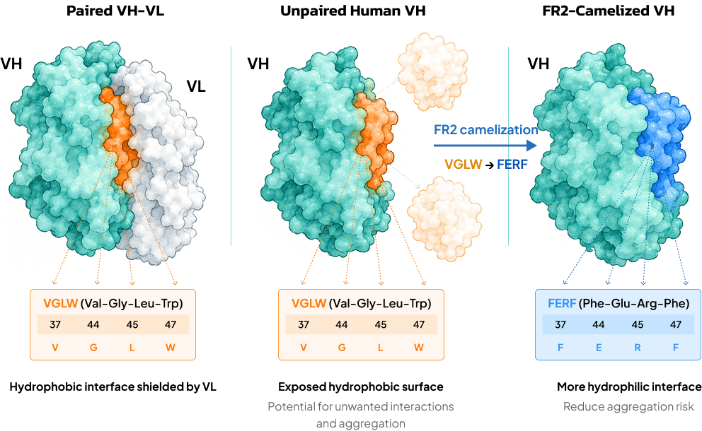
In human heavy chains, FR2 residues at positions 37, 44, 45, and 47 (Martin numbering) form part of the VH-VL interface. These residues are typically hydrophobic and are represented by the VGLW (Val-Gly-Leu-Trp) motif, which is normally buried through pairing with the light chain (Sang et al., 2022, Structure). In a heavy-chain-only or VHH format, however, these positions become more solvent-exposed. The exposed hydrophobic surface can increase unwanted interactions and aggregation, potentially reducing soluble expression and complicating candidate development.
FR2 camelization replaces the human VGLW motif with the camelid-associated FERF (Phe-Glu-Arg-Phe) motif, making the exposed interface more hydrophilic to reduce aggregation risk and improve candidate developability.
Comparative studies have shown that humanized libraries incorporating FERF can achieve higher screening success rates for viable lead candidates than libraries based on human VGLW (Arras et al., 2023, mAbs). Our in-house data further show that clinical-stage VHHs retaining camelid FR2 signatures exhibit higher expression yields and greater thermostability than those carrying the human VGLW motif.
Combining Fully Human and FR2-Camelized Human V Genes to Maximize HCAb Discovery Potential
Building on our proprietary RenNano® mouse platform for human heavy-chain-only antibody (HCAb) discovery, next-generation RenNano® mice retain the original, fully intact human V gene repertoire while incorporating dozens of engineered framework region 2 (FR2)-camelized human V gene segments.
This expanded repertoire increases sequence and framework diversity and is designed to improve discovery success and broaden epitope coverage.
Strategic Advantages of FR2-Camelized RenNano® Mice
The more hydrophilic FERF framework helps reduce unwanted hydrophobic interactions and aggregation risk, supporting improved solubility, lower nonspecific binding, better overall developability, and higher lead identification success.
Combines intact human V genes (VGLW) with engineered FR2-camelized human V genes (FERF), expanding candidate diversity across hard-to-target epitopes.
Retains the original human V gene repertoire, allowing discovery campaigns to continue generating fully human VHHs when unmodified human sequences are preferred.
Enables natural in vivo affinity maturation to generate high-affinity VHH leads, streamlining downstream optimization.
Immune Phenotype and Antibody Repertoire
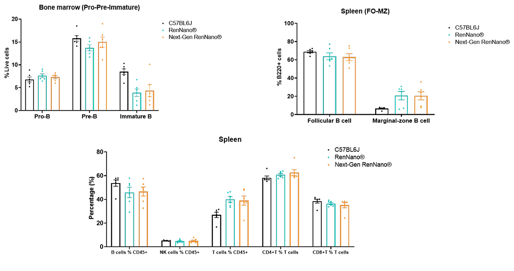
Preserved B-Cell Development and Immune Cell Profiles
FR2-camelized RenNano® mice showed B-cell development and splenic B-, T-, and NK-cell proportions comparable to those of the fully human RenNano® mice, supporting an expanded V gene repertoire while maintaining similar immune cell profiles.
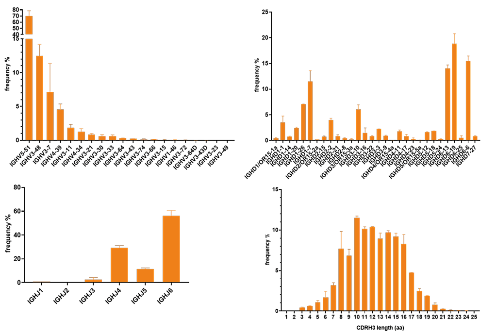
Diverse V(D)J Rearrangements and Broad CDR3 Length Distribution
NGS analysis of peripheral blood from unimmunized FR2-camelized RenNano® mice showed that nearly all FR2-camelized V gene segments participated in V(D)J recombination. D and J gene usage and CDR3 length distributions were similar to those observed in the fully human RenNano® mice.
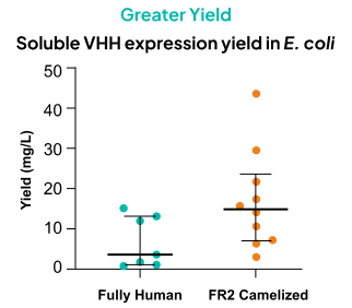
FR2 Camelization Improves Soluble VHH Expression
Higher Success Rate
Proportion of VHHs successfully expressed in soluble form in E. coli
FR2 camelization significantly improves soluble VHH expression in E. coli, achieving a 100% expression success rate and markedly higher expression yields.
VHHs derived from RenNano® mice were expressed in E. coli and purified in-house. Data are presented as the median and interquartile range.
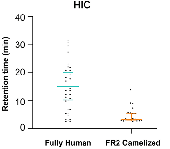
FR2-Camelized Human HCAbs Show Significantly Enhanced Hydrophilicity
Hydrophobic interaction chromatography (HIC) analysis demonstrates that FR2-camelized HCAbs exhibit significantly shorter retention times, indicating markedly enhanced surface hydrophilicity and a more favorable solubility profile. RenNano®-derived HCAbs were expressed in CHO cells and purified in-house. Data are presented as the median and interquartile range.
FR2-Camelized VHHs Exhibit Markedly Improved Monomer Purity
Percentage of VHHs Meeting Monomer Purity Thresholds
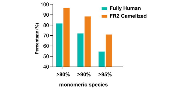
FR2-camelized VHHs demonstrate improved monomer purity, with a significantly higher proportion of clones meeting stringent purity thresholds (>80%, >90%, and >95%). RenNano®-derived VHHs were expressed in CHO cells or yeast and purified in-house. All 11 fully human VHHs were expressed in yeast; among the 60 FR2-camelized VHHs, 10 were expressed in yeast and 50 in CHO cells.
Case Studies: Expanding VHH Discovery with FR2 Camelization
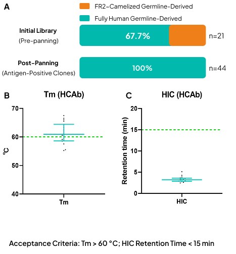
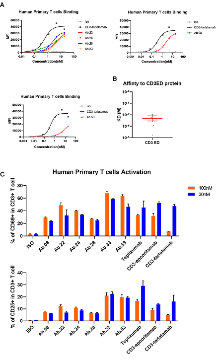
FR2-camelized RenNano® mice retain the intact fully human V gene repertoire while incorporating engineered FR2-camelized human V gene segments. Screening enriched FR2-camelized binders for CD28 and fully human binders for CD3, highlighting how the combined repertoires support candidate discovery across different targets.
Both case studies used FR2-camelized RenNano® mice for immunization.
Case Study 1 | CD28
CD28: Enrichment of FR2-Camelized Binders
FR2 Camelization Confers an Advantages in CD28 HCAb Discovery
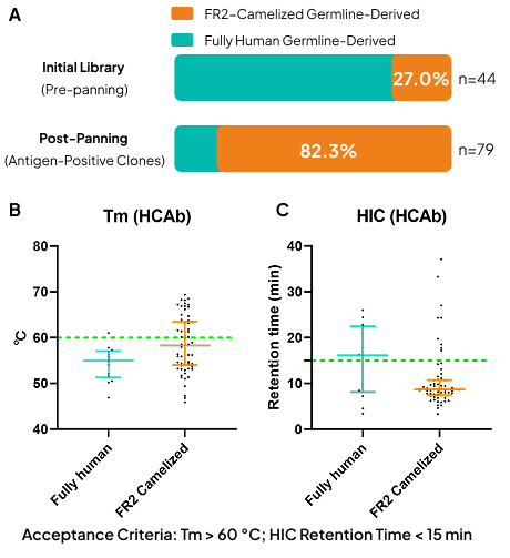
Phage display screening of libraries derived from FR2-camelized RenNano® mice revealed strong enrichment of FR2-camelized sequences among CD28 binders (genetic ratio: 1:1).
FR2-camelized sequences increased from 27.0% in the initial library to 82.3% among antigen-positive clones after panning, despite an initial 1:1 germline ratio. FR2-camelized binders showed generally higher thermal stability and favorable HIC profiles, supporting the value of FR2 camelization in obtaining stable, binding-competent VHHs.
Fully Human RenNano® Mice
Key Features of RenNano® Mice
1. Fully human heavy chain variable region and modified constant region
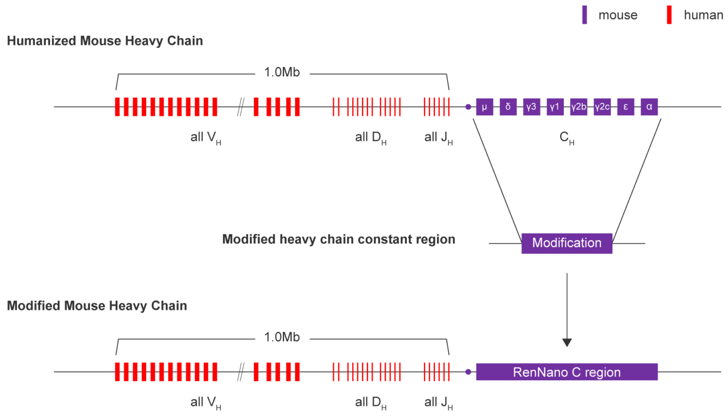
2. RenNano mice demonstrate robust immune responses against multiple antigens
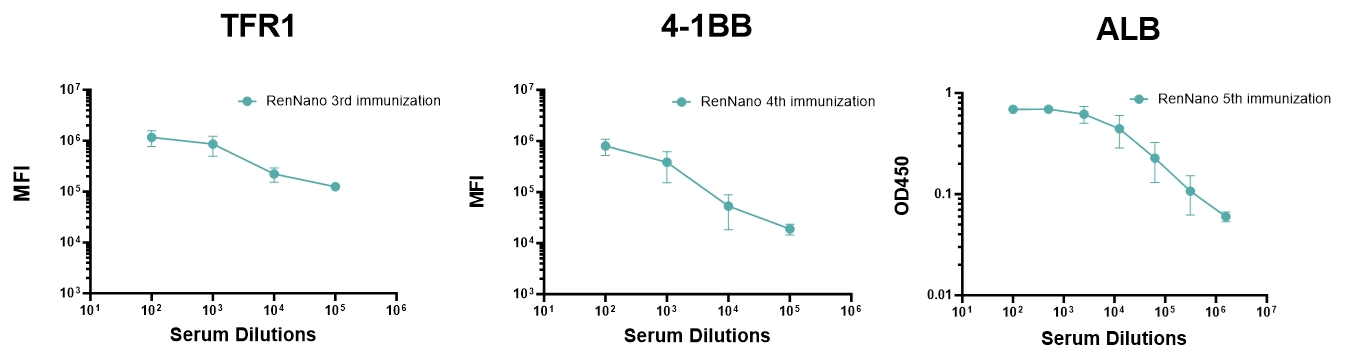
RenNano mice' immune responses against TFR1, 4-1BB, and ALB. Sera from RenNano mice immunized with TFR-1 and 4-1BB (membrane antigens) were diluted and incubated with antigen-expressing CHO cells. Fluorochrome-conjugated secondary antibodies were used to label CHO-bound HCAbs, and mean fluorescence intensity (MFI) was measured using flow cytometry to indicate the antigen-specific HCAb titer. Sera from RenNano mice immunized with ALB (secreted antigen) were diluted and incubated with fluorochrome-conjugated secondary antibodies. OD450 was measured using ELISA to indicate the antigen-specific HCAb titer.
3. RenNano-derived antigen-specific HCAbs showed great diversity
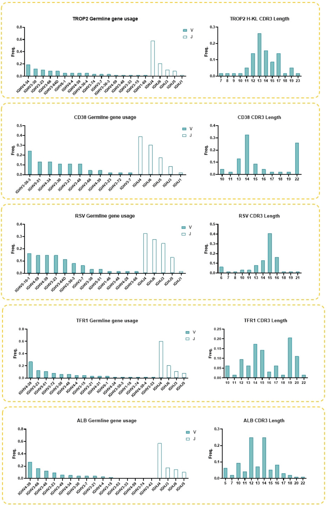
HCAbs derived from RenNano mice are diverse and many contain longer CDR3 regions. Antigen-specific HCAbs were analyzed by their heavy chain germline gene usage and CDR3 length. Analysis indicates broad IGHV germline diversity. Additionally, most CDR3 lengths observed were longer than 12 AA, with some longer than 17 AA.
Validation Data of RenNano® Mice
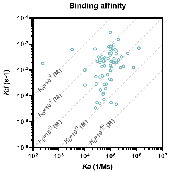
The affinity of 68 purified RenNano HCAbs for 8 targets (4-1BB, TROP2, IL3RA, OX40, Nectin-4, ROR1, PSMA and DLL3) were tested by SPR. For most HCAbs, KD were between 10-6 ~ 10-10 M.
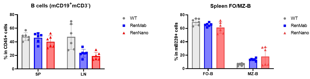
B cell development and differentiation patterns in RenNano mice. RenNano mice have comparable B cell proportions in peripheral lymphoid organs (spleen, SP and lymph nodes, LN) to WT mice or RenMab mice. Spleens from RenNano mice also have comparable percentages of follicular B (FO-B) cells and marginal zone B (MZ-B) cells to WT mice or RenMab mice.
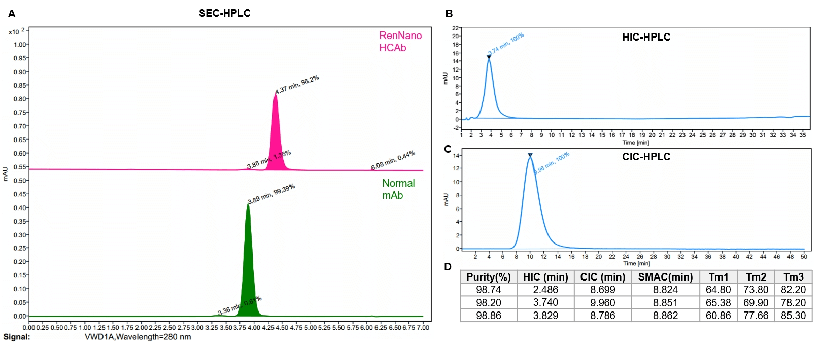
RenNano HCAbs' physiochemical property analysis. RenNano HCAbs show high purity after one-step purification (A). HCAbs exhibit short retention time in HIC-HPLC and CIC-HPLC, indicating superb hydrophilicity and specificity (B, C). RenNano HCAbs also exhibit favorable thermostability (D).