Description
- CD3 is composed of four protein chains, including CD3E, CD3D, CD3G, and CD3Z, which serve as critical components of the TCR/CD3 complex on T cells. The CD3 complex plays essential roles in T cell antigen recognition, signal transduction, immune activation, and T cell development.
- Cadherin 17 (CDH17) is a member of the 7D-cadherin superfamily characterized by seven cadherin repeats and a short cytoplasmic domain. CDH17 is primarily expressed in the gastrointestinal tract, especially in the small intestine and colon, where it contributes to epithelial integrity, intercellular adhesion, peptide transport, and calcium-dependent water absorption.
- In CD3EDG/CDH17 Humanized Mice, chimeric human CD3EDG genes were engineered while endogenous mouse Cd3edg genes are disrupted. In addition, the extracellular domain of mouse Cdh17 was replaced with the corresponding human CDH17 extracellular region, enabling evaluation of human-specific therapeutics targeting CDH17.
- Human CD3E was detected on T cells of homozygous CD3EDG/CDH17 Humanized Mice, while human CDH17 expression was confirmed in colon and small intestine tissues.
- This model is suitable for preclinical pharmacodynamic and toxicity studies of bispecific antibody-drug conjugates (BsADCs), T cell engagers, and other immunotherapies targeting CDH17-positive solid tumors, including colorectal cancer and gastrointestinal cancers.
Key Advantages
- Dual humanized CD3EDG and CDH17 platform
- Suitable for T cell engager and BsADC evaluation
- Supports gastrointestinal cancer immunotherapy studies
- Humanized CD3 signaling for translational immune activation studies
- Humanized CDH17 expression in gastrointestinal tissues
- Applicable for efficacy and toxicity assessment of human-specific therapeutics
Validation
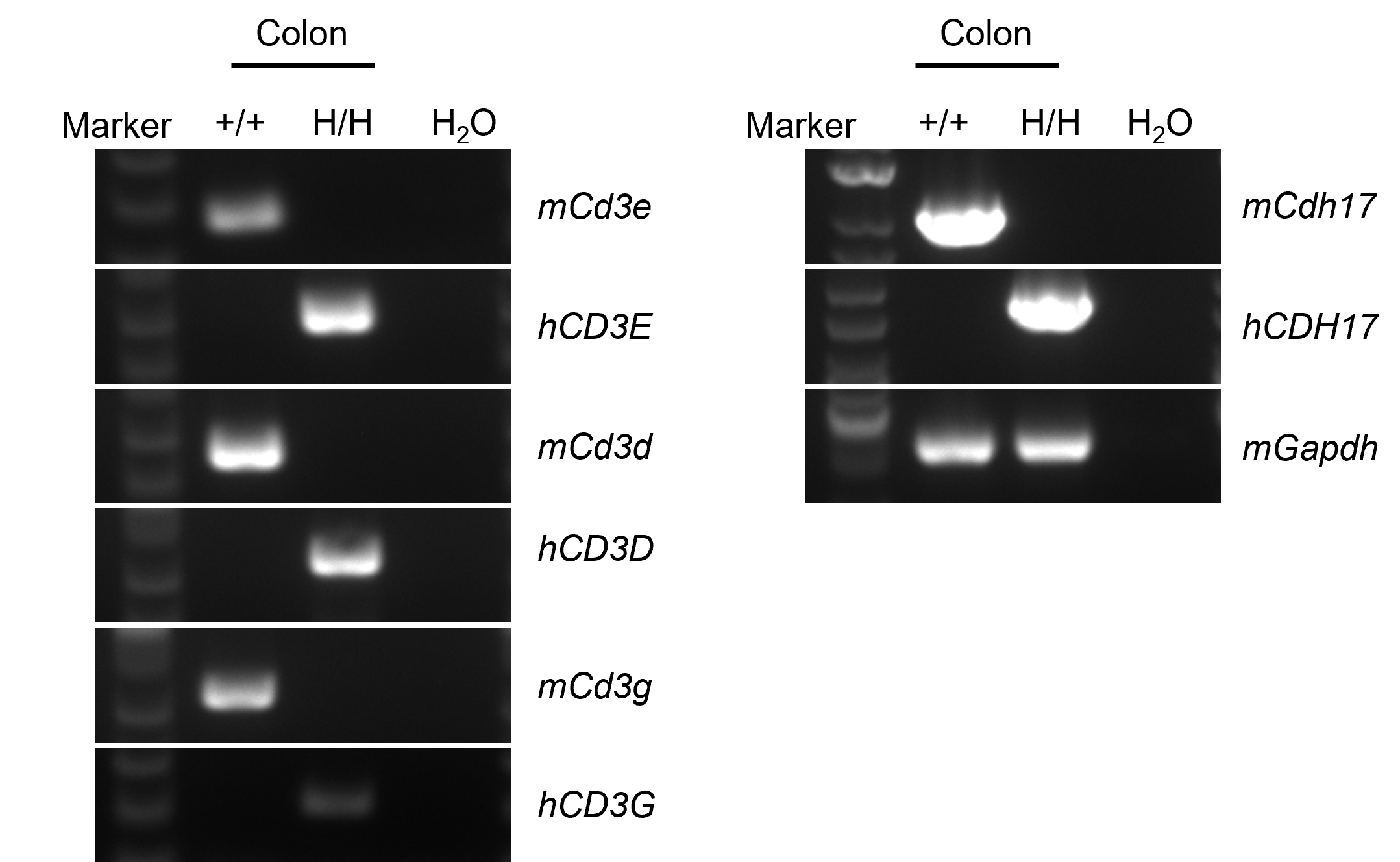
- Molecular Validation: Mouse Cd3e, Cd3d, Cd3g, and Cdh17 mRNA were detectable only in wild-type C57BL/6JNifdc mice, whereas human CD3E, CD3D, CD3G, and CDH17 mRNA were detectable in homozygous CD3EDG/CDH17 Humanized Mice by RT-PCR.
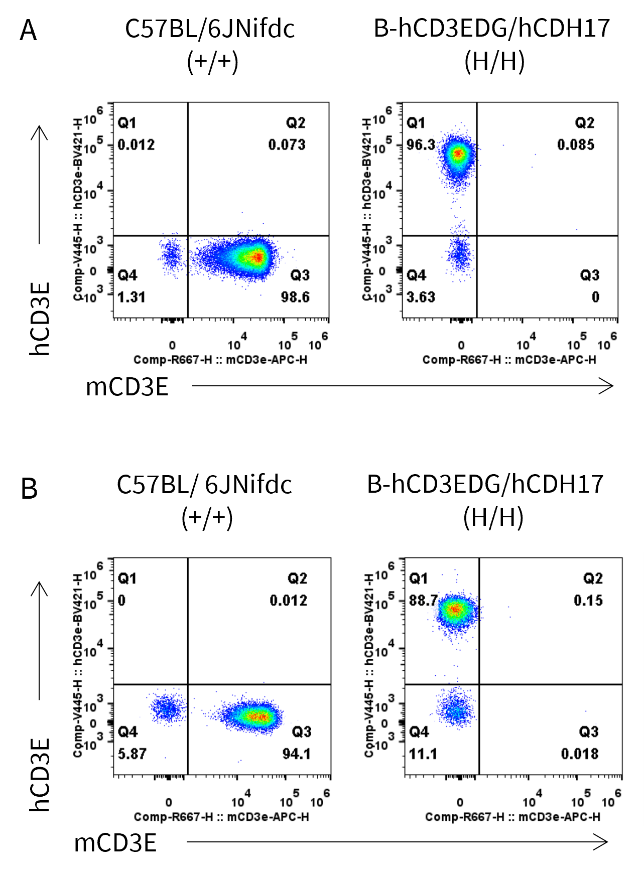
- Protein Validation: Human CD3E protein was detectable on splenic and peripheral blood T cells of homozygous CD3EDG/CDH17 Humanized Mice by flow cytometry.
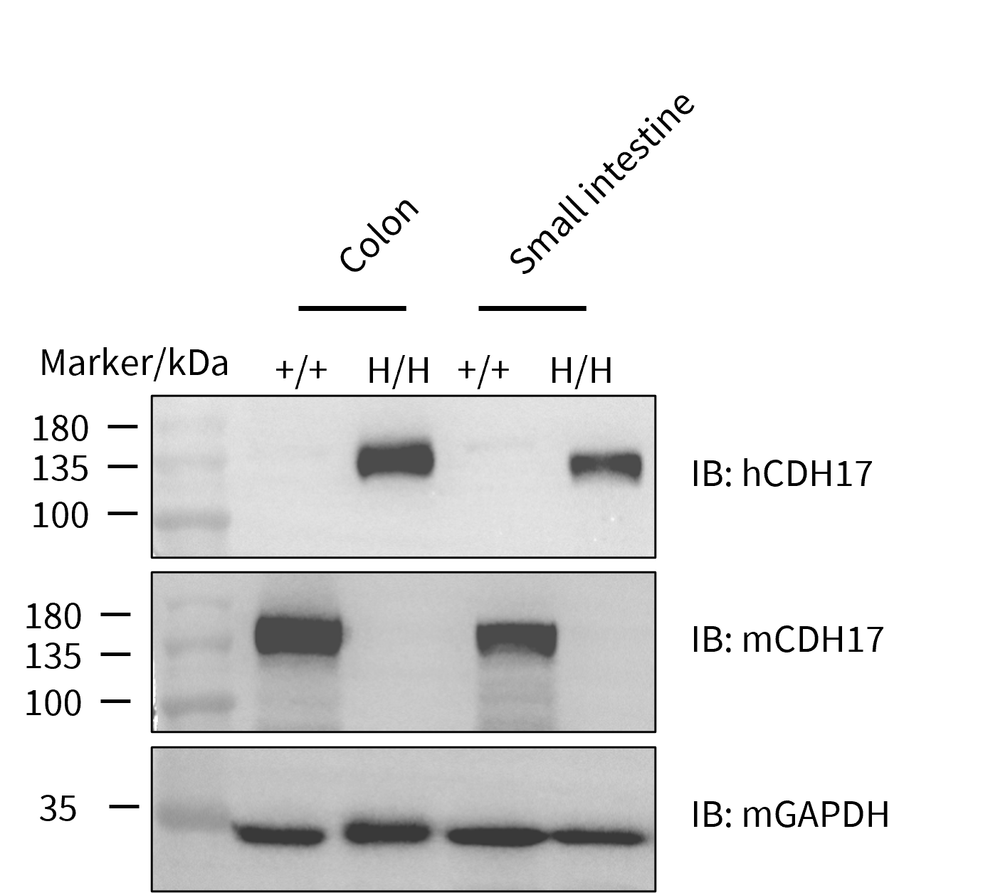
- Tissue Validation: Humanized CDH17 protein expression was confirmed in colon and small intestine tissues by western blot analysis.
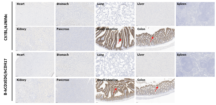
- Histopathological Validation: IHC analysis demonstrated CDH17 expression patterns in gastrointestinal tissues of CD3EDG/CDH17 Humanized Mice.
Application
- Bispecific antibody-drug conjugate evaluation
- T cell engager efficacy studies
- Colorectal cancer research
- Gastrointestinal cancer immunotherapy
- CDH17-targeted therapeutic development
- Preclinical toxicity and pharmacodynamic studies
Targeting Strategy
CD3EDG
- The chimeric human CD3EDG was expressed, while mouse Cd3edg were knocked out in B-hCD3EDG/hCDH17 mice.
CDH17
- The extracellular region (except signal peptide) of mouse Cdh17 was replaced by human CDH17 extracellular region sequences in B-hCD3EDG/hCDH17 mice.
mRNA Expression Analysis
- Mouse Cd3e, Cd3d, Cd3g and Cdh17 mRNA were only detectable in wild-type mice, but not in homozygous B-hCD3EDG/hCDH17 mice.
- Human CD3E, CD3D, CD3G and CDH17 mRNA were exclusively detectable in homozygous B-hCD3EDG/hCDH17 mice.
Strain specific analysis of CD3EDG/CDH17 mRNA expression in wild-type C57BL/6JNifdc mice and B-hCDH17 mice by RT-PCR. Colon RNA was isolated from C57BL/6JNifdc mice (+/+) and homozygous B-hCD3EDG/hCDH17 mice (H/H), and then cDNA libraries were synthesized by reverse transcription, followed by PCR with mouse or human CD3E, CD3D, CD3G and CDH17 primers.
CD3E Protein Expression Analysis
- Mouse CD3E was exclusively detectable on the T cells of wild-type mice, but not on homozygous B-hCD3EDG/hCDH17 mice. Human CD3E was exclusively detectable on the T cells of homozygous B-hCD3EDG/hCDH17 mice.
Strain specific CD3E expression analysis in wild-type mice and homozygous B-hCD3EDG/hCDH17 mice by flow cytometry. Splenocytes (A) and blood (B) were collected from wild-type C57BL/6JNifdc (+/+) mice and homozygous B-hCD3EDG/hCDH17 mice (H/H) and analyzed by flow cytometry with anti-mouse CD3E antibody (Biolegend, 100312) and anti-human CD3E antibody (BD Biosciences, 562426).
CDH17 Protein Expression Analysis
- Mouse CDH17 was detectable in colon and small intestine of wild-type mice, but not in homozygous B-hCD3EDG/hCDH17 mice.
- Human CDH17 was detectable in colon and small intestine of homozygous B-hCD3EDG/hCDH17 mice.
Western blot analysis of CDH17 protein expression in homozygous B-hCD3EDG/hCDH17 mice. Colon and small intestine tissue lysates were collected from wild-type C57BL/6JNifdc mice (+/+) and homozygous B-hCD3EDG/hCDH17 mice (H/H), and then analyzed by western blot with anti-human CDH17(abcam, ab109190) and anti-mouse CDH17 (R&D, AF8524) antibodys. 40 μg total proteins were loaded for western blot analysis.
- CDH17 was detectable in wild-type mice and homozygous B-hCD3EDG/hCDH17 mice due to the cross-reactivity of antibodies.
Immunohistochemical (IHC) analysis of CDH17 expression in wild-type C57BL/6JNifdc mice and homozygous B-hCD3EDG/hCDH17 mice. Various tissues were collected from wild-type C57BL/6JNifdc mice and B-hCD3EDG/hCDH17 mice and analyzed by IHC with anti-CDH17 antibody (abcam, ab109190).
* When publishing results obtained using this animal model, please acknowledge the source as follows: The animal model [B-hCD3EDG/hCDH17 mice] (Cat# 114256) was purchased from Biocytogen.