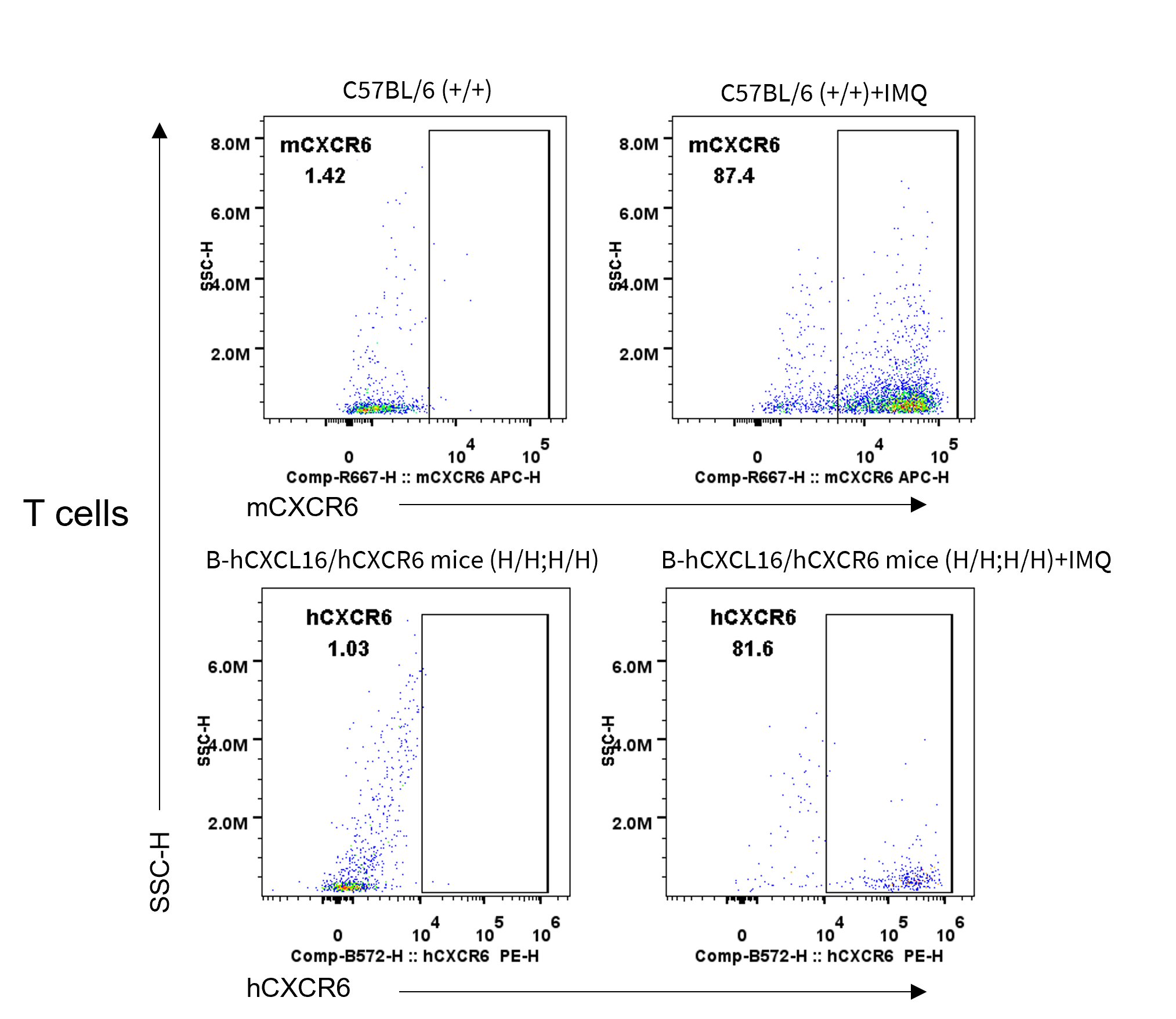
CXCR6 Protein Expression Analysis in Spleen
- Mouse CXCR6 was detected exclusively in wild-type C57BL/6N mice
- Human CXCR6 was detected in homozygous B-hCXCL16/hCXCR6 mice, but not in wild-type mice.
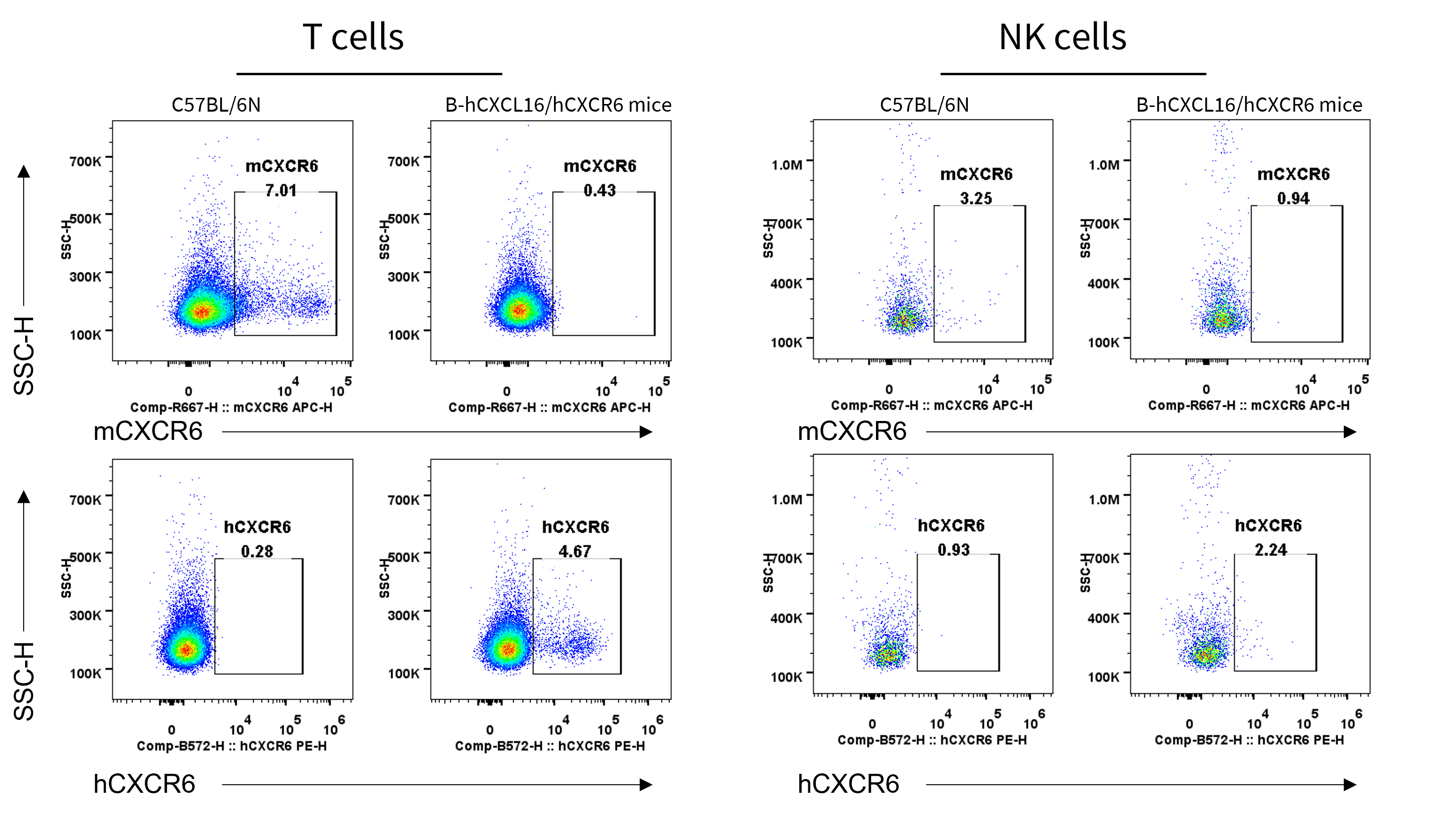
Strain specific CXCR6 expression analysis in homozygous B-hCXCL16/hCXCR6 mice by flow cytometry. Splenocytes were collected from wild-type C57BL/6N mice (+/+) and homozygous B-hCXCL16/hCXCR6 mice (H/H;H/H) and analyzed by flow cytometry with species-specific anti-CXCR6 antibody(Biolegend, 151105; Biolegend, 356003). mCXCR6 was detectable in T cells and NK cells of wild-type mice, hCXCR6 was detectable in T cells and NK cells of homozygous B-hCXCL16/hCXCR6 mice.
CXCR6 Protein Expression Analysis in Liver
- Mouse CXCR6 was detected exclusively in wild-type C57BL/6N mice
- Human CXCR6 was detected in homozygous B-hCXCL16/hCXCR6 mice, but not in wild-type mice.
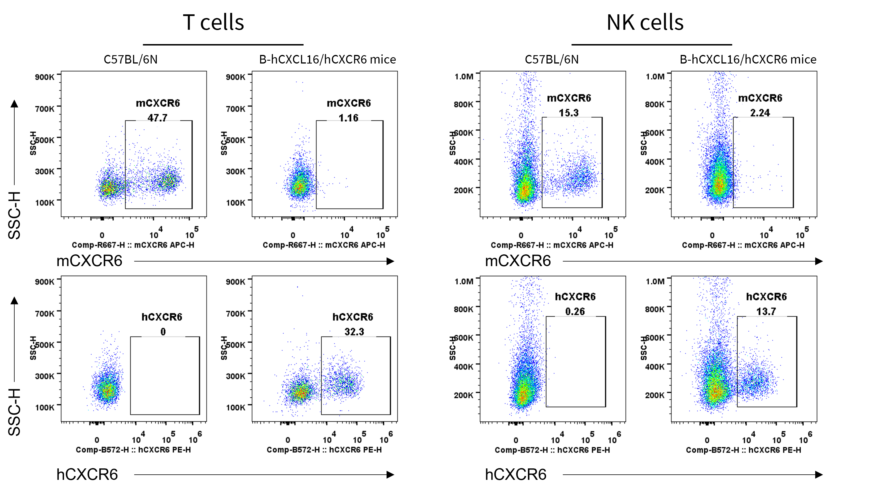
Strain specific CXCR6 expression analysis in homozygous B-hCXCL16/hCXCR6 mice by flow cytometry. Liver cells were collected from wild-type C57BL/6N mice (+/+) and homozygous B-hCXCL16/hCXCR6 mice (H/H;H/H) and analyzed by flow cytometry with species-specific anti-CXCR6 antibody(Biolegend, 151105; Biolegend, 356003). mCXCR6 was detectable in T cells and NK cells of wild-type mice, hCXCR6 was detectable in T cells of homozygous B-hCXCL16/hCXCR6 mice.
CXCR6 Protein Expression Analysis in Lung
- Mouse CXCR6 was detected exclusively in wild-type C57BL/6N mice
- Human CXCR6 was detected in homozygous B-hCXCL16/hCXCR6 mice, but not in wild-type mice.
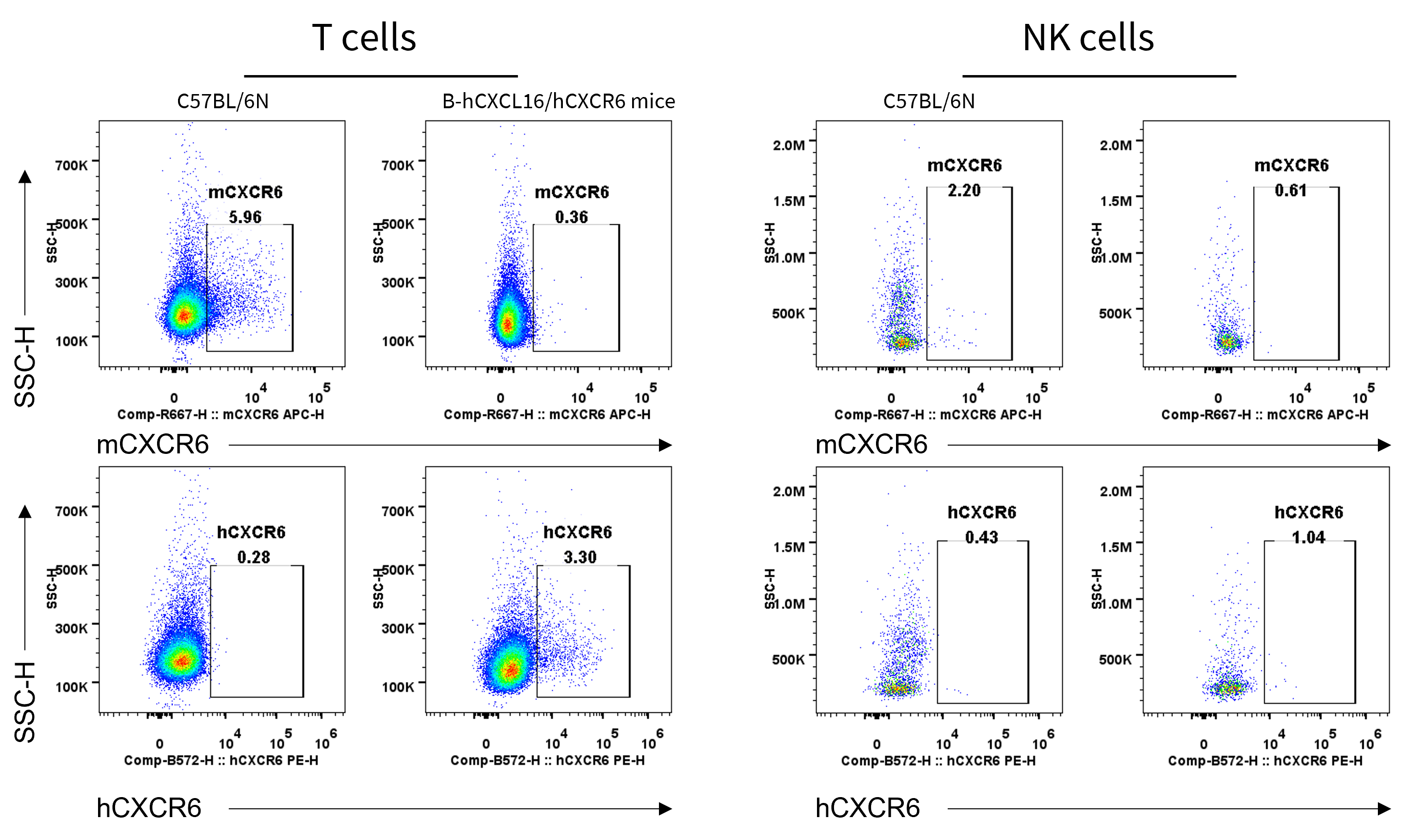
Strain specific CXCR6 expression analysis in homozygous B-hCXCL16/hCXCR6 mice by flow cytometry. Lung cells were collected from wild-type C57BL/6N mice (+/+) and homozygous B-hCXCL16/hCXCR6 mice (H/H;H/H) and analyzed by flow cytometry with species-specific anti-CXCR6 antibody(Biolegend, 151105; Biolegend, 356003). mCXCR6 was detectable in T cells and NK cells(weak expression) of wild-type mice, hCXCR6 was detectable in T cells and NK cells (weak expression) of homozygous B-hCXCL16/hCXCR6 mice.
CXCL16 Protein Expression Analysis in Serum
- Human CXCL16 was detectable in serum from B-hCXCL16/hCXCR6 mice but not on those cells of wild-type C57BL/6 mice.
Strain specific CXCL16 expression analysis in wild-type C57BL/6 mice and homozygous humanized B-hCXCL16/hCXCR6 mice by ELISA. Serum was collected from wild-type C57BL/6N mice (+/+) (male, n=3, 6-week-old) and homozygous B-hCXCL16/hCXCR6 mice (H/H;H/H) (male, n=3, 6-week-old). Expression level of mouse and human CXCL16 were analyzed by ELISA (Human CXCL16 ELISA Kit : Abcam, ab187397; Mouse CXCL16 ELISA Kit : Abcam, ab197744). Mouse CXCL16 was exclusively detectable in wild-type C57BL/6N mice (n=3). Human CXCL16 was exclusively detectable in homozygous B-hCXCL16/hCXCR6 mice (n=3). Values are expressed as mean ± SEM.
CXCR6 Protein Expression Analysis in Skin-IMQ Induced Psoriasis Model
- Human CXCL16 was detectable in skin of psoriasis model induced by IMQ from B-hCXCL16/hCXCR6 mice but not on those cells of wild-type C57BL/6 mice.
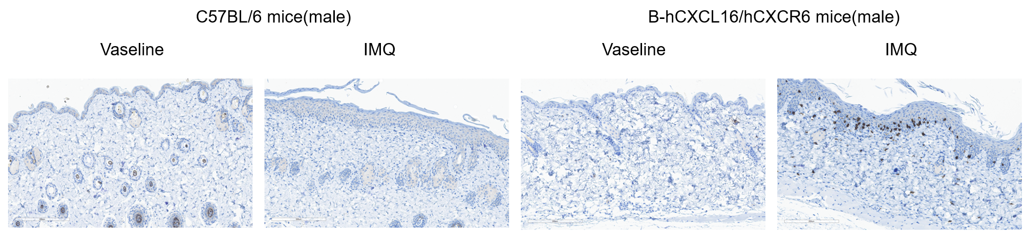
Immunohistochemical (IHC) analysis of CXCR6 protein expression in wild-type C57BL/6 mice and homozygous humanized B-hCXCL16/hCXCR6 mice. Mouse skins were collected from wild-type C57BL/6 mice (+/+) (male, 8-week-old, n=1) and homozygous B-hCXCL16/hCXCR6 mice (H/H;H/H)(male, 8-week-old, n=1). Psoriasis model was induced by IMQ. Protein expression was analyzed with anti-human CXCR6 antibody(Abcam, ab273116) by IHC. Human CXCR6 was detected in the skin of IMQ induced psoriasis model in B-hCXCL16/hCXCR6 mice.
- Human CXCL16 was detectable in skin of psoriasis model induced by IMQ from B-hCXCL16/hCXCR6 mice but not on those cells of wild-type C57BL/6 mice.
Immunohistochemical (IHC) analysis of CXCR6 protein expression in wild-type C57BL/6 mice and homozygous humanized B-hCXCL16/hCXCR6 mice. Mouse skins were collected from wild-type C57BL/6 mice (+/+) (male, 8-week-old, n=1) and homozygous B-hCXCL16/hCXCR6 mice (H/H;H/H)(male, 8-week-old, n=1). Psoriasis model was induced by IMQ. Protein expression was analyzed with anti-human CXCR6 antibody(Abcam, ab273116) by IHC. Human CXCR6 was detected in the skin of IMQ induced psoriasis model in B-hCXCL16/hCXCR6 mice.
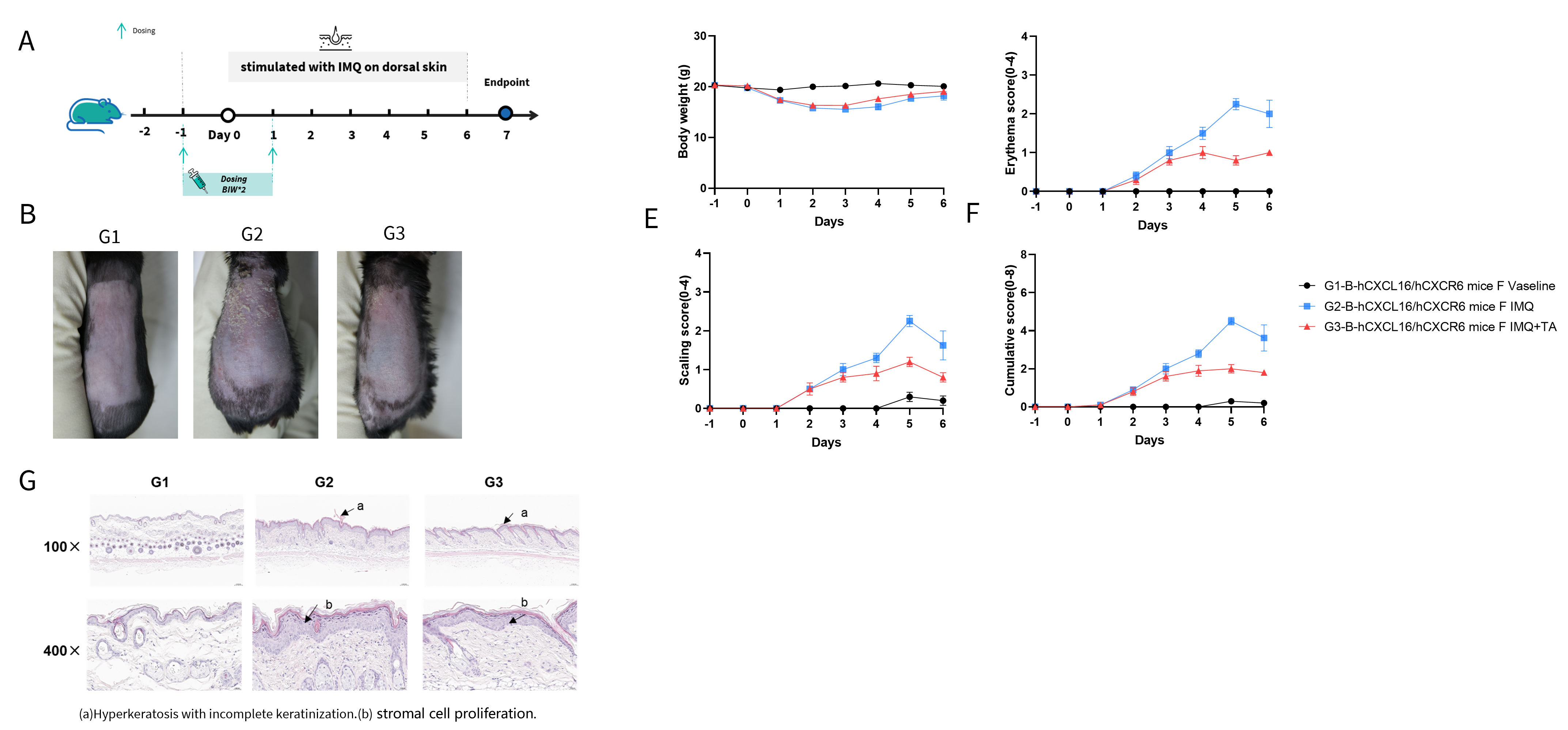
In Vivo Efficacy of Anti-human CXCR6 Antibody With Psoriasis Model
IMQ-induced skin inflammation in B-hCXCL16/hCXCR6 mice phenotypically resembles psoriasis. (A) Experimental schedule for induction of psoriasis-like skin lesions in B-hCXCL16/hCXCR6 mice. B-hCXCL16/hCXCR6 mice (Female, 8 week-old, n=5) were scored daily for up to 6 days for body weight and clinical signs of skin inflammation following treatment with imiquimod (IMQ) cream, Control mice were treated similarly with Vaseline cream. Mice in group 3 were treated with the monoclonal antibody TA targeting human CXCR6(Provided by the client). (B) Phenotypical presentation of mouse back skin after 6 days of treatment. (C) Body weight changes during treatment. (D-E) Erythema and scaling score of the back was scored daily on a scale from 0 to 4. Additionally, the cumulative score (F) (erythema plus scaling) is depicted. (G)H&E staining of the back skin of mice. Values are expressed as mean ± SEM.
* When publishing results obtained using this animal model, please acknowledge the source as follows: The animal model [B-hCXCL16/hCXCR6 mice] (Cat# 113754) was purchased from Biocytogen.