Description
TAU: plays an essential role in the pathogenesis of Alzheimer's disease (AD)
- Gene Information: Microtubule-Associated Protein Tau (MAPT) is a protein-coding gene located on chromosome 17q21.31. MAPT gene mutations have been associated with several neurodegenerative disorders such as Alzheimer's disease, Pick's disease et al.
- Protein Expression: TAU is mainly distributed in the central nervous system, most of it exists in the axons of neurons, and a small amount exists in oligodendrocytes. Under pathological conditions, tau undergoes hyperphosphorylation, which causes the protein to detach from microtubules and form insoluble intracellular aggregates.
- Signaling Pathway: Overactivation of kinases or inhibition of phosphatases triggers aberrant phosphorylation of Tau. This causes Tau to dissociate from microtubules and form aggregates, ultimately leading to synaptic dysfunction and neuronal cell death.
- Therapeutic Inhibition: Tau inhibitors are developed to treat Alzheimer’s disease and cognitive impairment. Tau antibodies modulate abnormal Tau aggregation and clearance through aggregate binding, lysosomal degradation after cellular internalization, and TRIM21-mediated proteasomal degradation.
Targeting strategy
TAU
- The exons 2~10 of mouse Mapt gene that encode the full-length protein were replaced by human MAPT exons 2~15 in B-hTAU mice. The 3’UTR region of the mouse gene are replaced by human counterparts.
- The chimeric MAPT expression is driven by endogenous mouse Mapt promoter, while mouse Mapt gene transcription and translation will be disrupted.
mRNA Expression Analysis
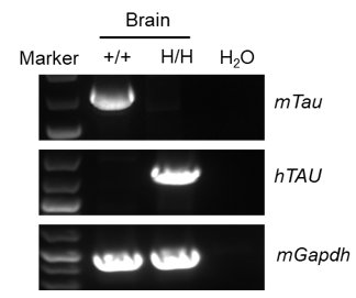
- Human Mapt mRNA is exclusively detectable in brain of homozygous B-hTAU mice, but not in wild-type C57BL/6 mice.
Strain-specific MAPT expression analysis in wild-type C57BL/6 mice and homozygous B-hTAU mice. Brain RNA was isolated from wild-type C57BL/6 mice (+/+) and homozygous B-hTAU mice (H/H), then cDNA libraries were synthesized by reverse transcription, followed by PCR with mouse or human MAPT primers. Human TAU mRNA was detectable only in homozygous B-hTAU mice but not in wild-type mice.
Protein Expression Analysis
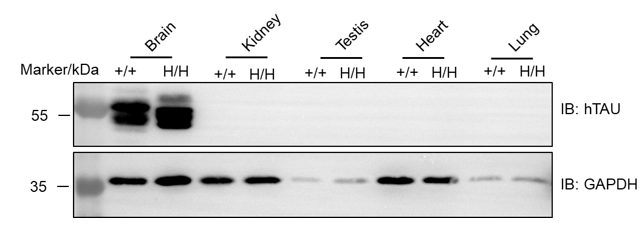
- TAU was detectable in the brain of both C57BL/6 mice and B-hTAU mice.
Protein expression analysis of TAU in homozygous B-hTAU mice. Various tissue lysates were collected from wild-type C57BL/6 mice (+/+) and homozygous B-hTAU mice (H/H), and then analyzed by western blot with anti-TAU antibody (CST, 46687S). 40 μg total protein was loaded for western blotting analysis. TAU was detected in brain from both wild-type C57BL/6 mice and homozygous B-hTAU mice, as the antibody was cross-reactive between human and mouse.
The inhibitory efficiency of the nucleic acid drugs against human MAPT
- The human MAPT mRNA in the treatment group (G2) was significantly reduced compared to the control group (G1), demonstrating that B-hTAU mice provide a powerful preclinical model for in vivo evaluation of human MAPT targeted nucleic acid drugs.
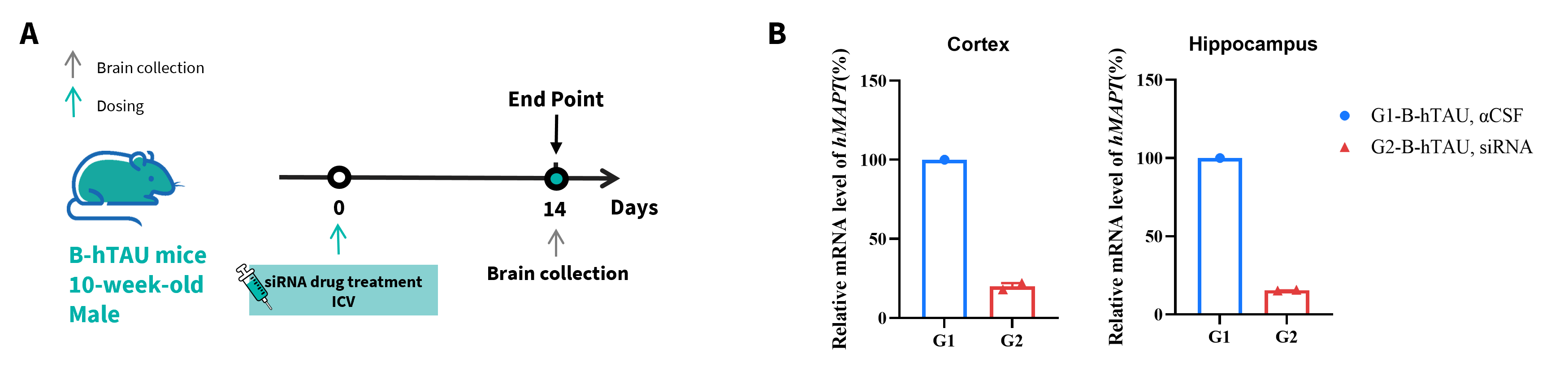
The inhibitory efficiency of the nucleic acid drugs against human MAPT in B-hTAU mice. B-hTAU mice were randomly divided into two groups (G1: n=1, 10 weeks old, male; G2: n=2, 10 weeks old, male). The human TAU targeted nucleic acid drugs (AD-1637701) and αCSF were administered to the mice individually by intra-cerebroventricular injection (ICV). The mice were sacrificed on day 14, and the brains were collected to detect the human MAPT mRNA expression by qRT-PCR. (A) The schematic diagram of experimental processing. (B) The expression of human MAPT mRNA in cortex and hippocampus. The human MAPT in the treatment group (G2) was significantly reduced compared to the control group (G1), demonstrating that B-hTAU mice provide a powerful preclinical model for in vivo evaluation of human MAPT targeted nucleic acid drugs. Values are expressed as mean ± SEM.
This experiment was conducted in collaboration with the client using B-hTAU mice.
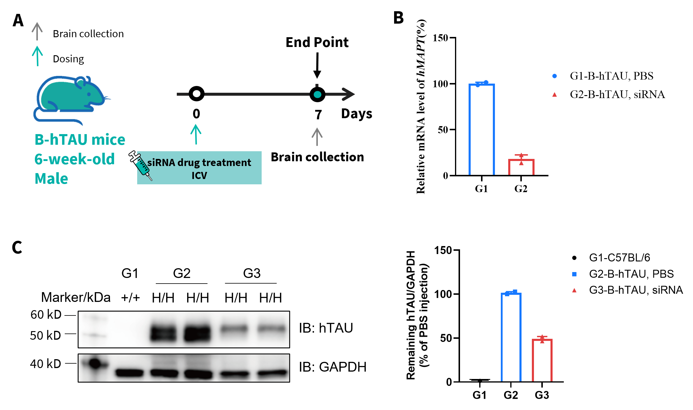
- Compared with the control group, the treatment group showed a significant decrease and the inhibition rate in the treatment group was 51.5%, demonstrating that B-hTAU mice provide a powerful preclinical model for in vivo evaluation of human TAU targeted nucleic acid drugs.
The inhibitory efficiency of the nucleic acid drugs against human TAU in B-hTAU mice. B-hTAU mice were randomly divided into two groups (n=2/group, 6 weeks old, male). The human MAPT targeted nucleic acid drugs (from client) and PBS were administered to the mice individually. The mice were sacrificed on day 7, and the brains were collected to detect the human TAU mRNA and protein expression by qRT-PCR and western blot, respectively. (A) The schematic diagram of experimental processing. (B) The expression of human TAU mRNA in brain. The human TAU in the treatment group (G2) was significantly reduced compared to the control group (G1). (C) The expression of human TAU protein in brain. Values are expressed as mean ± SEM.
* When publishing results obtained using this animal model, please acknowledge the source as follows: The animal model [B-hTAU mice] (Cat# 110953) was purchased from Biocytogen.