mRNA Expression Analysis
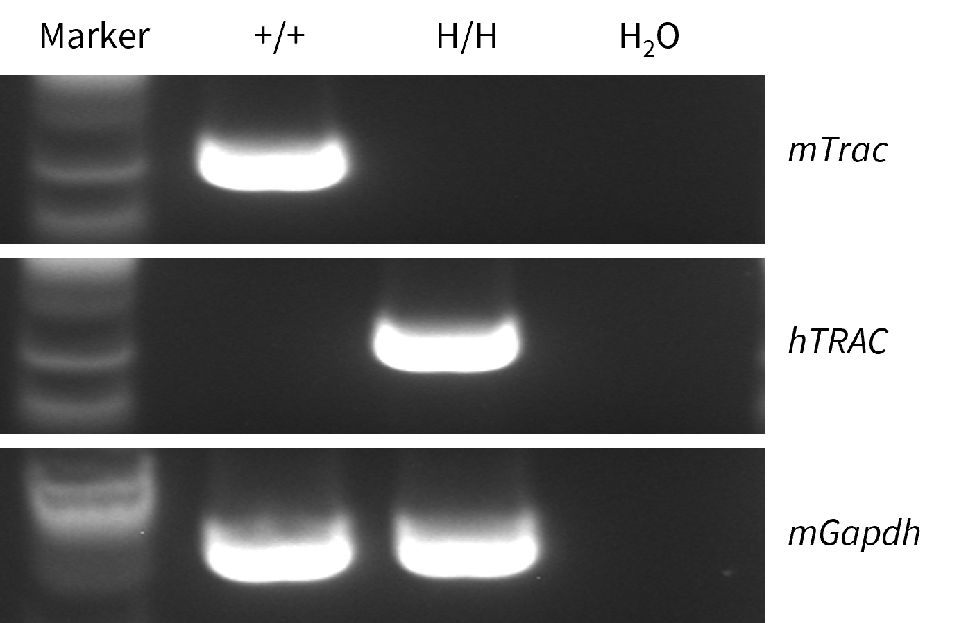
- Mouse Trac mRNA was detectable only in splenocytes of wild-type C57BL/6JNifdc mice (+/+), but not in B-hTRAC/hTRBC1/hTRBC2 mice (H/H).
- Human TRAC mRNA was detectable only in homozygous B-hTRAC/hTRBC1/hTRBC2 mice (H/H).
Strain specific analysis of TRAC gene expression in wild-type C57BL/6JNifdc mice and homozygous B-hTRAC/hTRBC1/hTRBC2 mice by RT-PCR. The splenocytes RNA was isolated from wild-type C57BL/6JNifdc mice (+/+) and homozygous B-hTRAC/hTRBC1/hTRBC2 mice (H/H), and then cDNA libraries were synthesized by reverse transcription, followed by PCR with mouse or human TRAC primers.
TRBC1 Protein Expression Analysis
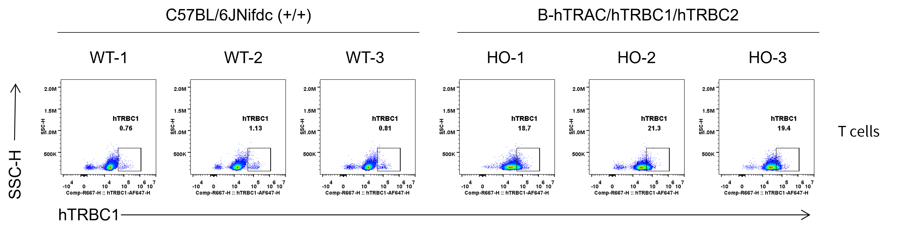
- Human TRBC1 was exclusively detectable in the in homozygous B-hTRAC/hTRBC1/hTRBC2 mice (H/H) but not in wild-type C57BL/6JNifdc mice (+/+).
Strain specific TRBC1 expression analysis in wild-type C57BL/6JNifdc mice and homozygous B-hTRAC/hTRBC1/hTRBC2 mice by flow cytometry. Splenocytes were collected from wild-type C57BL/6JNifdc mice (+/+) and homozygous B-hTRAC/hTRBC1/hTRBC2 mice (H/H), and analyzed by flow cytometry with species-specific anti-human TRBC1 antibody (Biolegend, 383502).
TRAC and TRBC1 Protein Expression Analysis
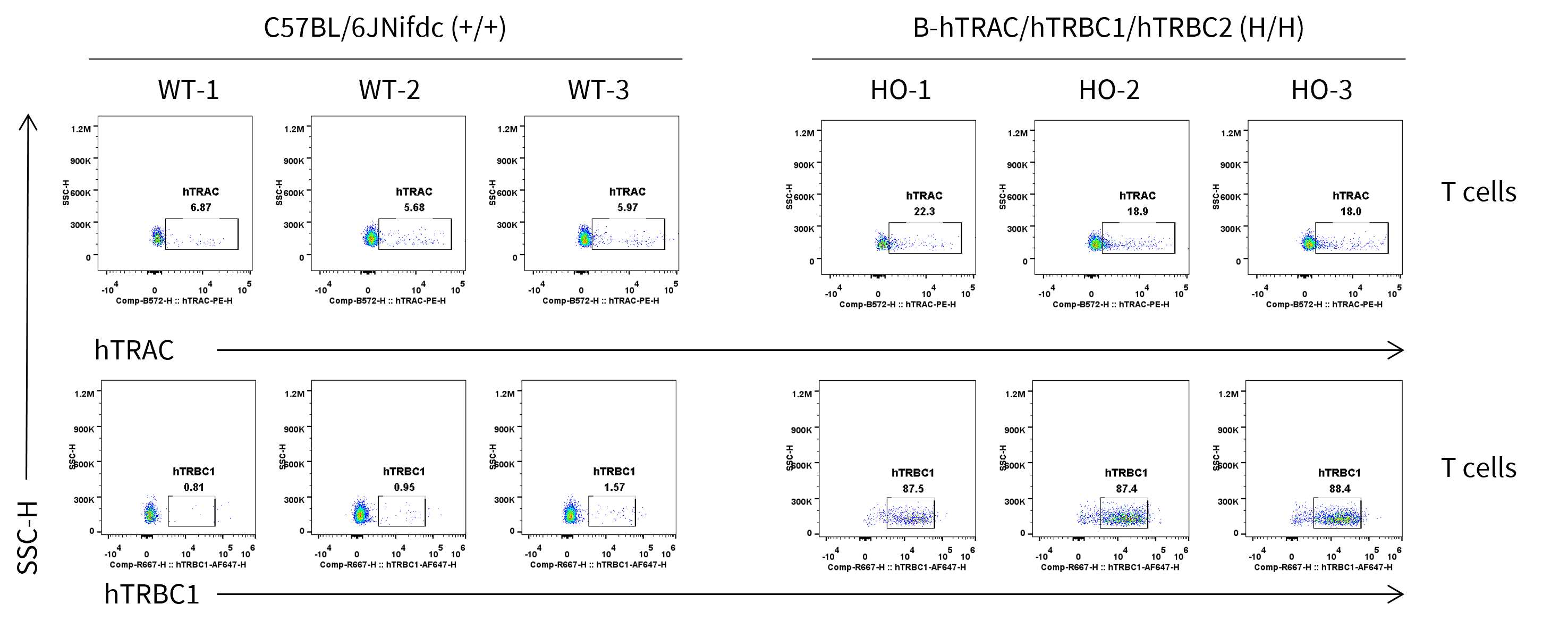
- Human TRAC was detectable in B-hTRAC/hTRBC1/hTRBC2 mice and weakly detectable in wild-type C57BL/6JNifdc mice because the antibody cross-recognizes human and mouse TRAC. Human TRBC1 was exclusively detectable in homozygous B-hTRAC/hTRBC1/hTRBC2 mice.
Strain specific TRAC and TRBC1 expression analysis in homozygous B-hTRAC/hTRBC1/hTRBC2 mice by flow cytometry. Blood cells were collected from wild-type C57BL/6JNifdc (+/+) and homozygous B-hTRAC/hTRBC1/hTRBC2 mice (H/H), and analyzed by flow cytometry with species-specific anti-human TRAC antibody (Invitrogen, PA5-95587) and anti-human TRBC1 antibody (Biolegend, 383502).
Analysis of Leukocyte Subpopulations
- The frequencies of T cells, B cells, NK cells, DCs, monocytes, macrophages, and neutrophils in homozygous B-hTRAC/hTRBC1/hTRBC2 mice were similar to those in C57BL/6JNifdc mice, demonstrating that humanization of TRAC, TRBC1, and TRBC2 does not change the frequency or distribution of these cell types in spleen, blood or lymph nodes.
Analysis of leukocyte subpopulations by flow cytometry in immune organs and blood. Splenocytes, peripheral blood, and lymph nodes were isolated from female C57BL/6JNifdc mice and B-hTRAC/hTRBC1/hTRBC2 mice (male, 8-week-old, n = 3). Single live cells were gated on the CD3⁺ T-cell population and analyzed by flow cytometry as indicated. Values are expressed as mean ± SEM.
Analysis of T Cell Subpopulations
- The frequencies of CD4+ T, CD8+ T, and Tregs in homozygous B-hTRAC/hTRBC1/hTRBC2 mice were comparable to those in C57BL/6JNifdc mice, demonstrating that introduction of human TRAC, TRBC1, and TRBC2 in place of its mouse counterpart does not change the overall development, differentiation or distribution of these T cell sub types in spleen, blood, or lymph nodes.
Analysis of T-cell subpopulations by flow cytometry in immune organs and blood. Splenocytes, peripheral blood, and lymph nodes were isolated from female C57BL/6JNifdc and B-hTRAC/hTRBC1/hTRBC2 Mice (male, 8-week-old, n=3). Single live cells were gated on the CD3⁺ T-cell population and analyzed by flow cytometry as indicated. Values are expressed as mean ± SEM.
The OVA Induced Immune Responses
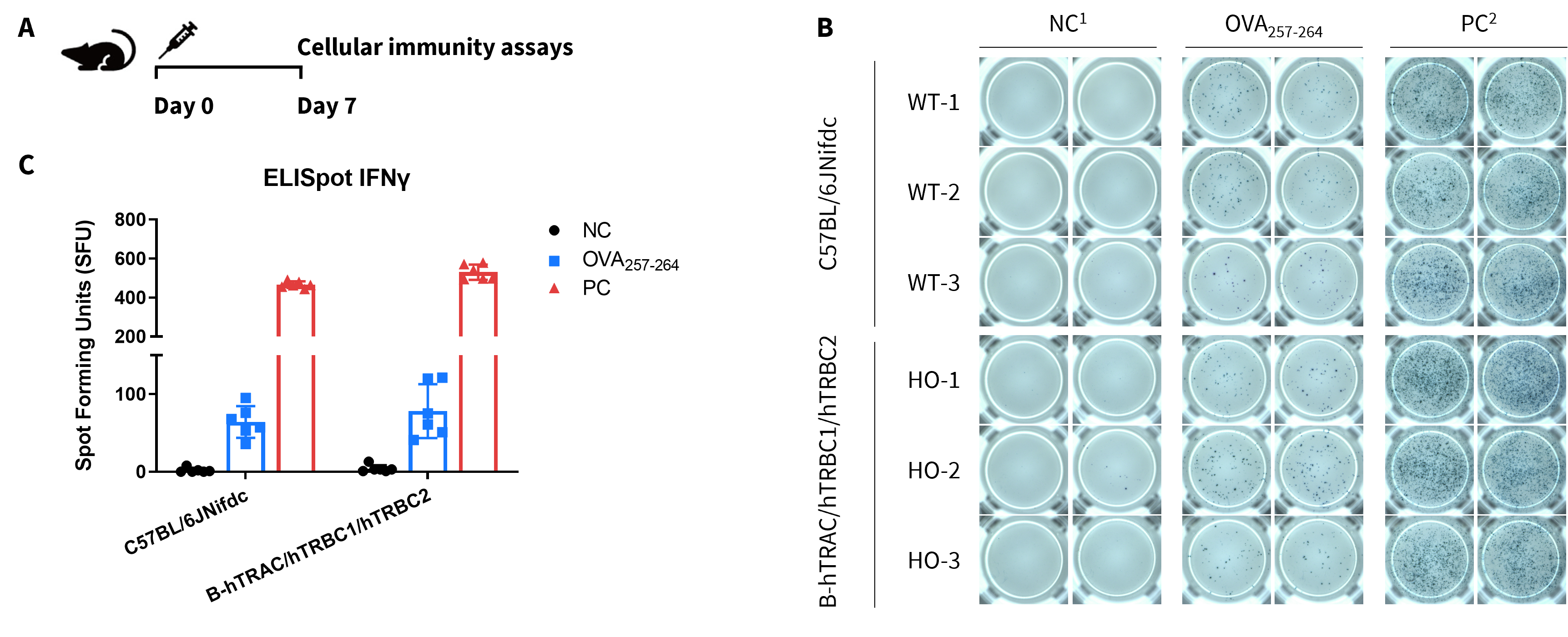
- OVA protein induced immune responses in B-hTRAC/hTRBC1/hTRBC2 mice. These data indicate that B-hTRAC/hTRBC1/hTRBC2 mice have normal T cell immunogenic function.
Detection of OVA-induced immune responses in B-hTRAC/hTRBC1/hTRBC2 mice by IFN-γ ELISpot assay. (A) Scheme of OVA immunization and testing. Female C57BL/6JNifdc (WT) and B-hTRAC/hTRBC1/hTRBC2 mice (9–10-week-old) received a single i.p. injection of 0.5 mg OVA protein + 50 μg poly(I:C). One week post-immunization, splenocytes were harvested and stimulated in vitro with negative control (1: NC/no peptide), OVA peptide257–264, or positive control (2: PC/Cell Activation Cocktail, (Biolegend, 42330) ) to measure IFN-γ secretion. (B) Representative results showing stimulation of splenocytes harvested from immunized mice with negative control, or OVA peptide257–264, or positive control in duplicates. (C) ELISpot Quantification: Statistical summary of IFN-γ-secreting cells.
Tetramer Staining for Analysis
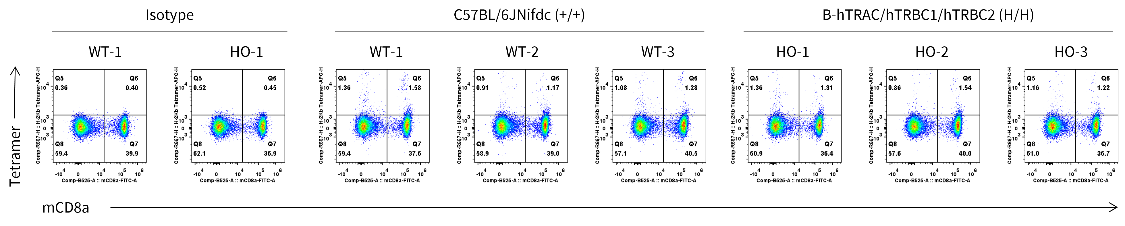
- The frequencies of Tetramer-positive CD8+ T cells were similar between the wild-type C57BL/6JNifdc mice and B-hTRAC/hTRBC1/hTRBC2 mice.
Frequency of OVA-specific T cells among spleen cells was assessed by flow cytometric analysis of SIINFEKL-MHC-I tetramer+ CD8+ T cells. Wild-type C57BL/6JNifdc mice and homozygous B-hTRAC/hTRBC1/hTRBC2 mice (Female, 9–10-week-old, n = 3) were immunized with intraperitoneal injection of 0.5 mg of OVA protein (Simga, A5503-25MG) and 50 μg poly (I:C) (InvivoGen, tlrl-pic). Mice were immunized with OVA one time. One week after the immunization, mice were sacrificed and spleen cells stained with Tetramers(MBL, TS-5001-2C). OVA-specific T cells were detected. The value on the upper right of each plot indicates the percentage of Tetramer-positive CD8+ T cells.
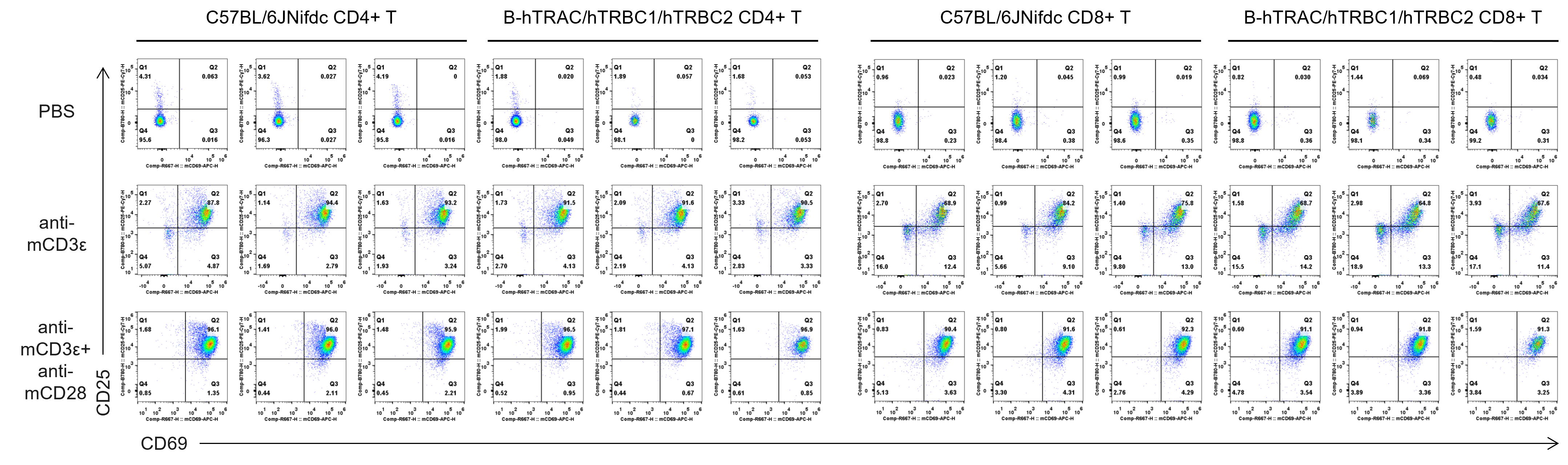
Analysis of T Cell Activation
- T cell activation in B-hTRAC/hTRBC1/hTRBC2 mice was significantly up-regulated by anti-mCD3ε antibody and anti-mCD28 antibody
In vitro T cell activation by anti-mCD3ε antibody with or without anti-mCD28 antibody in wild-type C57BL/6JNifdc mice and homozygous humanized B-hTRAC/hTRBC1/hTRBC2 mice (24h). T cells were isolated from splenocytes of C57BL/6JNifdc and B-hTRAC/hTRBC1/hTRBC2 mice (female, 13-week-old, n = 3), and incubated in the presence of anti-mCD3ε antibody (2 ug/ml, BioXcell, BE0001-2), with or witnout anti-mCD28 antibody (5 ug/ml, BioXcell, BE0015-1) for 24h.
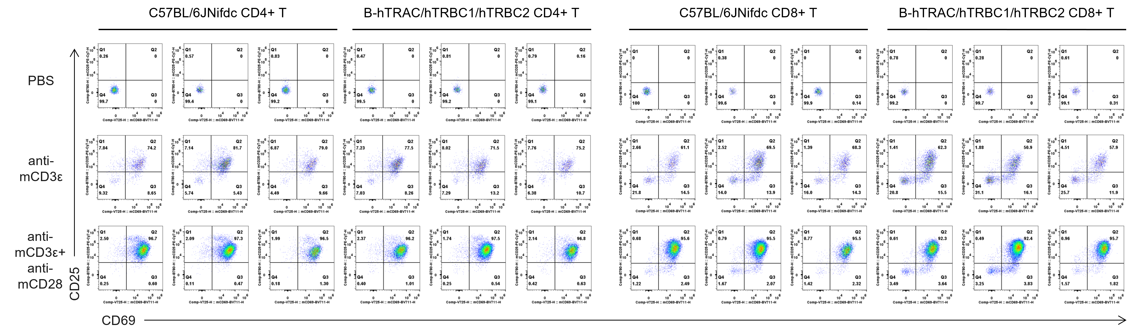
- T cell activation in B-hTRAC/hTRBC1/hTRBC2 mice was significantly up-regulated by anti-mCD3ε antibody and anti-mCD28 antibody.
In vitro T cell activation by anti-mCD3ε antibody with or without anti-mCD28 antibody in wild-type C57BL/6JNifdc mice and homozygous humanized B-hTRAC/hTRBC1/hTRBC2 mice (48h). T cells were isolated from splenocytes of C57BL/6JNifdc and B-hTRAC/hTRBC1/hTRBC2 mice (female, 13-week-old, n = 3), and incubated in the presence of anti-mCD3ε antibody (2 ug/ml, BioXcell, BE0001-2), with or witnout anti-mCD28 antibody (5 ug/ml, BioXcell, BE0015-1) for 48h.
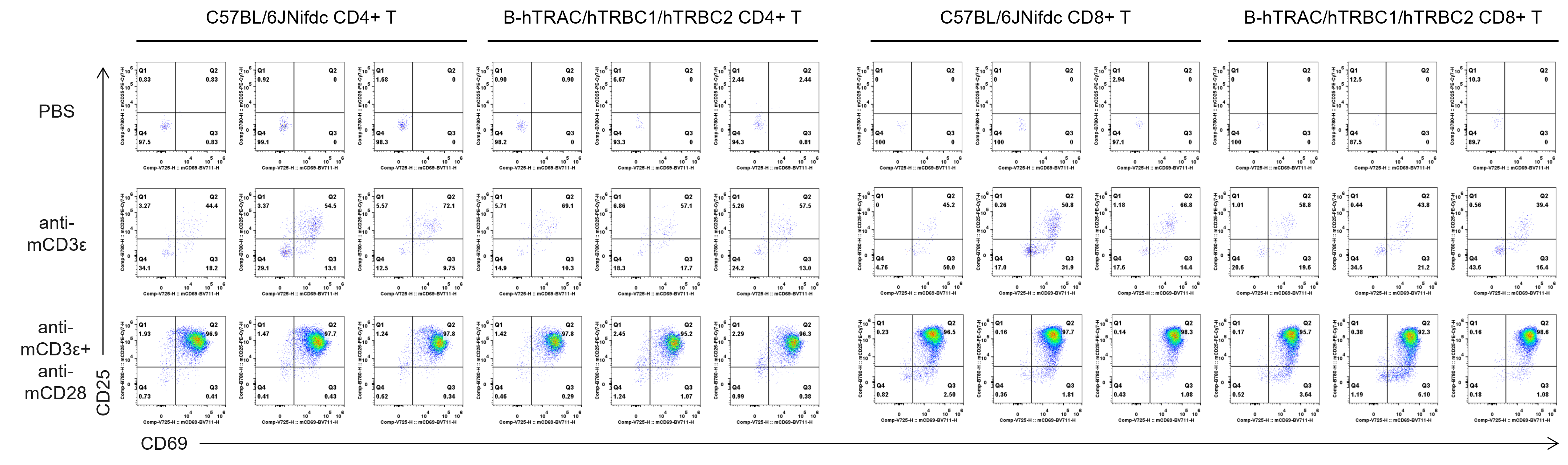
- T cell activation in B-hTRAC/hTRBC1/hTRBC2 mice was significantly up-regulated by anti-mCD3ε antibody and anti-mCD28 antibody.
In vitro T cell activation by anti-mCD3ε antibody with or without anti-mCD28 antibody in wild-type C57BL/6JNifdc mice and homozygous humanized B-hTRAC/hTRBC1/hTRBC2 mice (72h). T cells were isolated from splenocytes of C57BL/6JNifdc and B-hTRAC/hTRBC1/hTRBC2 mice (female, 13-week-old, n = 3), and incubated in the presence of anti-mCD3ε antibody (2 ug/ml, BioXcell, BE0001-2), with or witnout anti-mCD28 antibody (5 ug/ml, BioXcell, BE0015-1) for 72h.
Analysis of T Cell Proliferation
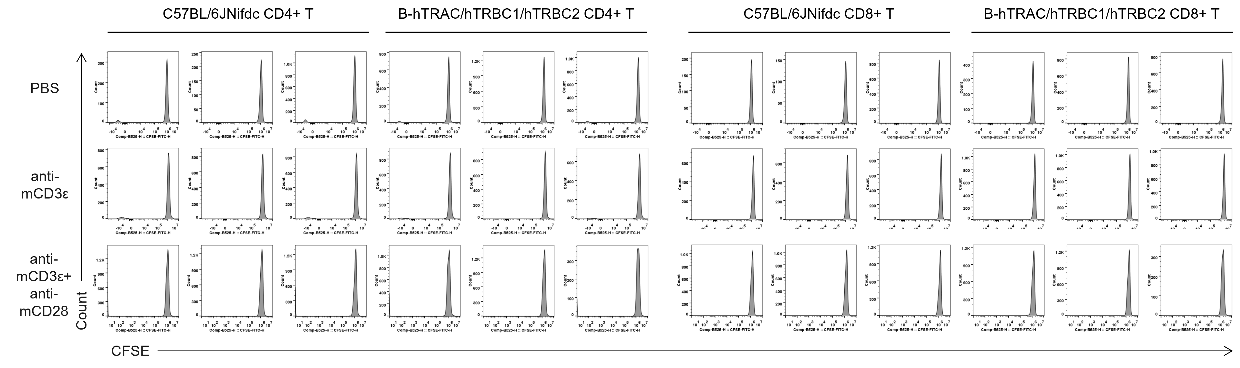
- T cell proliferation was tested by flow cytometry.
Quantification of T cell proliferation in vitro by anti-CD3ε antibody with or without anti-mCD28 antibody in wild-type C57BL/6JNifdc mice and homozygous humanized B-hTRAC/hTRBC1/hTRBC2 mice (24h). T cells were isolated from splenocytes of C57BL/6JNifdc and B-hTRAC/hTRBC1/hTRBC2 mice (female,13-week-old, n = 3), and incubated in the presence of anti-mCD3ε antibody (2 ug/ml, BioXcell, BE0001-2), with or witnout anti-mCD28 antibody (5 ug/ml, BioXcell, BE0015-1) for 24h.
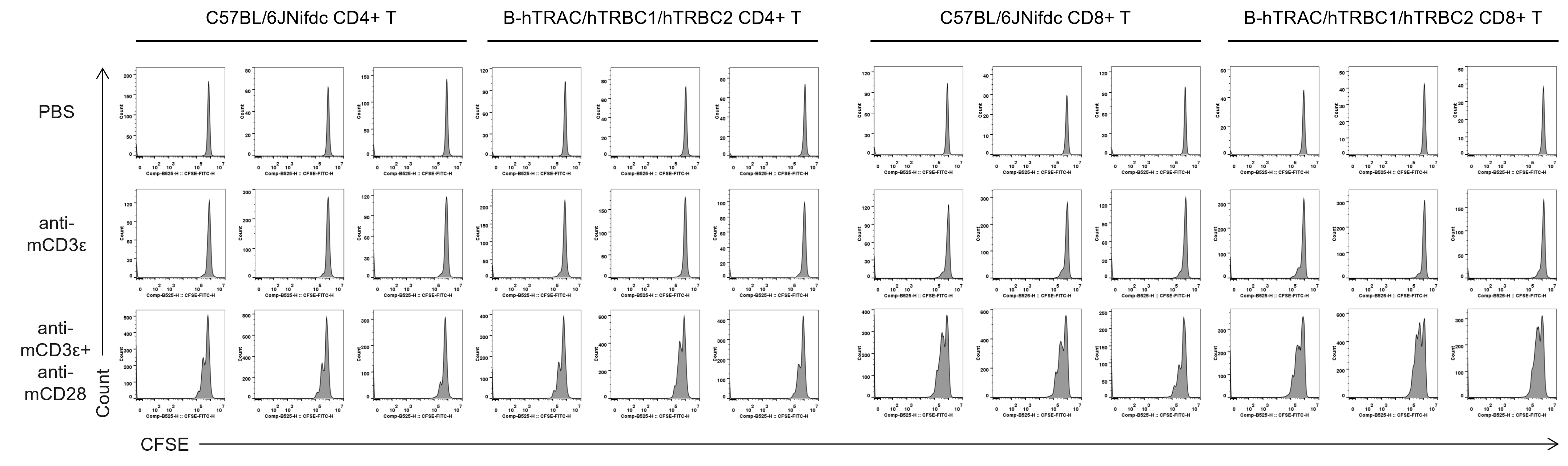
- T cell proliferation was tested by flow cytometry.
Quantification of T cell proliferation in vitro by anti-CD3ε antibody with or without anti-mCD28 antibody in wild-type C57BL/6JNifdc mice and homozygous humanized B-hTRAC/hTRBC1/hTRBC2 mice (48h). T cells were isolated from splenocytes of C57BL/6JNifdc and B-hTRAC/hTRBC1/hTRBC2 mice (female,13-week-old, n = 3), and incubated in the presence of anti-mCD3ε antibody (2 ug/ml, BioXcell, BE0001-2), with or witnout anti-mCD28 antibody (5 ug/ml, BioXcell, BE0015-1) for 48h.
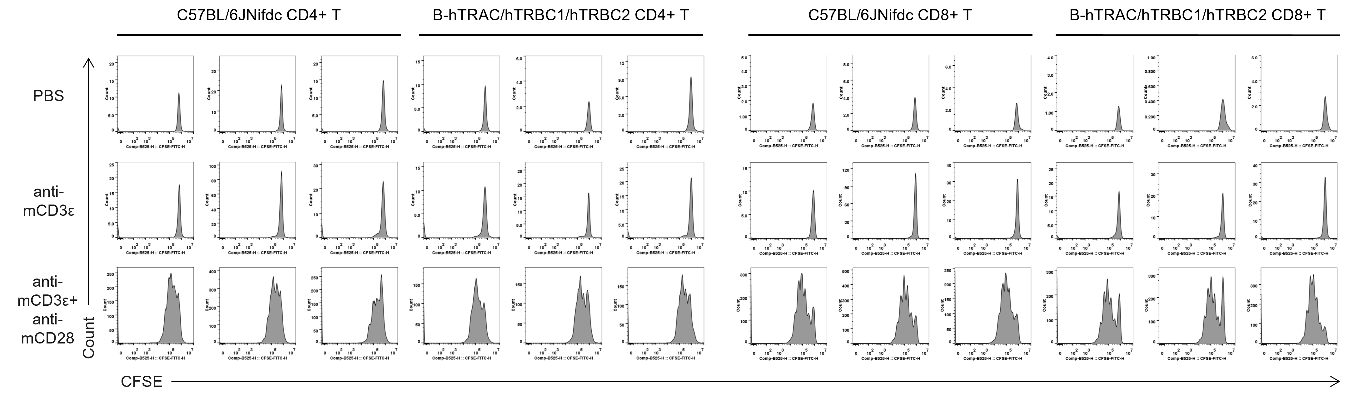
- T cell proliferation was tested by flow cytometry.
Quantification of T cell proliferation in vitro by anti-CD3ε antibody with or without anti-mCD28 antibody in wild-type C57BL/6JNifdc mice and homozygous humanized B-hTRAC/hTRBC1/hTRBC2 mice (72h). T cells were isolated from splenocytes of C57BL/6JNifdc and B-hTRAC/hTRBC1/hTRBC2 mice (female, 13-week-old, n = 3), and incubated in the presence of anti-mCD3ε antibody (2 ug/ml, BioXcell, BE0001-2), with or witnout anti-mCD28 antibody (5 ug/ml, BioXcell, BE0015-1) for 72h.
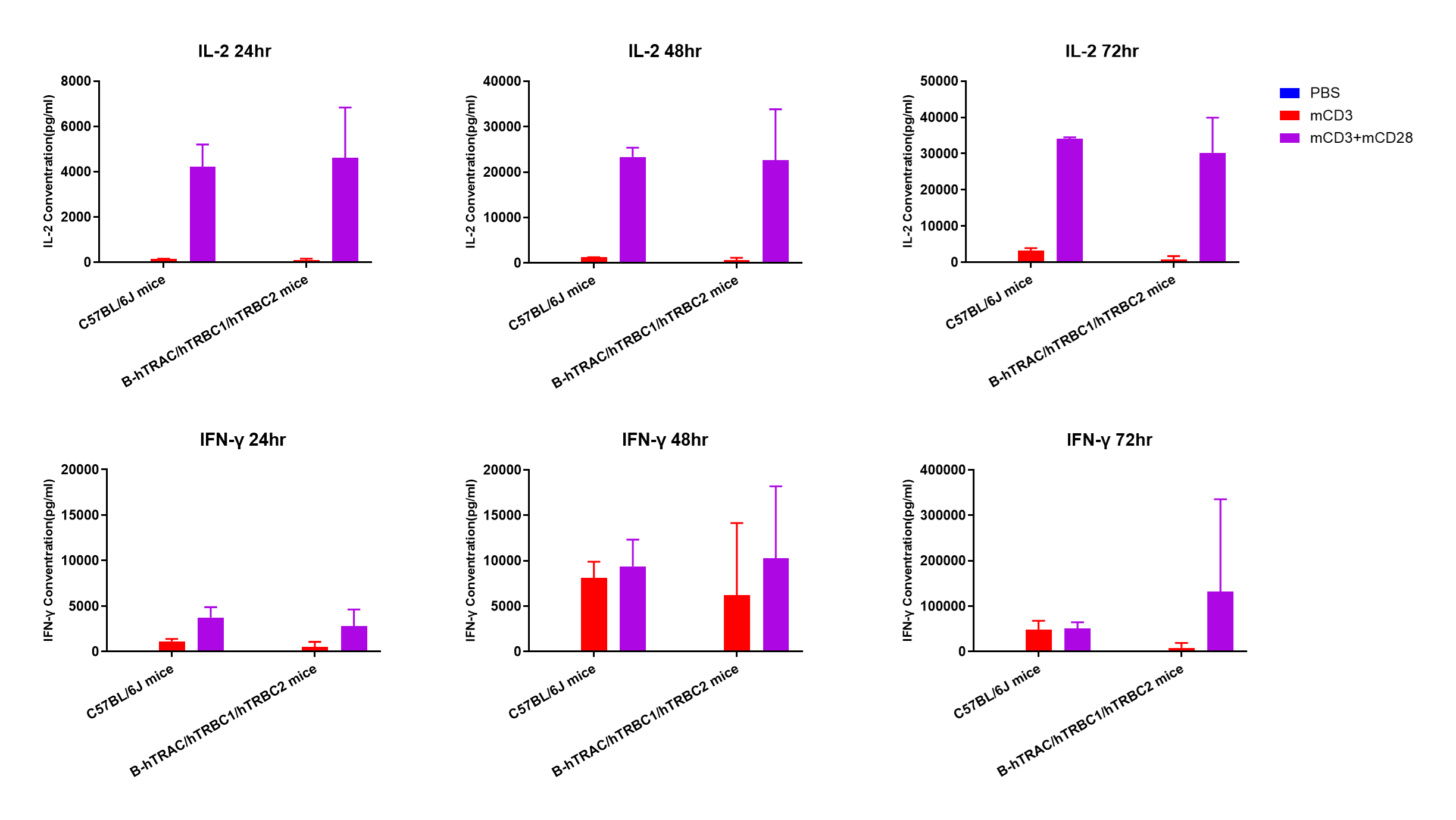
- Under in vitro anti-CD3 and anti-CD28 antibody co-stimulation, wild-type C57BL/6JNifdc and homozygous B-hTRAC/hTRBC1/hTRBC2 mice mice showed comparable secretion levels of IL-2 and IFN-γ.
In vitro cytokine production (IFN-γ and IL-2) in B-hTRAC/hTRBC1/hTRBC2 humanized mice. T cells (2×10⁵) were isolated from the splenocytes of C57BL/6JNifdc and B-hTRAC/hTRBC1/hTRBC2 mice (female, 13-week-old, n = 3), incubated in the presence of anti-mouse CD3ε antibody (BioXCell, BE0001-1, clone 145-2C11, 2 ug/ml) and anti-mCD28 antibody (BioXCell, BE0015-1, clone 37.51, 5 ug/ml) for 24h, 48h and 72h. IFN-γ and IL-2 productions were then tested using ELISA method.
* When publishing results obtained using this animal model, please acknowledge the source as follows: The animal model [B-hTRAC/hTRBC1/hTRBC2 mice] (Cat# 113570) was purchased from Biocytogen.