Description
TSLP: A key cytokine in inflammation and its therapeutic intervention
- Gene Information: Thymic stromal lymphopoietin (TSLP) is a protein-coding gene located on chromosome 5q22.1. It encodes a hemopoietic cytokine that is a member of the interleukin 7-like cytokine family.
- Protein Expression: TSLP is primarily expressed by activated epithelial cells (lung, gut), skin keratinocytes, and fibroblasts. Two main isoforms exist: the short form (sfTSLP) is constitutively expressed and plays a homeostatic role, while the long form (lfTSLP) is induced during inflammation.
- Signaling Pathway: TSLP exerts its effects by binding to a high-affinity heterodimeric receptor complex composed of the TSLP receptor chain (TSLPR) and the IL-7 receptor alpha chain (IL-7Rα).
- Therapeutic Inhibition: By blocking TSLP binding to its receptor, tezepelumab inhibits downstream inflammation and improves clinical outcomes, including reduced serum IgE levels, decreased airway eosinophils, reduced mucus production, and lowered cytokine secretion.
Targeting strategy
TSLP
- Exons 1–5 of the mouse Tslp gene, which encode the entire protein (from ATG to stop codon), are replaced with the corresponding human sequences.
- The endogenous mouse promoter, 5′ UTR, and 3′ UTR regions are retained, allowing human TSLP expression to be driven by the native mouse Tslp promoter, while endogenous mouse Tslp transcription and translation are abolished.
TSLPR
- A chimeric CDS encoding the human TSLPR extracellular and transmembrane domains fused to the mouse TSLPR cytoplasmic domain, followed by the mouse 3′ UTR and stop codon, is inserted immediately downstream of the mouse Tslpr signal peptide to replace part of exon 2 of the endogenous Tslpr gene.
- Expression of the chimeric TSLPR protein is driven by the native mouse Tslpr promoter, while endogenous mouse Tslpr transcription and translation are disrupted.
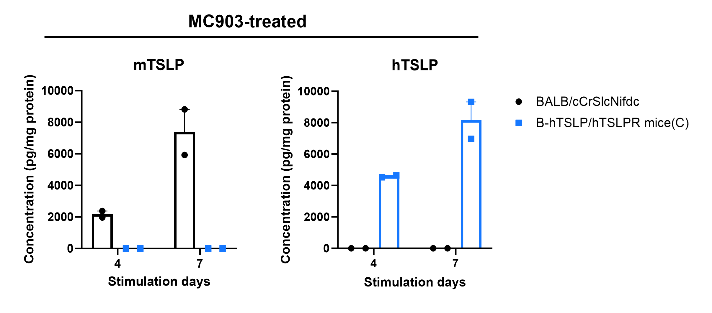
TSLP Protein Expression Analysis in Ear
- Mouse TSLP was detected exclusively in wild-type BALB/cCrSlcNifdc mice
- Human TSLP was detected in homozygous B-hTSLP/hTSLPR mice(C), but not in wild-type mice.
Strain specific TSLP expression analysis in wild-type BALB/cCrSlcNifdc mice and homozygous B-hTSLP/hTSLPR mice(C) by ELISA. Calcipotriol (MC903) was dissolved in ethanol and applied on ears of wild-type BALB/cCrSlcNifdc mice and homozygous B-hTSLP/hTSLPR mice(C) for 4 or 7 days. The ear grinding supernatant collected from the two strains of mouse were analyzed by ELISA (anti-mouse TSLP antibody: Biolegend, 434104; anti-human TSLP antibody: Biolegend, 434204). Mouse TSLP was only detectable in wild-type BALB/cCrSlcNifdc mice. Human TSLP was only detectable in homozygous B-hTSLP/hTSLPR mice(C) but not in wild-type mice. The expression level of TSLP continues to increase from day 4 to day 7 during stimulation.
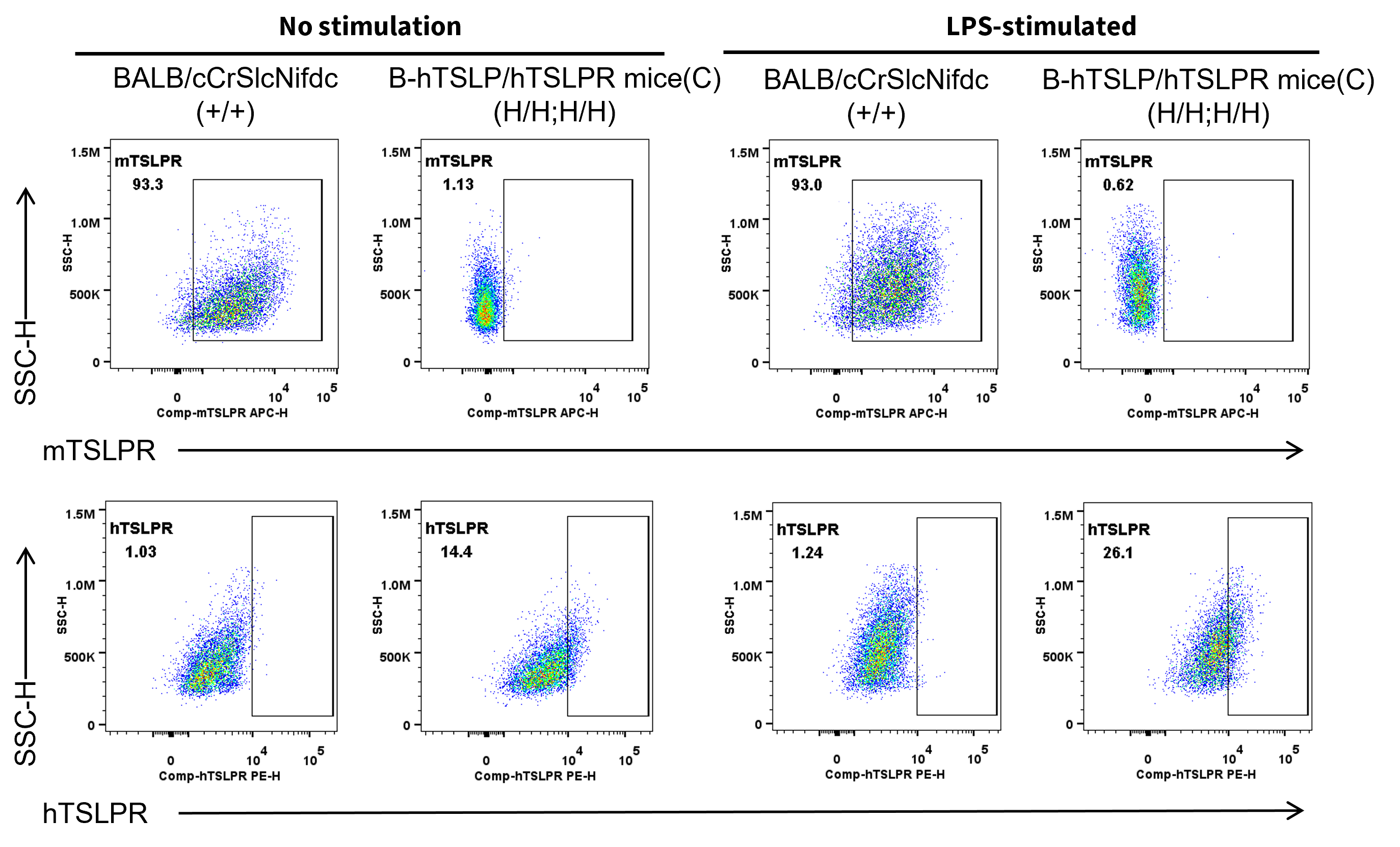
TSLPR Protein Expression in Bone marrow
- Mouse TSLPR was detected on DCs populations in wild-type BALB/cCrSlcNifdc mice.
- Human TSLPR was detected on DCs populations in B-hTSLP/hTSLPR mice(C), but not in wild-type BALB/cCrSlcNifdc mice.
Strain specific TSLPR expression analysis in wild-type BALB/cCrSlcNifdc mice and homozygous B-hTSLP/hTSLPR mice(C) by flow cytometry. To generate DCs in vitro, bone marrow cells were isolated and induced with 200 ng/mL FLT3L (acrobiosystems, FLL-H5218) for 6 days. Purified DCs were respectively stimulated with 1 μg/mL LPS (sigma, L4391). Protein expression was analyzed with anti-mouse TSLPR antibody (Biolegend, 151805) and anti-human TSLPR antibody (Biolegend, 322805) by flow cytometry. Mouse TSLPR was exclusively detectable in wild-type BALB/cCrSlcNifdc mice. Human TSLPR was exclusively detectable in homozygous B-hTSLP/hTSLPR mice(C), but not in wild-type BALB/cCrSlcNifdc mice.
Functional Validation
- In B-hTSLP/hTSLPR mice(C): Mouse TARC was induced by human TSLP, but not by mouse TSLP.
- In wild-type BALB/cCrSlcNifdc mice: Mouse TARC was induced by mouse TSLP, but not by human TSLP.
- These results demonstrate species-specific TSLP–TSLPR signaling and confirm functional activation of dendritic cells by human TSLP in B-hTSLP/hTSLPR mice(C).
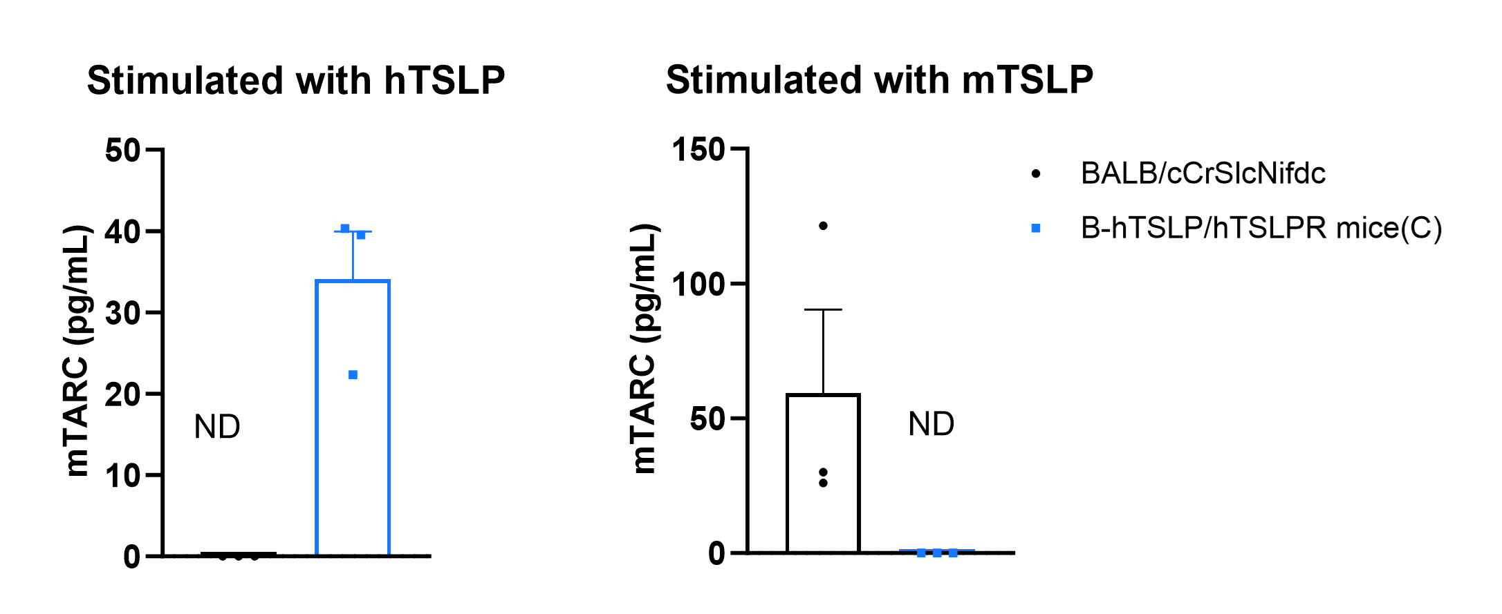
Mouse TARC was induced with mouse TSLP and human TSLP in wild-type BALB/cCrSlcNifdc mice and homozygous B-hTSLP/hTSLPR mice(C). To generate DCs in vitro, bone marrow cells were isolated from wild-type BALB/cCrSlcNifdc mice and B-hTSLP/hTSLPR mice(C) (male, 8-week-old, n=3), and induced with 200 ng/mL FLT3L (acrobiosystems, FLL-H5218) for 6 days. Purified DCs were respectively stimulated with 400 ng/mL mouse TSLP (novoprotein, CJ69) or 400 ng/mL human TSLP (acrobiosystems, TSP-H52Hb). Concentration of mouse TARC secreted from DCs was assayed with anti-mouse TARC antibody (R&D, MCC170) by ELISA. Mouse TARC was successfully induced with mouse TSLP in wild-type BALB/c mice, but not in homozygous B-hTSLP/hTSLPR mice(C). Meanwhile, mouse TARC was successfully induced with human TSLP in B-hTSLP/hTSLPR mice(C), but not in wild-type BALB/c mice. These results indicate that humanized TSLP receptor can only recognize human TSLP ligand to activate DCs from B-hTSLP/hTSLPR mice(C). Values are expressed as mean ± SEM. Significance was determined by two-way ANOVA test. *P < 0.05, **P < 0.01, ***P < 0.001.
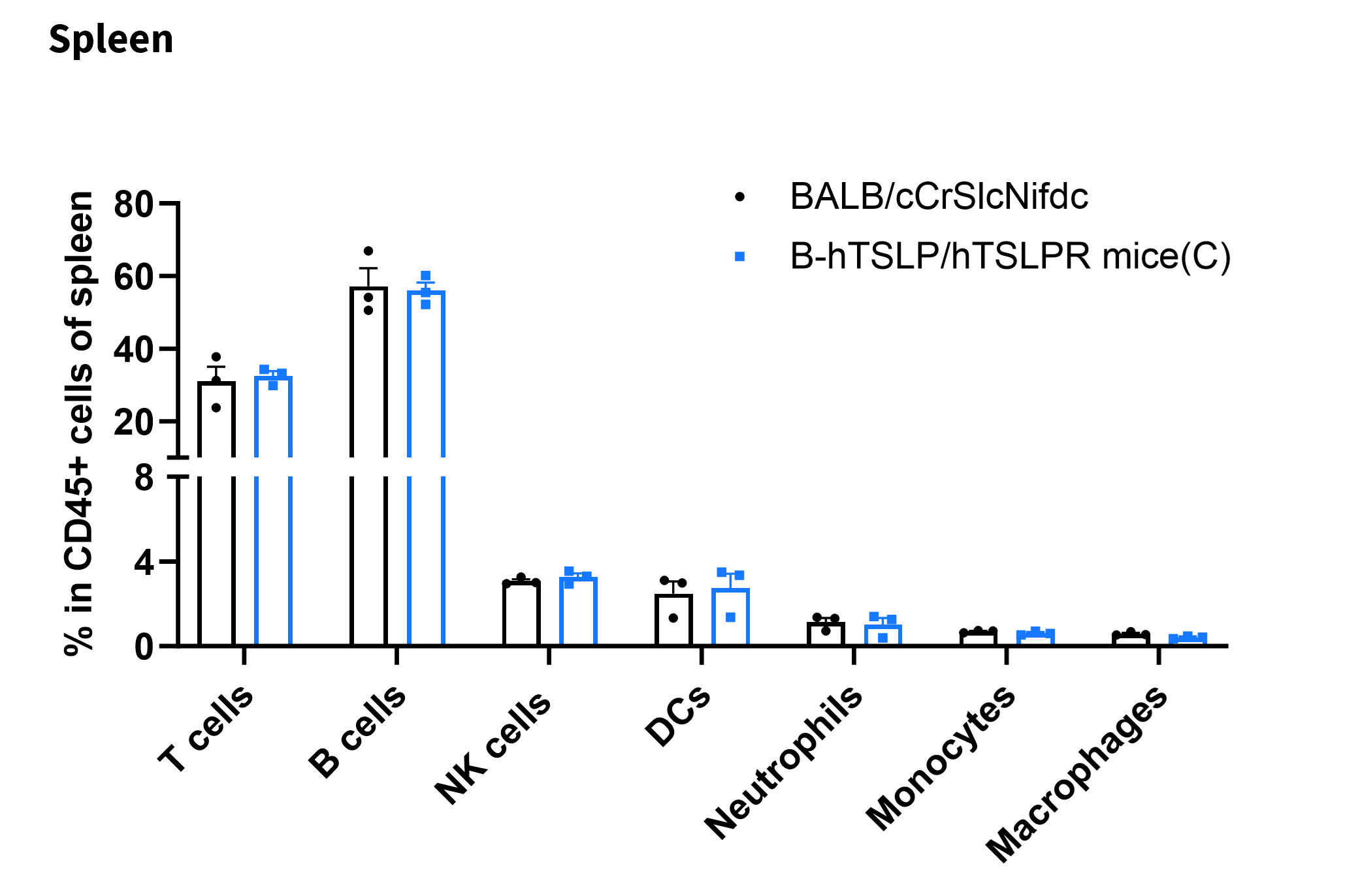
Analysis of Leukocyte Subpopulations in Spleen
- The frequencies of T cells, B cells, NK cells, DCs, neutrophils, monocytes, and macrophages in homozygous B-hTSLP/hTSLPR mice(C) were similar to those in BALB/cCrSlcNifdc mice
- Humanization of TSLP and TSLPR does not affect normal immune cell development or splenic distribution.
Analysis of leukocyte subpopulations by flow cytometry in spleen. Splenocytes were isolated from BALB/cCrSlcNifdc mice and B-hTSLP/hTSLPR mice(C) (male, 8-week-old, n = 3). Single live cells were gated on the CD45⁺ population and analyzed by flow cytometry as indicated. Values are expressed as mean ± SEM.
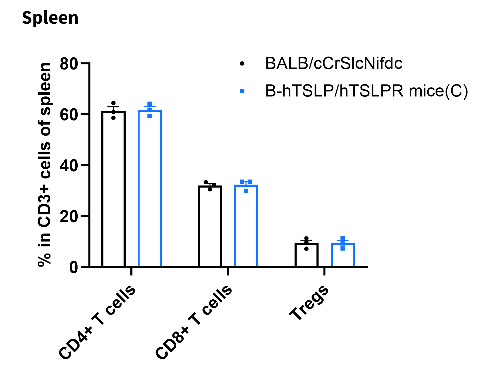
Analysis of T Cell Subpopulations in Spleen
- The proportions of CD4⁺ T cells, CD8⁺ T cells, and Tregs in homozygous B-hTSLP/hTSLPR mice were comparable to those in BALB/cCrSlcNifdc mice
- Humanization of TSLP and TSLPR does not affect normal T cell development, differentiation, or splenic distribution.
Analysis of T-cell subpopulations by flow cytometry in spleen. Splenocytes were isolated from BALB/cCrSlcNifdc mice and B-hTSLP/hTSLPR mice(C) (male, 8-week-old, n = 3). Single live cells were gated on the CD3⁺ T-cell population and analyzed by flow cytometry as indicated. Values are expressed as mean ± SEM.
Induction of Asthma Model and In Vivo Efficacy of Anti–Human TSLP Antibody
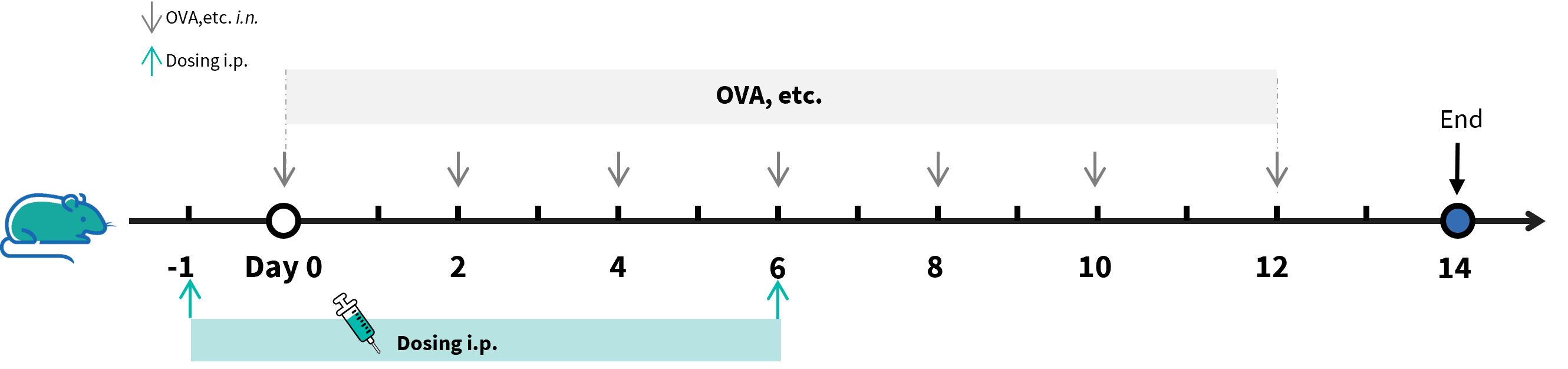
Experimental schedule for the induction of asthma model and in vivo efficacy of anti-human TSLP antibody in B-hTSLP/hTSLPR mice(C). B-hTSLP/hTSLPR mice(C) (Female, 10-week-old, n=6) were immunized with OVA etc. inducer to induce asthma.. The anti-human TSLP antibody tezepelumab analog (in-house) was administered by intraperitoneal injection (n = 6).
In Vivo Efficacy of Anti-Human TSLP Antibody in an Asthma Model
- CD45⁺ cells, eosinophils were significantly reduced in the anti–human TSLP antibody–treated group (G4,G5) compared with the isotype control group (G2).

Analysis of immune cells in BALF by flow cytometry. B-hTSLP/hTSLPR mice(C) (female, 10-week-old, n=6) were stimulated with inducer to induce asthma-like symptoms. Anti-human TSLP antibody (tezepelumab analog, synthesized in-house) was intraperitoneally injected. Broncheoalveolar fluid (BALF) was collected at the end of the experiment to detect inflammatory cell infiltration in lung tissue. The results showed that the number and frequency of eosinophils induced in the untreated modeling group (G2) was significantly higher than that in the un-modeling group (G1), while the number and frequencies of these cells in the treated group (G3-G5) decreased significantly when compared with the untreated modeling group (G2). Values are expressed as mean ± SEM. Significance was determined by one-way ANOVA test. *P < 0.05, **P < 0.01, ***P < 0.001.
The overage of this mouse model is 0%.
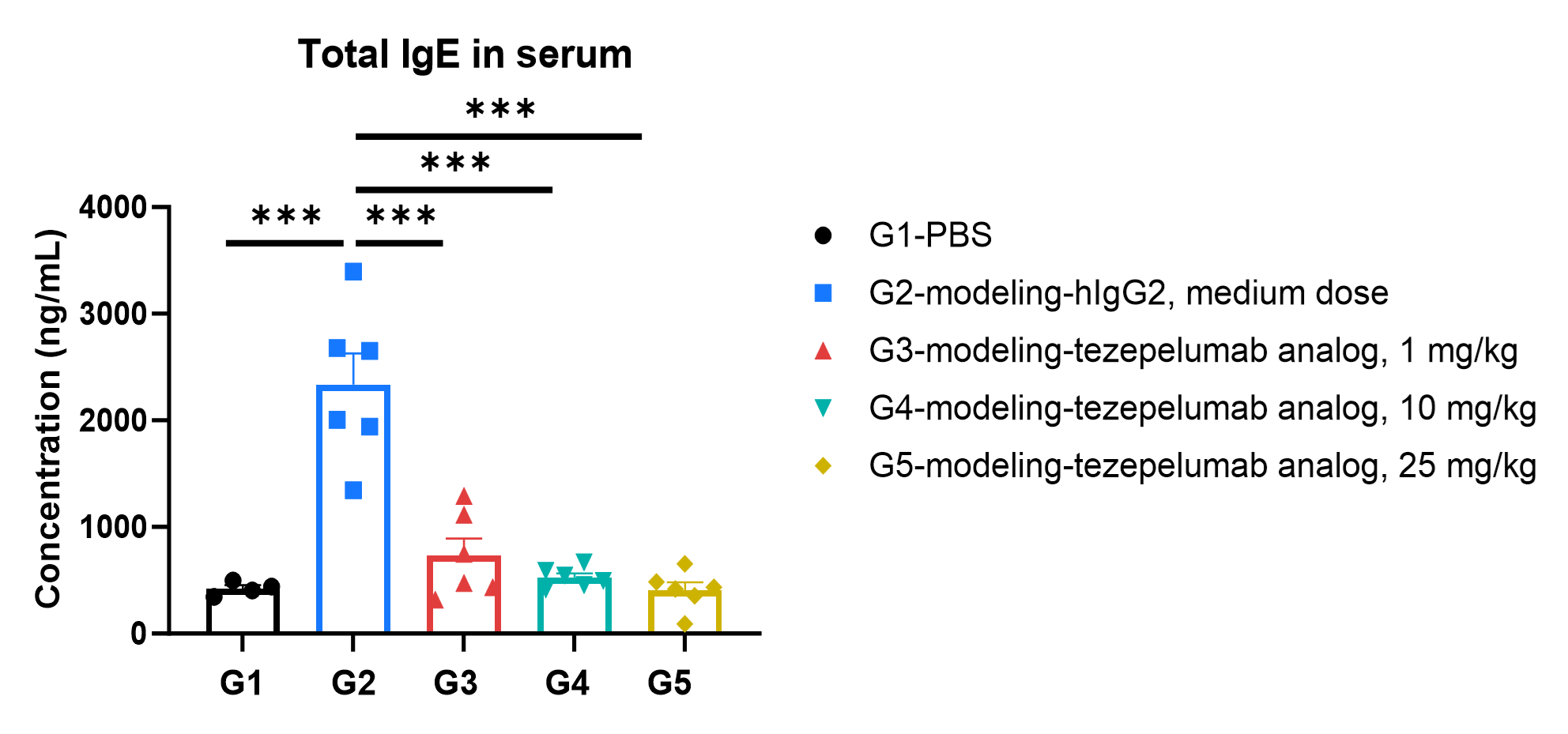
- Tezepelumab analog treatment reduced serum IgE levels compared with untreated controls.
Total IgE in serum were significantly reduced in the mouse asthma model treated with anti-TSLP antibody. Serum was collected at the study endpoint. IgE levels were analyzed by ELISA. The results showed that the levels of total IgE in the groups (G3-G5) treated with tezepelumab analog (in-house) was significantly lower than that in untreated group (G2). Values are expressed as mean ± SEM. Significance was determined by one-way ANOVA test. *P < 0.05, **P < 0.01, ***P < 0.001.
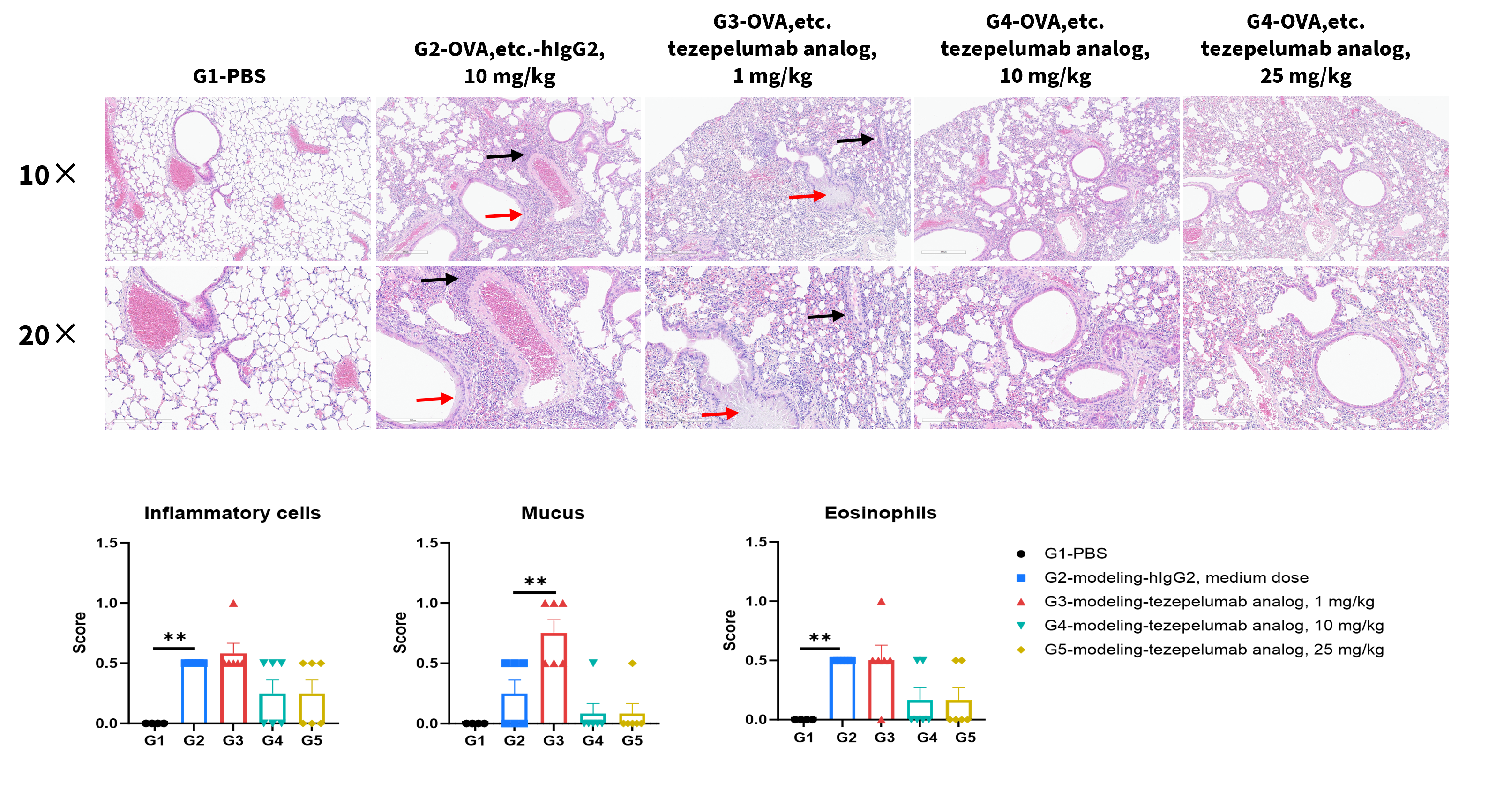
- Tezepelumab analog significantly reduced inflammatory infiltration and mucus secretion in lung tissue compared with untreated controls (G2).
- B-hTSLP/hTSLPR mice(C) provide a robust in vivo preclinical model for evaluating anti–human TSLP antibodies.
H&E staining of asthma-like model in B-hTSLP/hTSLPR mice(C). Lung tissues were collected at the study endpoint and analyzed with H&E staining. The results showed that compared to the unmodeled group (G1), the modeled group (G2) showed a significant reduction in inflammatory infiltration and mucus secretion in lung tissue. The groups treated with higher doses of tezepelumab analog (in-house) showed a downward trend in inflammatory infiltration and mucus secretion in lung tissue. Black arrow: inflammatory cells; Red arrow: mucus. Values are expressed as mean ± SEM. Significance was determined by one-way ANOVA test. *P < 0.05, **P < 0.01, ***P < 0.001.
* When publishing results obtained using this animal model, please acknowledge the source as follows: The animal model [B-hTSLP/hTSLPR mice(C)] (Cat# 112923) was purchased from Biocytogen.