mRNA Expression Analysis in Spleen
- Mouse Il10 mRNA was not detectable in homozygous B-Il10 KO mice(C).
Species specific analysis of Il10 gene expression in wild-type BALB/cCrSlcNifdc mice and homozygous B-Il10 KO mice(C) by RT-PCR. Spleen was collected from wild-type BALB/cCrSlcNifdc mice (+/+) and homozygous B-Il10 KO mice(C) (-/-). Mouse Il10 mRNA was only detectable in wild-type BALB/cCrSlcNifdc mice, but not in homozygous B-Il10 KO mice(C).
DSS-Induced Acute Colitis
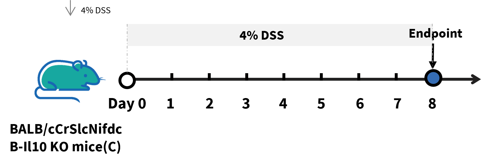
Experimental schedule for DSS induced acute colitis in B-Il10 KO mice(C). The BALB/cCrSlcNifdc mice and B-Il10 KO mice(C) (female, 8-9 weeks-old, n=8) were divided into two groups. The normal controls were administered drinking water for 8 consecutive days, the treatment groups were administered 4% DSS solution for 8 consecutive days.
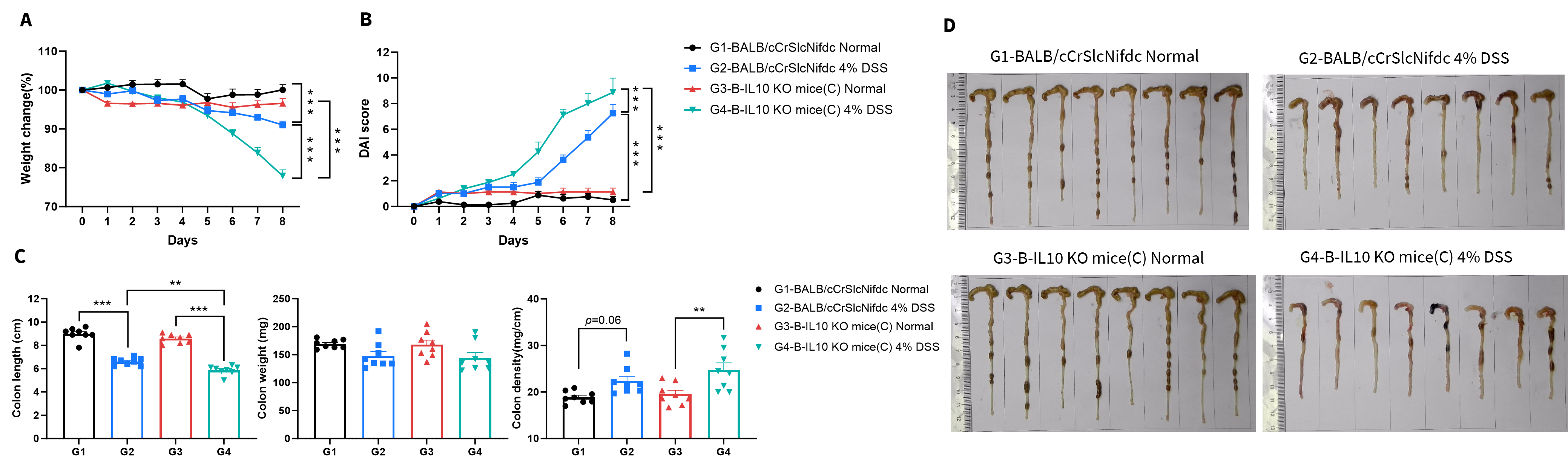
- B-IL10 KO mice(C) were more susceptible to DSS than wild-type BALB/cCrSlcNifdc mice.
Wild-type BALB/cCrSlcNifdc mice and B-IL10 KO mice(C) were administered DSS in drinking water for 8 consecutive days. The BALB/cCrSlcNifdc mice and B-Il10 KO mice(C) (female, 8-9 weeks-old, n=8) were divided into two groups. The normal controls were administered drinking water for 8 consecutive days, the treatment groups were administered 4% DSS solution for 8 consecutive days. (A) Body weight change. (B) DAI score. (C) Colon index. (D) Colon photo. Values are expressed as mean ± SEM. Significance was determined by two-way ANOVA test. *P < 0.05, **P < 0.01, ***P < 0.001.
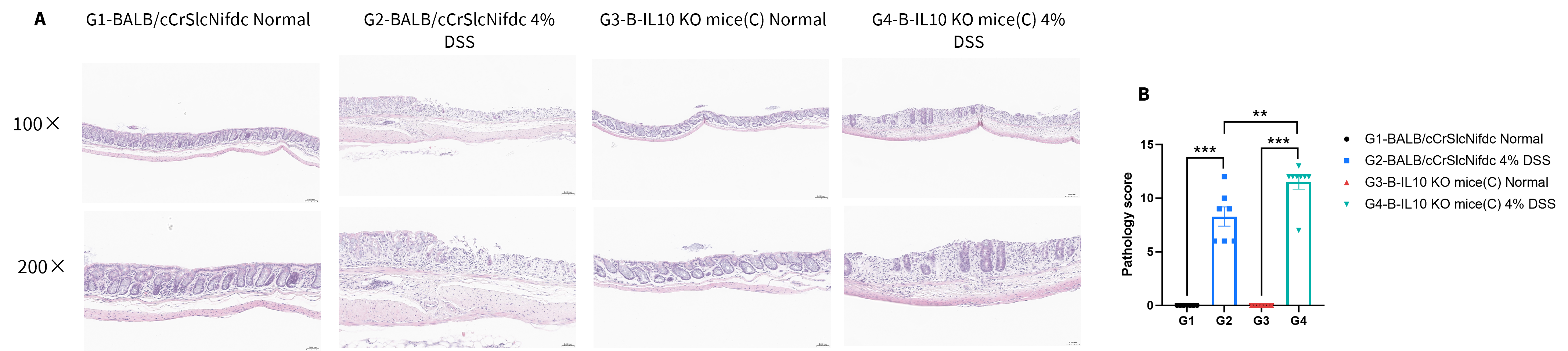
- B-IL10 KO mice(C) exhibited more severe inflammatory infiltration and epithelial damage phenotypes in response to DSS compared to wild-type BALB/cCrSlcNifdc mice.
Wild-type BALB/cCrSlcNifdc mice and B-IL10 KO mice(C) were administered DSS in drinking water for 8 consecutive days. The BALB/cCrSlcNifdc mice and B-Il10 KO mice(C) (female, 8-9 weeks-old, n=8) were divided into two groups. The normal controls were administered drinking water for 8 consecutive days, the treatment groups were administered 4% DSS solution for 8 consecutive days. (A) H&E staining of colon tissue. (B) Pathological score. Values are expressed as mean ± SEM. Significance was determined by two-way ANOVA test. *P < 0.05, **P < 0.01, ***P < 0.001.
Spontaneous Colitis
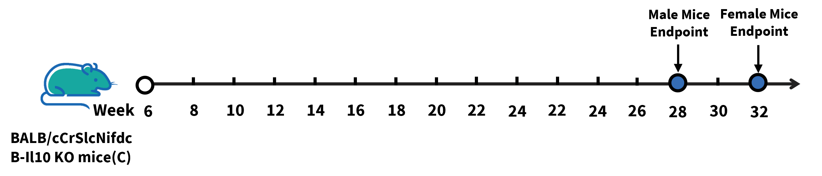
Experimental schedule for Spontaneous colitis in female and male B-Il10 KO mice(C). The female and male BALB/cCrSlcNifdc mice, as well as female and male B-Il10 KO mice(C) were grouped at 6 weeks. The body weight and DAI score of the mice were recorded weekly. The endpoint for male mice was 28 weeks, and the endpoint for female mice was 32 weeks.
- Compared with BALB/cCrSlcNifdc mice, B-Il10 KO mice(C) exhibited obvious spontaneous intestinal inflammation.
- Male B-Il10 KO mice(C) developed the disease earlier than female B-Il10 KO mice(C), and the disease symptoms in male B-Il10 KO mice(C) were more severe than those in female mice.
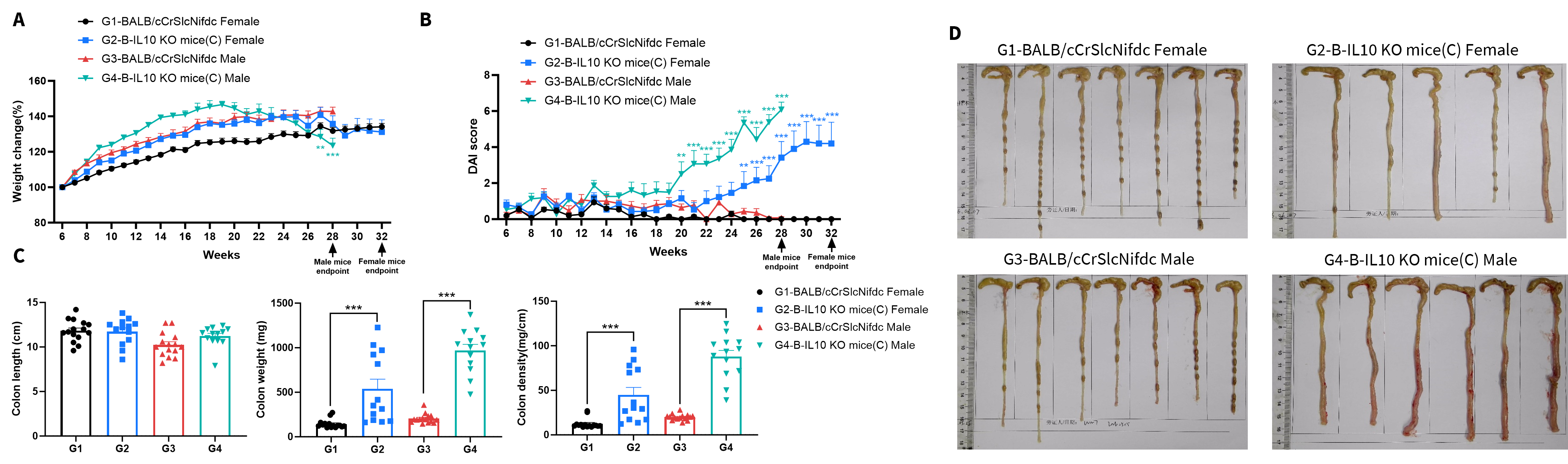
Spontaneous colitis in female and male B-Il10 KO mice(C). The female and male BALB/cCrSlcNifdc mice, as well as female and male B-Il10 KO mice(C) were grouped at 6 weeks. The body weight and DAI score of the mice were recorded weekly. The endpoint for male mice was 28 weeks, and the endpoint for female mice was 32 weeks. (A) Body weight change. (B) DAI score. (C) Colon index. (D) Colon photo. Values are expressed as mean ± SEM. Significance was determined by two-way ANOVA test. *P < 0.05, **P < 0.01, ***P < 0.001.
Note: Due to individual differences and environmental factors, the disease onset in these mice may vary. The data presented here were obtained under the internal facility conditions of Biocytogen and are for reference only. The actual disease onset in mice should be based on the actual situation.
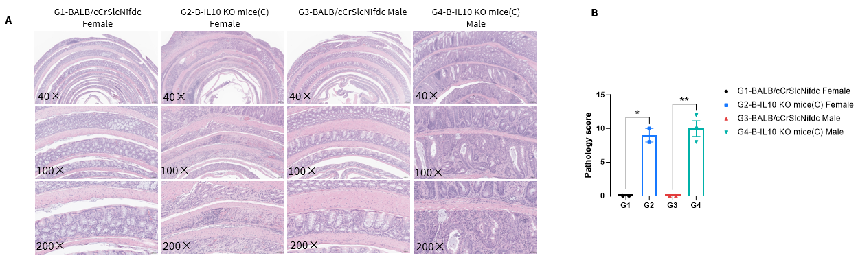
- In the colon tissue of B-Il10 KO mice(C), extensive inflammatory cell infiltration and epithelial damage were observed.
Spontaneous colitis in female and male B-Il10 KO mice(C). The female and male BALB/cCrSlcNifdc mice, as well as female and male B-Il10 KO mice(C) were grouped at 6 weeks. The body weight and DAI score of the mice were recorded weekly. The endpoint for male mice was 28 weeks, and the endpoint for female mice was 32 weeks. (A) H&E staining of colon tissue. (B) Pathological score. Values are expressed as mean ± SEM. Significance was determined by two-way ANOVA test. *P < 0.05, **P < 0.01, ***P < 0.001.
Note: Due to individual differences and environmental factors, the disease onset in these mice may vary. The data presented here were obtained under the internal facility conditions of Biocytogen and are for reference only. The actual disease onset in mice should be based on the actual situation.
* When publishing results obtained using this animal model, please acknowledge the source as follows: The animal model [B-Il10 KO mice(C)] (Cat# 112269) was purchased from Biocytogen.