Description
The B-NDG MHC I/II DKO mice plus model was generated by knocking out β2-microglobulin (B2m) as well as both MHC class I (H-2Kᵇ/H-2Dᵇ) and MHC class II (I-Aᵏ) molecules on the NOD background. This produces a highly immunodeficient strain lacking functional T cells, B cells, and NK cells, with complete abolition of murine antigen presentation.
These features significantly reduce xenogeneic graft-versus-host disease (GvHD) induced by human PBMC engraftment and extend the experimental window for long-term human immune-system reconstitution, tumor modeling, and therapeutic antibody evaluation.
This strain enables more stable and longer-lasting human PBMC engraftment, supports robust T-cell reconstitution, and reduces GvHD severity compared with conventional B-NDG or B-NDG B2m KO Mice Plus.
Key Advantages
- Complete loss of MHC I and MHC II antigen presentation, minimizing murine immune-mediated GvHD.
- Enables long-term survival after human PBMC engraftment, significantly extending the experimental window.
- Supports robust human T-cell reconstitution, including CD4⁺, CD8⁺, and Treg lineages.
- Reduced severity of xenogeneic GvHD allows higher PBMC doses and long-term tracking.
- Ideal for humanized immune-system studies, CDX models, immuno-oncology research, and bispecific antibody evaluation.
- More stable than B-NDG and B-NDG B2m KO mice plus for PBMC-driven immune-response research.
Validation
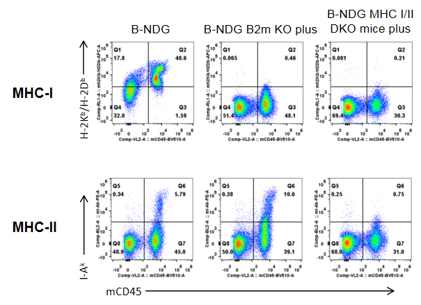
- Genetic Validation: Complete knockout of MHC class I (H-2Kb/H-2Db) and MHC class II (I-Ak) expression was confirmed by flow cytometry. Neither molecule was detectable on splenocytes of B-NDG MHC I/II DKO mice plus, validating successful double knockout on the NOD background.
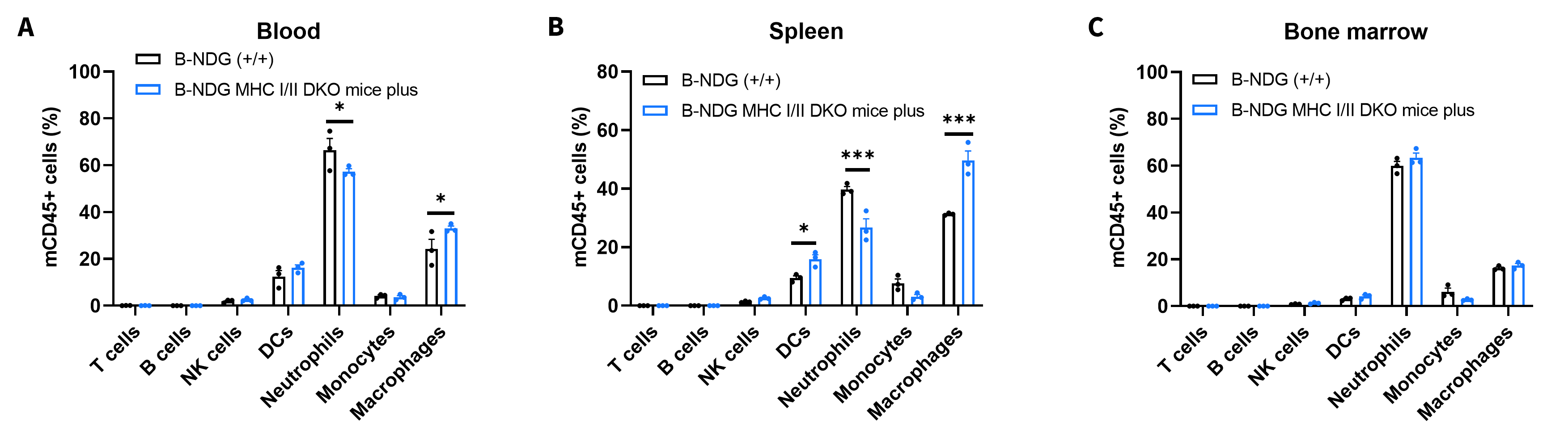
- Immunophenotyping Validation: Flow cytometry analysis of spleen, blood, and bone marrow demonstrated absence of T cells, B cells, and NK cells, confirming deep immunodeficiency and alignment with the NDG platform.
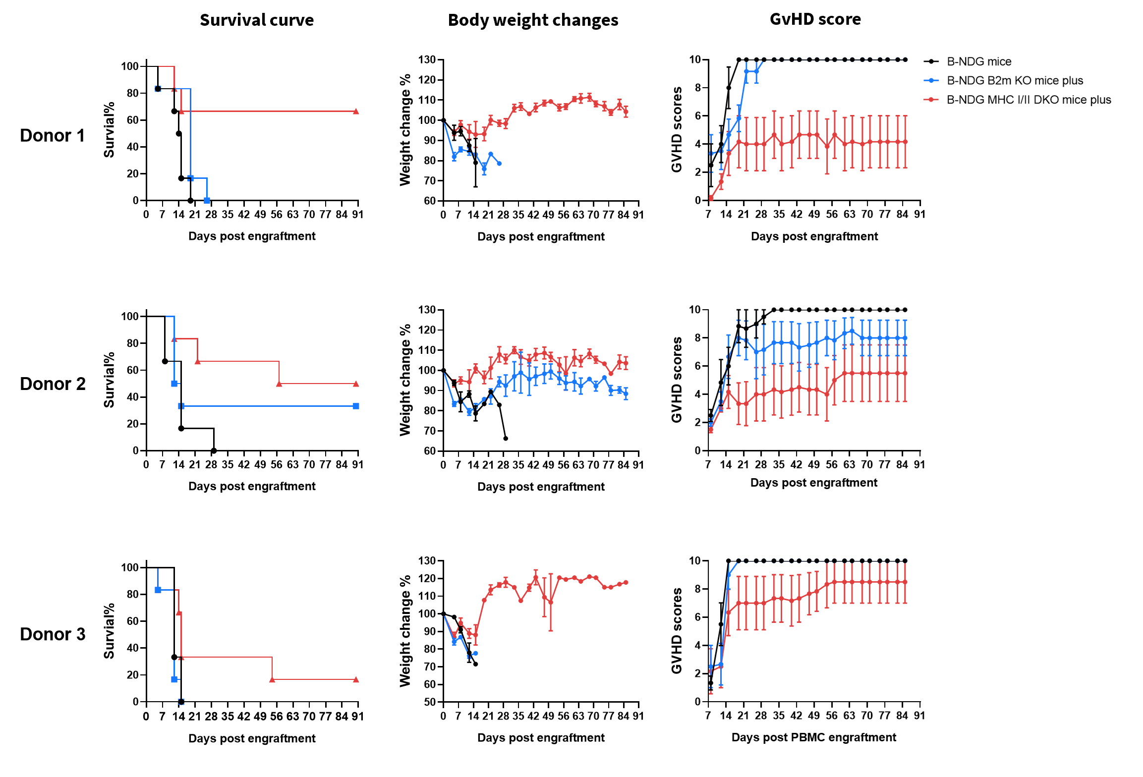
- GvHD Reduction Validation: After human PBMC engraftment, B-NDG MHC I/II DKO mice plus showed significantly reduced GvHD severity, extended survival, and improved body-weight stability compared with B-NDG and B-NDG B2m KO mice plus, confirming the functional impact of MHC-I/II deletion.
- Human PBMC Engraftment Validation: Human PBMCs successfully reconstituted in B-NDG MHC I/II DKO mice plus, supporting long-term engraftment for 96–112 days with stable human CD45⁺ leukocyte and T-cell reconstitution, including CD4⁺, CD8⁺, and Tregs, as well as detectable dendritic cells (DCs).
- Functional Human Immune Response Validation: Reconstituted human T cells expressed PD-1 and expanded robustly in vivo, indicating functional activation and suitability for immunotherapy testing.
- Preclinical Therapeutic Validation: In a CDX model (NCI-N87), anti-hCD3/hHER2 bispecific antibodies showed dose-dependent tumor-growth inhibition, confirming the model's value for evaluating T-cell–dependent human biologics.
Applications
- Human PBMC engraftment and long-term immune-system reconstitution
- GvHD modeling and mitigation studies
- Immuno-oncology research, including CDX and immune–tumor interaction studies
- Evaluation of bispecific antibodies, T-cell engagers, and immune-modulating biologics
- Checkpoint and immune-activation pathway research using human T cells (PD-1, CD4⁺, CD8⁺, Tregs)
- Pharmacology and efficacy studies with reduced mouse-driven GvHD interference
- Translational immune-response research requiring long experimental windows
MHC-I and MHC-II Protein Expression Analysis
- Mouse H-2Kb/H-2Db was only detectable in B-NDG mice but not in B-NDG B2m KO plus mice and B-NDG MHC I/II DKO Mice Plus.
- Mouse I-Ak was only detectable in B-NDG mice and B-NDG B2m KO plus mice but not in B-NDG MHC I/II DKO Mice Plus.
Strain specific H-2Kb/H-2Db (MHC-I) and I-Ak (MHC-II) expression analysis in B-NDG mice, B-NDG B2m KO plus mice and B-NDG MHC I/II DKO mice plus by flow cytometry. Splenocytes were collected from the three mice and analyzed by flow cytometry.
Analysis of Leukocyte Subpopulations
- T cells, B cells and NK cells were not detectable in all tissues of B-NDG mice and B-NDG MHC I/II DKO mice plus.
Frequency of leukocyte subpopulations in spleen, blood and bone marrow by flow cytometry. Blood, spleen and bone marrow were collected from B-NDG mice and B-NDG MHC I/II DKO mice plus (male, 9-week-old, n=3). Leukocyte subpopulations were analyzed by flow cytometry analysis. Values are expressed as mean ± SEM. Significance was determined by two-way ANOVA test. *P < 0.05, **P < 0.01, ***p < 0.001.
Reduced GvHD Severity after hPBMC Engraftment
- B-NDG MHC I/II DKO mice plus provide a GvHD-reduced immunodeficient model for human PBMC engraftment studies.
Comparison of GvHD severity after human PBMC engraftment in B-NDG mice, B-NDG B2m KO mice plus, and B-NDG MHC I/II DKO mice plus. Five-week-old female B-NDG mice, B-NDG B2m KO mice plus, and B-NDG MHC I/II DKO mice plus were irradiated with 1.0 Gy and intravenously engrafted with human PBMCs from three healthy donors on day 0 (5×10⁶ cells/mouse, n=6). Survival, body weight, and clinical GvHD scores were monitored. Compared with B-NDG mice and B-NDG B2m KO mice plus, B-NDG MHC I/II DKO mice plus showed prolonged survival, improved body-weight maintenance, and reduced GvHD scores across donors. Values are expressed as mean ± SEM.
Prolonged Survival with Reduced GvHD after hPBMC
- B-NDG MHC I/II DKO mice plus can extend the study window for human PBMC engraftment studies.
B-NDG MHC I/II DKO mice plus supports an extended experimental window after human PBMC engraftment. B-NDG MHC I/II DKO mice plus were intravenously engrafted with human PBMCs on day 0 (1×10⁷ cells/mouse, n=6). Survival, body weight, and clinical GvHD scores were monitored over time. Mice survived up to 96 days after engraftment and maintained body weight for an extended period. Clinical GvHD scores increased gradually but remained moderate, with no obvious GvHD-related clinical symptoms other than body-weight loss during most of the observation period. Values are expressed as mean ± SEM..
Sustained Human Immune-Cell Reconstitution after hPBMC Engraftment
- Sustained human immune-cell reconstitution was maintained for up to 16 weeks, dominated by human T cells.
Human PBMCs were successfully reconstituted in B-NDG MHC I/II DKO mice plus. Mice were intravenously engrafted with human PBMCs on day 0 (1×10⁷ cells/mouse, n=6). Peripheral blood was collected at the indicated time until week 16. (A) Frequency and (B) absolute numbers of reconstituted human immune-cell subsets. Human CD45+ cells increased from week 2 and remained detectable through week 16. Human T cells were the dominant population, with CD4+ T cells, CD8+ T cells, Tregs, and DCs subsets also detected. Values are expressed as mean ± SEM.
Anti-hCD3/hHER2 BsAb analog Efficacy in a huPBMC-Engrafted CDX Model
- Anti-hCD3/hHER2 BsAb analog showed strong antitumor efficacy in the huPBMC-engrafted NCI-N87 CDX model.
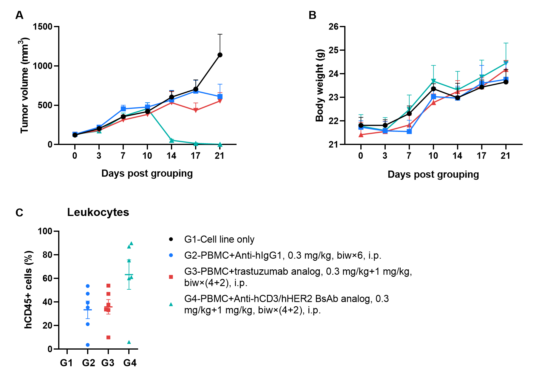
Efficacy evaluation of anti-hCD3/hHER2 bispecific antibody in a huPBMC-engrafted NCI-N87 CDX model using B-NDG MHC I/II DKO mice plus. NCI-N87 human gastric cancer cells were subcutaneously inoculated into female B-NDG MHC I/II DKO mice plus. Human PBMCs were intravenously engrafted after tumor inoculation. When tumor volume reached approximately 100 mm³, mice were randomized and treated intraperitoneally with control antibody, trastuzumab analog, or anti-hCD3/hHER2 BsAb analog. (A) Tumor volume. (B) Body weight. (C) Frequency of human CD45+ cells in peripheral blood at the endpoint. Anti-hCD3/hHER2 bispecific antibody markedly inhibited tumor growth, while body weight was maintained during treatment. These results demonstrate that huPBMC-engrafted B-NDG MHC I/II DKO mice plus can be used to evaluate T cell–engaging bispecific antibody efficacy in CDX models. Values are expressed as mean ± SEM.
AMG509 Analog Antitumor Efficacy and Tolerability
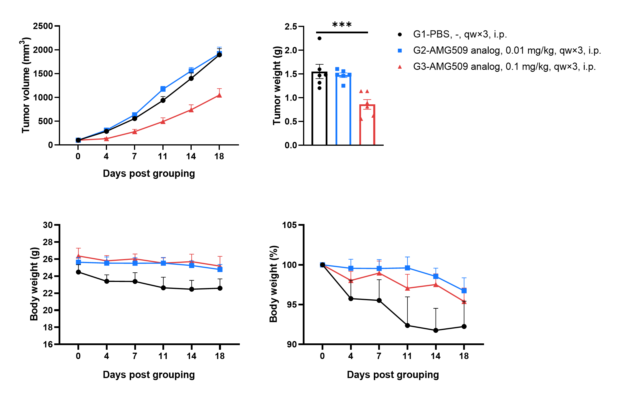
- AMG509 showed strong antitumor efficacy in the huPBMC-engrafted 22Rv1 CDX model.
Tumor growth, body weight change, and terminal tumor weight in 22Rv1 tumor-bearing huPBMC-B-NDG MHC I/II DKO mice plus. Female B-NDG MHC I/II DKO mice plus were first engrafted with human PBMCs (1×10⁷), followed 8 days later by subcutaneous inoculation of 22Rv1 cells (5 × 10⁶). When tumors reached 80–120 mm³, mice were randomized and treated intraperitoneally with PBS or AMG509 analog at 0.01 or 0.1 mg/kg once weekly for three doses (n=6). AMG509 analog at 0.1 mg/kg significantly inhibited tumor growth and reduced terminal tumor weight, while 0.01 mg/kg showed no meaningful antitumor activity. No obvious body-weight toxicity was observed at 0.1 mg/kg. Values are expressed as mean ± SEM. Significance was determined by one-way ANOVA test. *P < 0.05, **P < 0.01, ***P < 0.001.
Cytokine Profile after AMG509 Analog Treatment
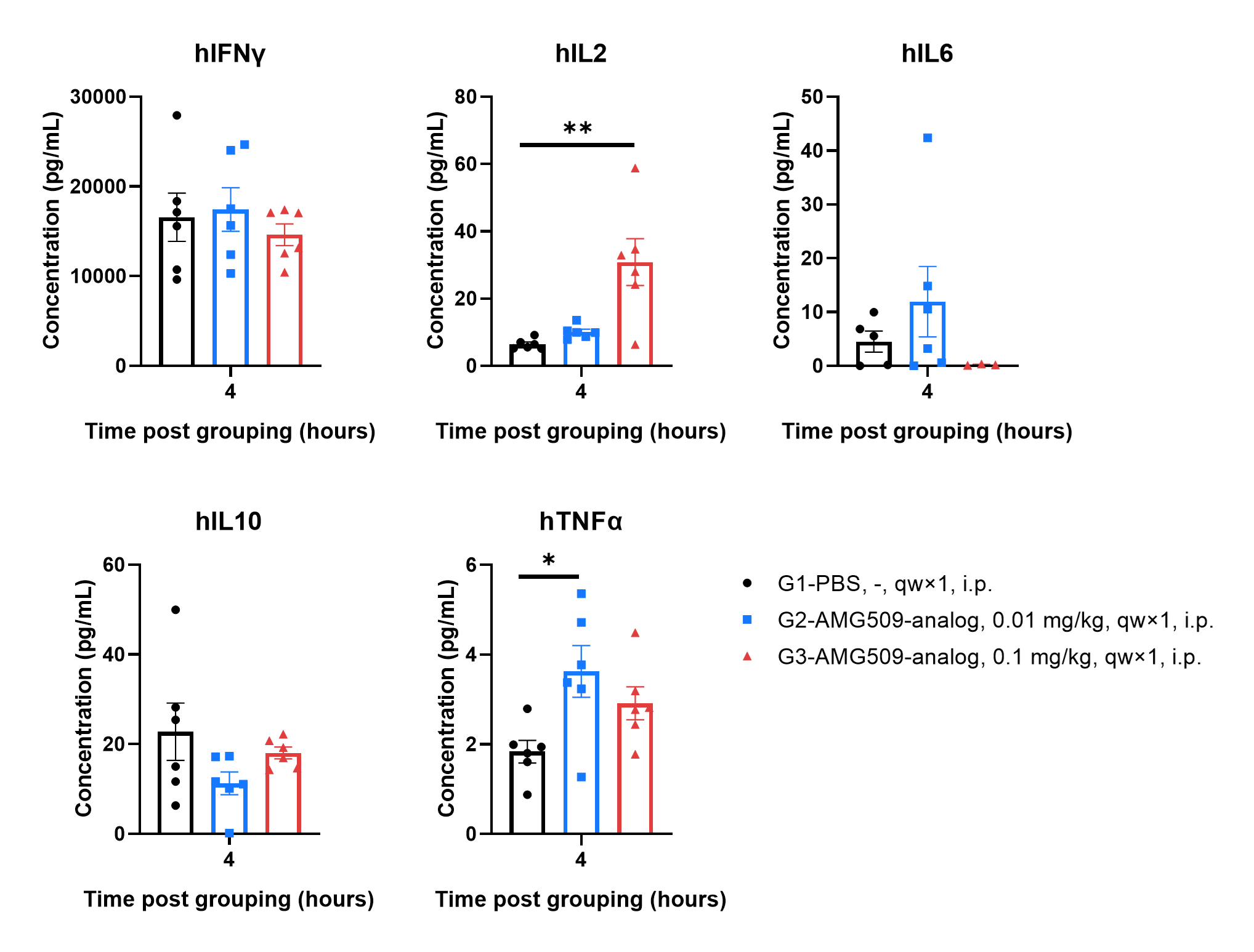
- AMG509 analog at the efficacious dose increased human IL-2, indicating T-cell activation, without inducing broad CRS-like cytokine elevation.
Serum cytokine profiling after AMG509 analog treatment in 22Rv1 tumor-bearing huPBMC-engrafted B-NDG MHC I/II DKO mice plus. Serum cytokines were measured 4 hours after dosing with PBS or AMG509 analog. AMG509 analog at 0.1 mg/kg significantly increased human IL-2, indicating T-cell activation, while IFNγ, IL-6, IL-10 and TNFα were not broadly elevated at this efficacious dose. These results suggest no overt systemic CRS-like cytokine response under the tested conditions. Values are expressed as mean ± SEM.
The Frequency Profile of Human Immune Cells in AMG509 Efficacy Study
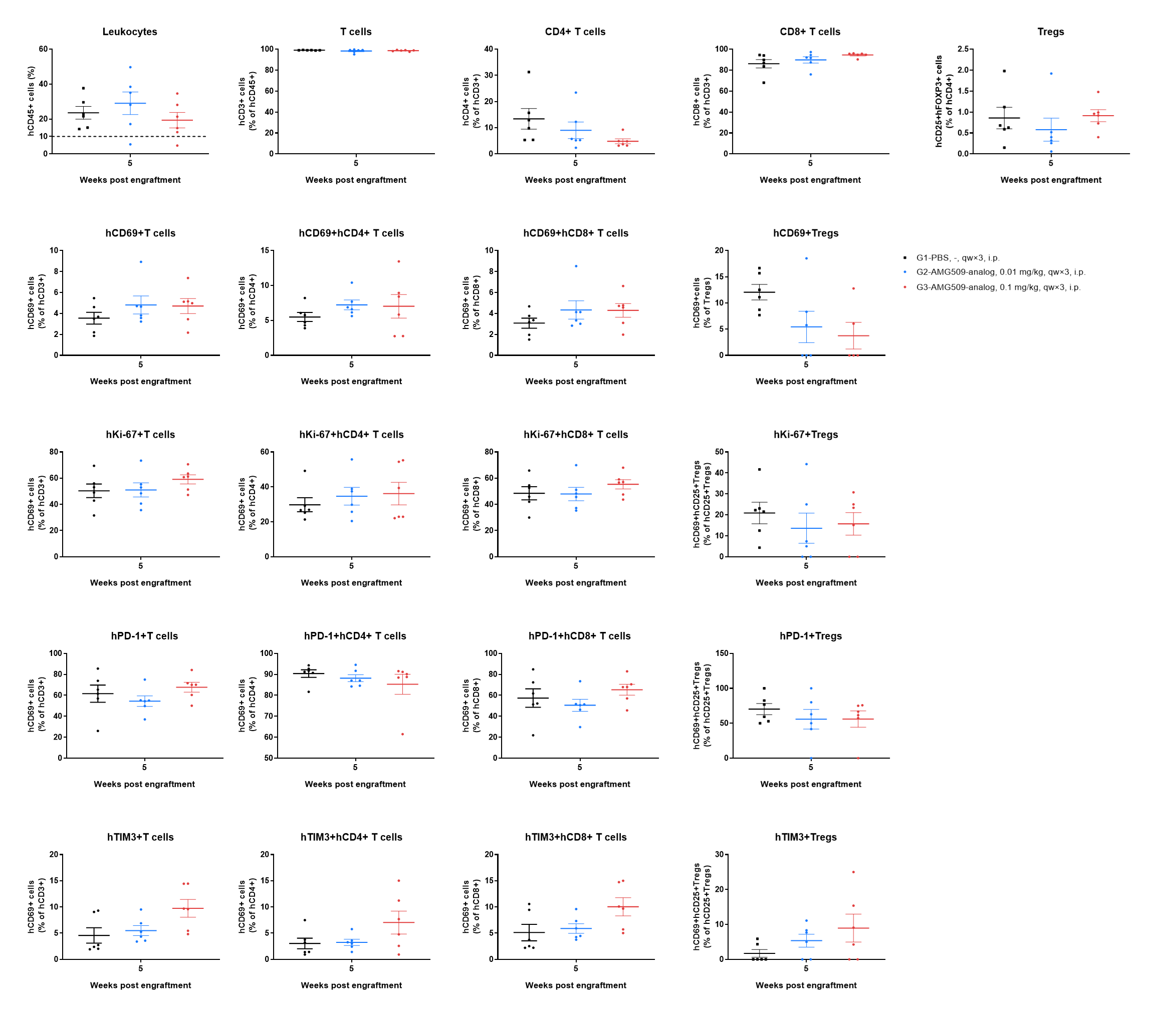
- AMG509 analog increased the frequencies of Ki-67+ and TIM-3+ human T-cell subsets.
The peripheral blood human immune-cell profiling in the AMG509 analog efficacy study using 22Rv1 tumor-bearing B-NDG MHC I/II DKO mice plus. Peripheral blood was collected at the efficacy-study endpoint, 5 weeks after human PBMC engraftment, and analyzed by flow cytometry. Human CD45+ leukocytes were detected in all groups, and CD3+ T cells were the dominant human immune-cell population. The T-cell compartment showed a CD8+ T-cell–dominant profile, with relatively low CD4+ T-cell and Treg frequencies. AMG509 analog at 0.1 mg/kg was associated with increased Ki-67+ and TIM-3+ T-cell subsets, particularly in CD8+ T cells, indicating enhanced peripheral T-cell proliferation and checkpoint/exhaustion-associated activation. Values are expressed as mean ± SEM.
The Absolute Number Profile of Human Immune Cells in AMG509 Efficacy Study
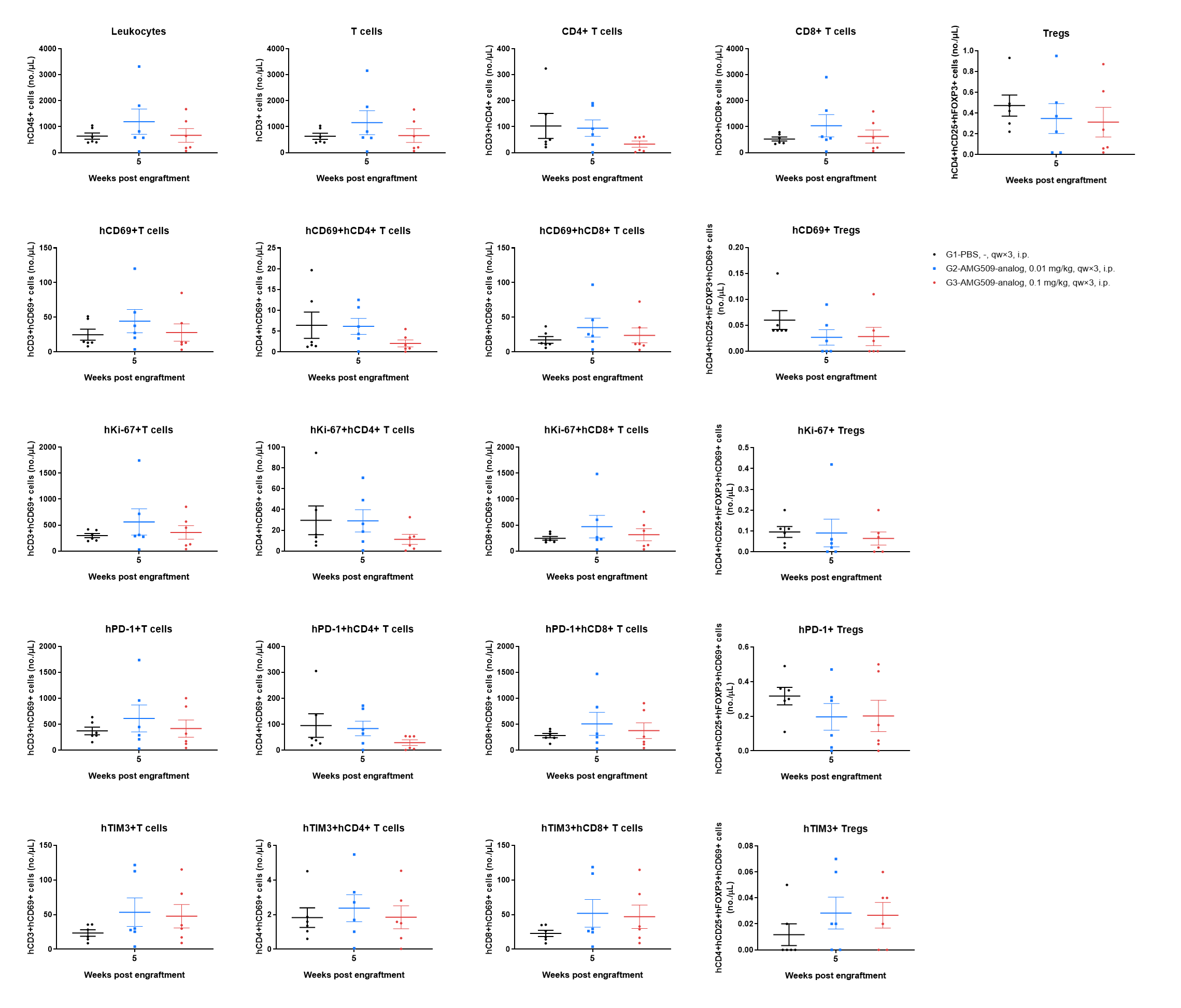
- Ki-67+, PD-1+ and TIM-3+ T-cell subsets were detectable, supporting the presence of proliferating and activated/checkpoint-associated human T cells in peripheral blood during antitumor efficacy evaluation.
The absolute number analysis of peripheral blood human immune-cell subsets in the AMG509 analog efficacy study using 22Rv1 tumor-bearing B-NDG MHC I/II DKO mice plus. Peripheral blood was collected at the efficacy-study endpoint, 5 weeks after human PBMC engraftment, and analyzed by flow cytometry. Human CD45+ leukocytes and CD3+ T cells were detected in all groups, confirming successful human PBMC reconstitution. The human T-cell compartment was mainly driven by CD8+ T cells, while CD4+ T cells and Tregs remained relatively low. Values are expressed as mean ± SEM.
The Frequency Profile of Human TILs in AMG509 Efficacy Study
- AMG509 analog increased the frequencies of Ki-67+ and TIM-3+ T-cell subsets, indicating enhanced intratumoral T-cell proliferation and activation/checkpoint-associated phenotypes.
Human tumor-infiltrating lymphocytes in 22Rv1 tumor-bearing huPBMC-B-NDG MHC I/II DKO mice plus were analyzed by flow cytometry at the study endpoint. Tumor tissues were collected at the efficacy-study endpoint and analyzed by flow cytometry. Human CD45+ immune cells were detectable in tumors, and CD3+ T cells were the predominant human immune-cell population. AMG509 analog at 0.1 mg/kg did not markedly increase total T-cell infiltration, but increased Ki-67+ and TIM-3+ cells within CD3+, CD4+ and CD8+ T-cell subsets, with increased CD69+ and PD-1+ subsets also observed. Values are expressed as mean ± SEM. Significance was determined by one-way ANOVA test. *P < 0.05, **P < 0.01, ***P < 0.001.
Tissue Tolerability Assessment in AMG509 Efficacy Study
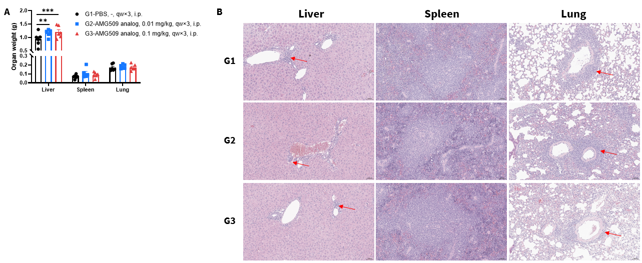
- No overt AMG509 analog–specific severe organ toxicity was observed in liver, spleen, or lung tissues.
Organ weight and H&E histopathology in the AMG509 analog efficacy study using 22Rv1 tumor-bearing huPBMC-engrafted B-NDG MHC I/II DKO mice plus. Liver, spleen, and lung tissues were collected for analysis. (A) Organ weights. (B) Representative H&E staining images. Liver weight was slightly increased in AMG509 analog–treated groups, while spleen and lung weights showed no obvious treatment-related changes. Inflammatory cell infiltration was observed in liver and lung tissues, as indicated by red arrows. Similar findings were also present in PBS controls, suggesting these changes were mainly related to the huPBMC-engrafted model background rather than AMG509 analog–specific organ toxicity. Values are expressed as mean ± SEM. Significance was determined by one-way ANOVA test. *P < 0.05, **P < 0.01, ***P < 0.001.
CRS-like Tolerability Assessment of AMG509 Analog
- High-dose AMG509 analog induced a transient decrease in rectal temperature, suggesting a CRS-like response under high-dose treatment.
CRS-like tolerability assessment of AMG509 analog in 22Rv1 tumor-bearing huPBMC-engrafted B-NDG MHC I/II DKO mice plus. Mice were treated intravenously with PBS or AMG509 analog at 0.1, 1, or 10 mg/kg as a single dose (n=3). Rectal temperature was monitored before and after dosing. AMG509 analog at 10 mg/kg induced a transient decrease in rectal temperature at 6 hours, suggesting a high-dose CRS-like response. Values are expressed as mean ± SEM.
CRS-like Cytokine Response after AMG509 Analog Treatment
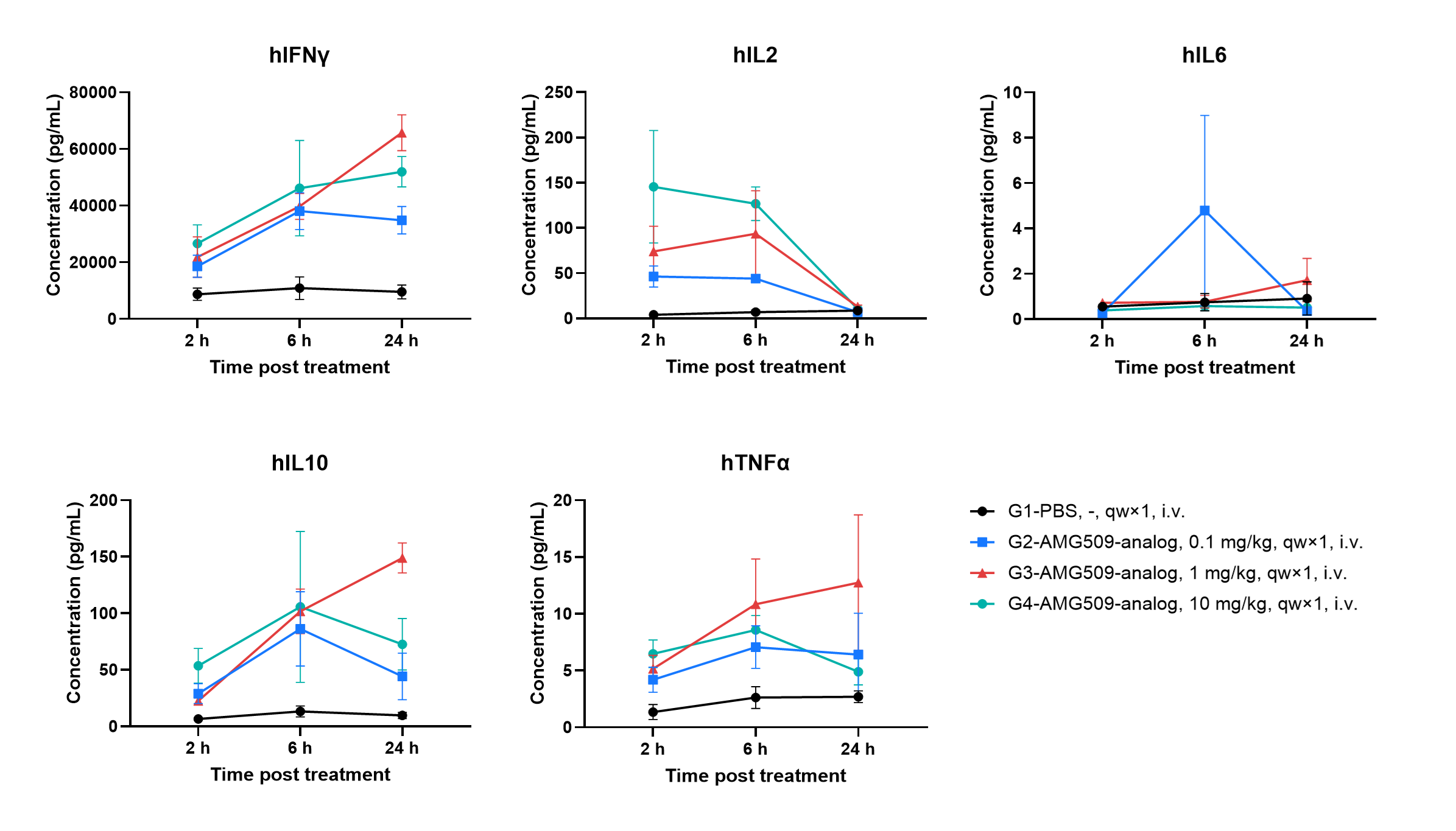
- AMG509 analog induced acute cytokine responses, including increased IFNγ, IL2, IL10, and TNFα, supporting the use of B-NDG MHC I/II DKO mice plus for CRS-like cytokine response assessment.
Serum cytokine profiling after single-dose AMG509 analog treatment in 22Rv1 tumor-bearing huPBMC-B-NDG MHC I/II DKO mice plus. Mice were treated intravenously with PBS or AMG509 analog at 0.1, 1, or 10 mg/kg, and serum cytokines were measured at 2, 6, and 24 hours after dosing. AMG509 analog induced acute cytokine responses, including increased IFNγ, IL2, IL10, and TNFα, mainly at 1 and/or 10 mg/kg. IL2 peaked early and declined by 24 hours, whereas IFNγ remained elevated at 24 hours in high-dose groups. IL6 did not show a consistent dose-dependent increase. Values are expressed as mean ± SEM.
* When publishing results obtained using this animal model, please acknowledge the source as follows: The animal model [B-NDG MHC I/II DKO mice plus] (Cat# 111895) was purchased from Biocytogen.