Description
TREM2: As a prominent therapeutic target in neurodegeneration research
- Gene Information: The human TREM2 gene maps to chromosomal band 6p21.1 with a full-length sequence of ~4676 bp across five exons, which translates to a polypeptide containing 230 amino acids.
- Protein Expression: TREM2 expression is largely restricted to myeloid cells. Microglia constitute the primary cell type expressing TREM2 in the central nervous system, and this receptor can also be detected in peripheral myeloid cells including macrophages, monocytes and osteoclasts.
- Signaling Pathway: Ligand engagement of TREM2 triggers tyrosine phosphorylation of the DAP12 ITAM motif. Activated Syk kinase is then recruited to launch a panel of downstream signaling pathways (ERK, PI3K, PLCγ, Vav), which collectively govern immune cell viability, motility, phagocytosis and cellular function.
- Therapeutic Inhibition: Agonistic monoclonal antibodies and small-molecule chemicals represent the two dominant branches of TREM2 drug development. Other modalities such as CAR-T, nanobodies and recombinant polypeptides have also been deployed for TREM2-targeted therapy, yet none have advanced beyond preclinical or drug discovery phases.
Targeting strategy
5XFAD
- The Tg(5XFAD) mouse line was introduced from Northwest University. A transgene was designed with a mutant human amyloid beta precursor protein (APP) cDNA sequence, including Swedish (K670N/M671L), Florida (I716V), and London (V717I) mutations, inserted into exon 2 of the mouse Thy1 gene. A second transgene was designed with a mutant human presenilin 1 (PSEN1) cDNA sequence, including M146L and L286V mutations, inserted into exon 2 of the mouse Thy1 gene. Both transgenes were added together in equal proportions and co-injected into the pronuclei of embryos to generate the Tg(5XFAD) mice.
TREM2
- Exons 1-5 of the mouse Trem2 gene that encode the full-length protein were replaced by human counterparts in B-hTREM2 mice. The promoter and 5' UTR of the mouse Trem2 gene were retained, and the 3' UTR region of the mouse gene was replaced by human counterparts.
- TREM2 expression is driven by the endogenous mouse Trem2 promoter, while mouse Trem2 gene transcription and translation will be disrupted.
- Tg(5XFAD)/B-hTREM2 mice were obtained by mating Tg(5XFAD) mice (114651) and B-hTREM2 mice (111176).
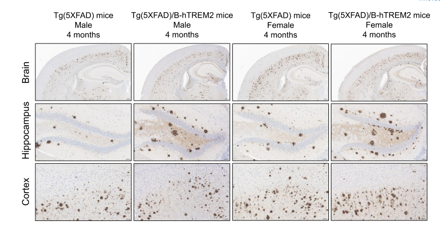
Histopathological Analysis-Aβ in Tg(5XFAD)/B-hTREM2 mice
Histopathological analysis of human Aβ in Tg(5XFAD)/B-hTREM2 mice. Brain was collected from 4-month-old Tg(5XFAD) mice and Tg(5XFAD)/B-hTREM2 mice and processed into paraffin sections. The Aβ plaque in the brain was detected by IHC with anti-human β-Amyloid antibody. Aβ deposition in brain tissues is comparable between age-matched Tg(5XFAD) mice and Tg(5XFAD)/B-hTREM2 mice. Female mice presented more severe Aβ plaques relative to males. Scale bar: 200 μm.
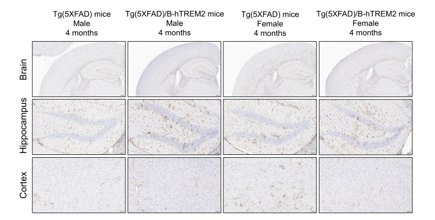
Histopathological Analysis-Astrocyte in Tg(5XFAD)/B-hTREM2 mice
Histopathological analysis of astrocytes in Tg(5XFAD)/B-hTREM2 mice. Brain was collected from 4-month-old Tg(5XFAD) mice and Tg(5XFAD)/B-hTREM2 mice and processed into paraffin sections. The astrocytes in the brain were detected by IHC with anti-GFAP antibody. No significant difference in astrocytes was observed between Tg(5XFAD) mice and Tg(5XFAD)/B-hTREM2 mice. Scale bar: 50 μm.
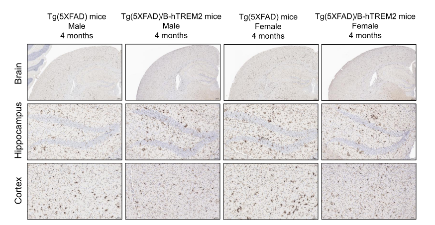
Histopathological Analysis-Microglia in Tg(5XFAD)/B-hTREM2 mice
Histopathological analysis of astrocytes in Tg(5XFAD)/B-hTREM2 mice. Brain was collected from 4-month-old Tg(5XFAD) mice and Tg(5XFAD)/B-hTREM2 mice and processed into paraffin sections. The microglia cells in the brain were detected by IHC with anti-Iba1 antibody. No significant difference in microglia cells was observed between Tg(5XFAD) mice and Tg(5XFAD)/B-hTREM2 mice. Scale bar: 50 μm.
* When publishing results obtained using this animal model, please acknowledge the source as follows: The animal model [Tg(5XFAD)/B-hTREM2 mice] (Cat# 114908) was purchased from Biocytogen.