Description
- CD3 consists of four protein chains (CD3E, CD3D, CD3G and CD3Z), which are important biological markers on the T cell membrane. CD3 can form a TCR/CD3 complex with the T cell receptor, participating in the regulation of T cell antigen recognition, signal transduction and T cell development. EGFR is expressed in various tissues. Upon binding of ligands like EGF to the EGFR receptor, EGFR dimers are formed. The activation of EGFR dimers stimulates intracellular tyrosine kinases, resulting in phosphorylation that triggers downstream signaling cascades, leading to cancer cell proliferation and the proliferation of new blood vessels within cancer cells. The mechanism of action of EGFR-targeting drugs primarily involves blocking or inhibiting the activity of EGFR, thereby preventing the growth and spread of cancer cells.
- B-hCD3EDG/hEGFR mice were obtained by mating B-hCD3EDG mice(110039) and B-hEGFR mice(120771). In B-hCD3EDG/hEGFR mice, chimeric human CD3EDG was expressed, while mouse Cd3edg were knocked out. The extracellular region of mouse Egfr was replaced by human counterparts in B-hCD3EDG/hEGFR mice.
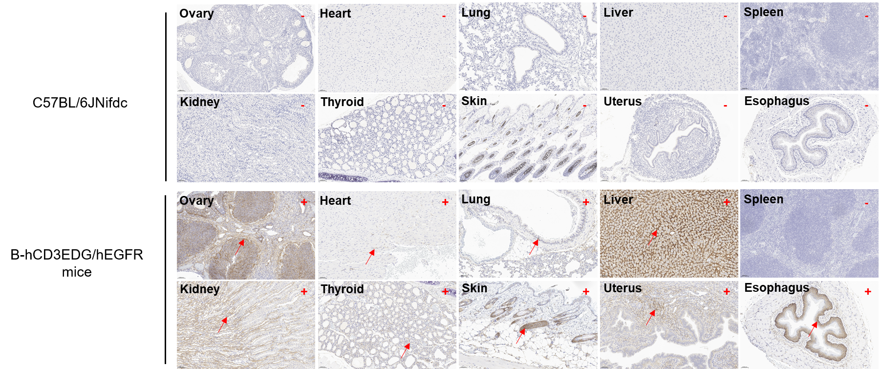
- Human EGFR was detectable in heart, lung, liver, kidney, thyroid gland, skin, uterus, esophagus, ovary, and testis of B-hCD3EDG/hEGFR mice but not in C57BL/6JNifdc mice.
- This product is used for tumor pharmacology and safety evaluation of TCE targeting CD3 and EGFR
Targeting strategy
Gene targeting strategy for B-hCD3EDG/hEGFR mice. The exons 2-17 of mouse Egfr gene that encode extracellular domain were replaced by human counterparts in B-hCD3EDG/hEGFR mice. The genomic region of mouse Egfr gene that encodes transmembrane domain and cytoplasmic portion was retained. The promoter, 5’UTR, signal peptide and 3’UTR region of the mouse gene were also retained.
Protein expression profile of EGFR
Immunohistochemical (IHC) analysis of EGFR expression in B-hCD3EDG/hEGFR mice. The ovary, heart, lung, liver, spleen, kidney, thyroid gland, skin, uterus, and esophagus were collected from wild-type C57BL/6JNifdc mice and B-hCD3EDG/hEGFR mice (female, 6-week-old), analyzed by IHC with anti-EGFR (Invitrogen, MA5-49312). Human EGFR was detectable in B-hCD3EDG/hEGFR mice but not in C57BL/6JNifdc mice. The arrow indicates tissue cells with positive EGFR staining (brown). “+” indicates that the tissue is positive, and “-” indicates that the tissue is negative.
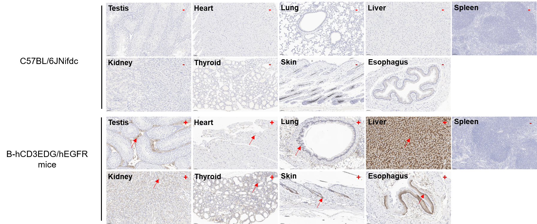
Immunohistochemical (IHC) analysis of EGFR expression in B-hCD3EDG/hEGFR mice. The testis, heart, lung, liver, spleen, kidney, thyroid gland, skin and esophagus were collected from wild-type C57BL/6JNifdc mice and B-hCD3EDG/hEGFR mice (male, 6-week-old), analyzed by IHC with anti-EGFR (Invitrogen, MA5-49312). Human EGFR was detectable in B-hCD3EDG/hEGFR mice but not in C57BL/6JNifdc mice. The arrow indicates tissue cells with positive EGFR staining (brown). “+” indicates that the tissue is positive, and “-” indicates that the tissue is negative.
* When publishing results obtained using this animal model, please acknowledge the source as follows: The animal model [B-hCD3EDG/hEGFR mice] (Cat# 113562) was purchased from Biocytogen.