C57BL/6-Il33tm1(IL33)Tslptm1(TSLP)Crlf2tm2(CRLF2)/Bcgen • 111867
on this page
Key Advantages
Validation
Application
In B-hIL33/hTSLP/hTSLPR mice, the exons 1-5 of the mouse Tslp gene encoding the full-length protein were replaced with human TSLP exons 1-4. The exons 2-8 of the mouse Il33 gene were replaced with human IL33 exons 2-8.
For TSLPR humanization, a chimeric CDS encoding the human TSLPR signal peptide, extracellular region, and transmembrane region together with the mouse Tslpr cytoplasmic region was inserted into mouse Tslpr exon 2. This strategy enables B-hIL33/hTSLP/hTSLPR mice to express human IL33, human TSLP, and chimeric humanized TSLPR while disrupting endogenous mouse Il33, Tslp, and Tslpr expression.
Strain-specific IL33 and TSLP protein expression was analyzed in homozygous B-hIL33/hTSLP/hTSLPR mice by ELISA.
Calcipotriol (MC903) was dissolved in ethanol and topically applied to the ears of wild-type C57BL/6 mice (+/+) and homozygous B-hIL33/hTSLP/hTSLPR mice (H/H) for 7 days (n=3). Ear grinding supernatants were collected and analyzed with species-specific ELISA kits. Mouse IL33 and mouse TSLP were detectable in wild-type C57BL/6 mice. Human IL33 and human TSLP were detectable in homozygous B-hIL33/hTSLP/hTSLPR mice but not in wild-type mice. ND: not detectable.
Strain-specific TSLPR protein expression was analyzed in homozygous B-hIL33/hTSLP/hTSLPR mice by flow cytometry.
Bone marrow cells were collected from wild-type C57BL/6 mice (+/+) and homozygous B-hIL33/hTSLP/hTSLPR mice (H/H). Dendritic cells were induced from bone marrow cells in culture and analyzed with species-specific anti-TSLPR antibodies. Mouse TSLPR was detectable in wild-type C57BL/6 mice. Human TSLPR was detectable in homozygous B-hIL33/hTSLP/hTSLPR mice but not in wild-type mice.
BALF immune-cell infiltration was analyzed in an OVA/hIL33/hTSLP-induced asthma model using B-hIL33/hTSLP/hTSLPR mice.
Male B-hIL33/hTSLP/hTSLPR mice (11-week-old, n=8) were immunized with OVA/hIL33/hTSLP to induce asthma-like inflammation. Anti-human IL33 antibody (Itepekimab analog, synthesized in-house) and anti-human TSLP antibody (Tezepelumab analog, synthesized in-house) were intraperitoneally injected from Day -1 and Day 6. CD45+ cells and eosinophils in BALF were significantly reduced in the Itepekimab group and the Itepekimab plus Tezepelumab combination group compared with the OVA/hIL33/hTSLP-induced PBS group. Values are expressed as mean ± SEM. Significance was determined by two-way ANOVA. *P < 0.05, **P < 0.01, ***P < 0.001.
Serum total IgE was analyzed in an OVA/hIL33/hTSLP-induced asthma model using B-hIL33/hTSLP/hTSLPR mice.
Male B-hIL33/hTSLP/hTSLPR mice (11-week-old, n=8) were immunized with OVA/hIL33/hTSLP to induce asthma-like inflammation. Anti-human IL33 antibody (Itepekimab analog) and anti-human TSLP antibody (Tezepelumab analog) were intraperitoneally administered, and serum was collected at the study endpoint for total IgE ELISA analysis. Total IgE levels were significantly reduced in mice treated with Itepekimab, Tezepelumab, or the Itepekimab plus Tezepelumab combination therapy compared with untreated mice. Values are expressed as mean ± SEM. Significance was determined by two-way ANOVA. *P < 0.05, **P < 0.01, ***P < 0.001.
Lung histopathology was evaluated by H&E staining in OVA/hIL33/hTSLP-induced B-hIL33/hTSLP/hTSLPR mice.
Lung tissues were collected at the study endpoint and analyzed by H&E staining. Inflammatory infiltration and mucus secretion in lung tissue were lower in mice treated with Itepekimab and in the Itepekimab plus Tezepelumab combination therapy group than in untreated mice. These results indicate that B-hIL33/hTSLP/hTSLPR mice provide a powerful preclinical model for in vivo evaluation of anti-human IL33 antibodies, anti-human TSLP antibodies, and IL33/TSLP combination therapies. Values are expressed as mean ± SEM. Significance was determined by unpaired t-test. *P < 0.05, **P < 0.01, ***P < 0.001.
Q1: What are B-hIL33/hTSLP/hTSLPR mice?
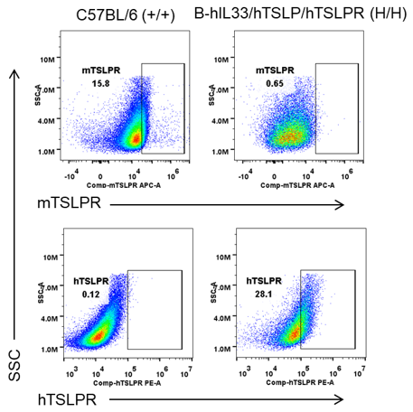
B-hIL33/hTSLP/hTSLPR mice are triple target-humanized mice expressing human IL33, human TSLP, and chimeric humanized TSLPR in a C57BL/6 background for allergic inflammation and asthma research.
Q2: Why are IL33, TSLP, and TSLPR important in asthma and allergic disease?
IL33 and TSLP are epithelial cytokines that initiate and amplify type 2 inflammation, while TSLPR mediates TSLP signaling in immune cells involved in allergic airway inflammation.
Q3: How was human IL33 and human TSLP expression validated in this model?
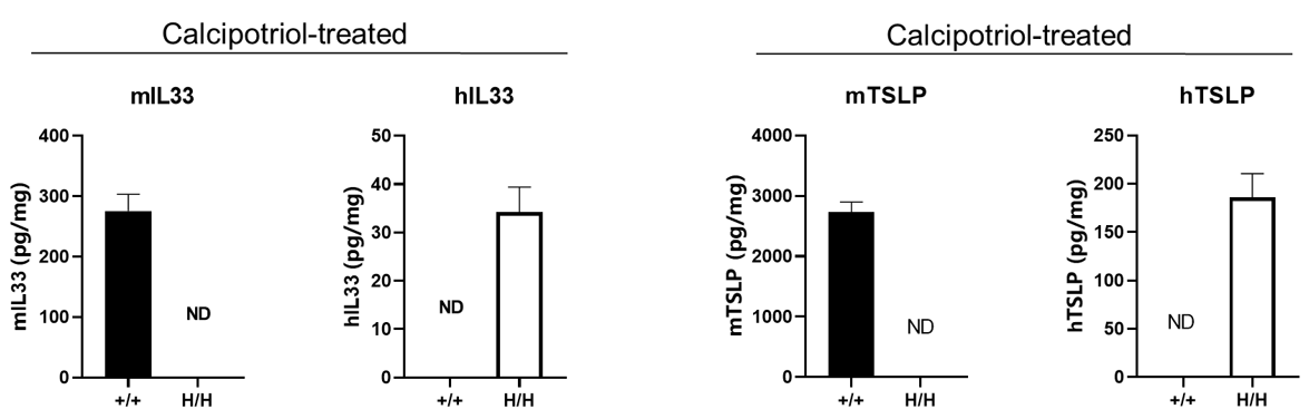
After MC903 stimulation, human IL33 and human TSLP were detected in homozygous B-hIL33/hTSLP/hTSLPR mice by ELISA, while mouse IL33 and mouse TSLP were detected in wild-type mice.
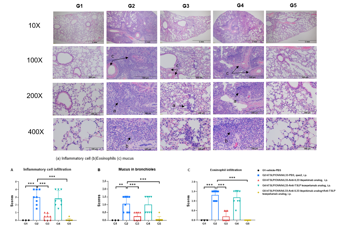
Q4: Can B-hIL33/hTSLP/hTSLPR mice be used for anti-human antibody efficacy studies?
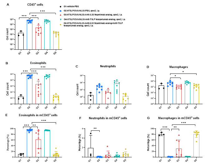
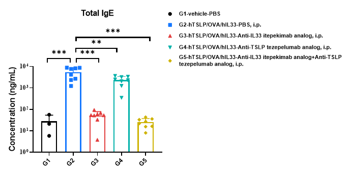
Yes. Anti-human IL33 antibody and anti-human TSLP antibody reduced BALF immune-cell infiltration, serum total IgE, and lung inflammation in an OVA/hIL33/hTSLP-induced asthma model.
Q5: What are the main applications of B-hIL33/hTSLP/hTSLPR mice?
Applications include anti-human IL33 antibody evaluation, anti-human TSLP antibody studies, IL33/TSLP combination therapy research, asthma models, allergic airway inflammation, and type 2 inflammation studies.