

C57BL/6-Pdcd1tm1(PDCD1)BcgenCd274tm1(CD274)BcgenVegfatm1(VEGFA)Bcgen/Bcgen • 112706
on this page

VEGFA: a master regulator of angiogenesis

PD-1
PD-L1
VEGFA
The B-hPD-1/hPD-L1/hVEGFA three knock-in model, was developed by breeding the B-hPD-1 mice, the B-hPD-L1 mice and the B-hVEGFA mice

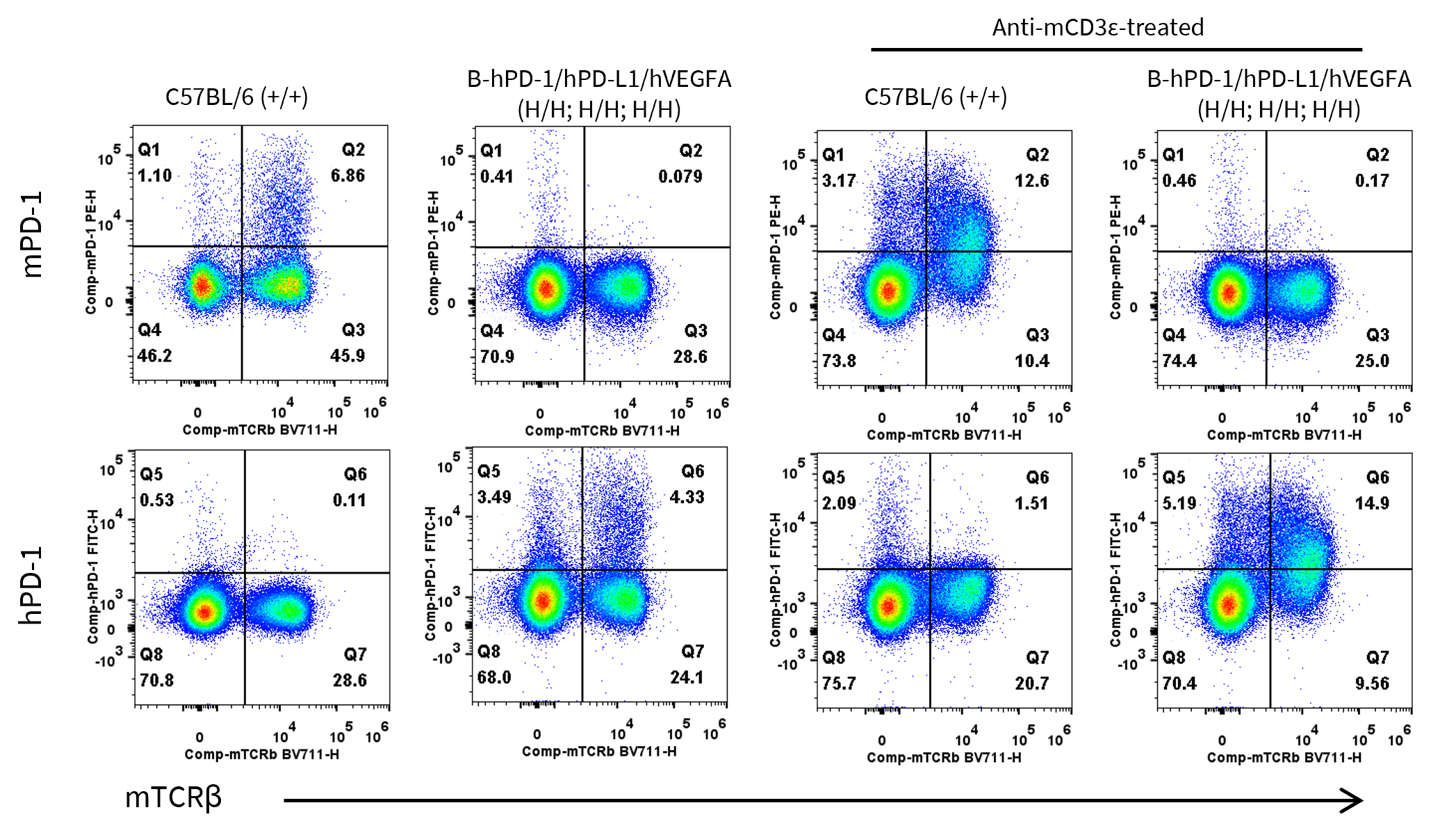
Strain specific PD-1 expression analysis in wild-type C57BL/6 and homozygous B-hPD-1/hPD-L1/hVEGFA mice by flow cytometry. Splenocytes were collected from wild-type C57BL/6 mice (+/+) and homozygous B-hPD-1/hPD-L1/hVEGFA mice (H/H) after stimulated with anti-mouse CD3ε antibody (7.5 μg, i.p.) in vivo for 24 hrs (female, 6-week-old, n=1) or not. Protein expression was analyzed with anti-mouse PD-1 antibody (Biolegend, 109104) and anti-human PD-1 antibody (Biolegend, 329904) by flow cytometry.
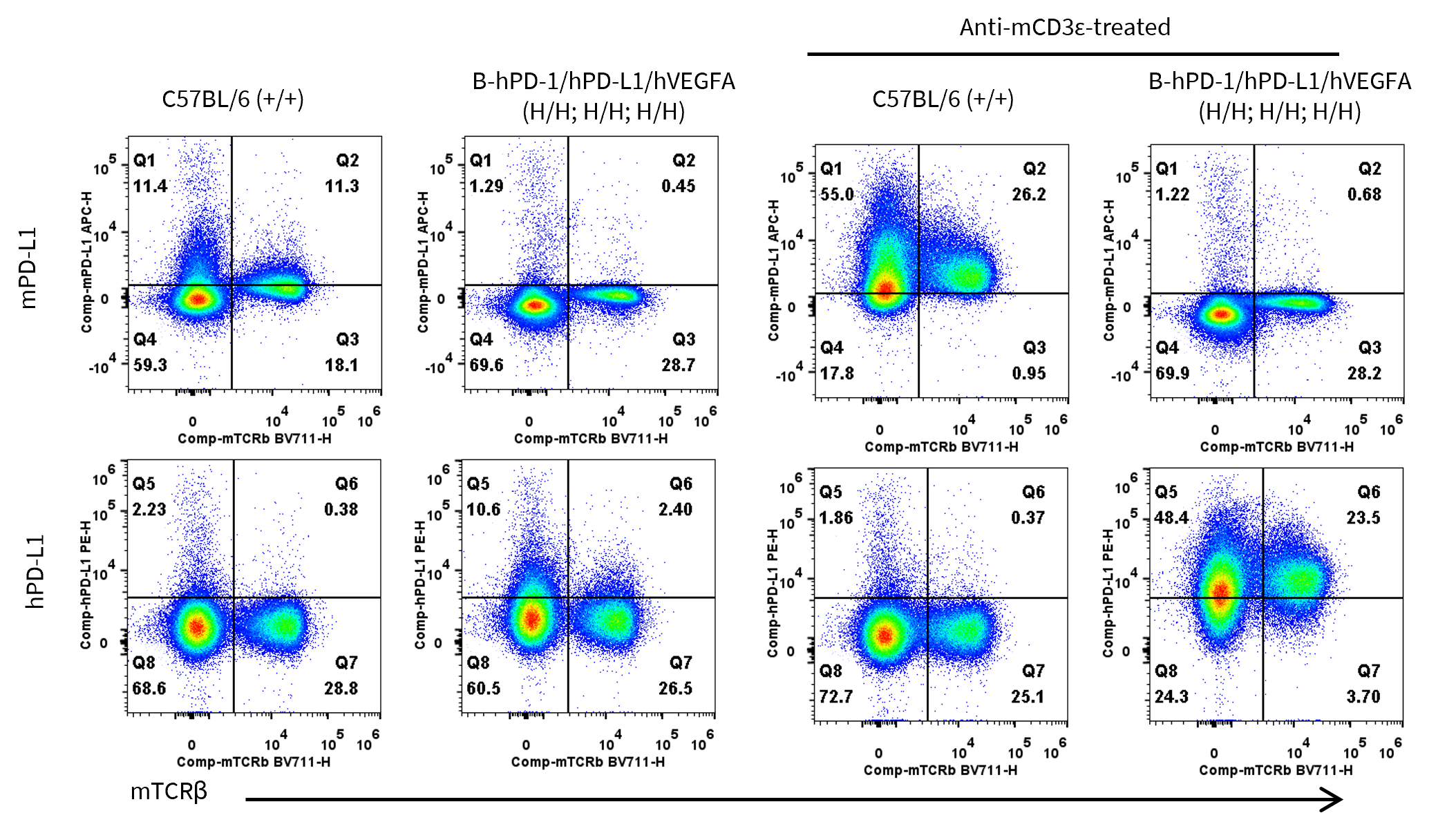
Strain specific PD-L1 expression analysis in wild-type C57BL/6 and homozygous B-hPD-1/hPD-L1/hVEGFA mice by flow cytometry. Splenocytes were collected from wild-type C57BL/6 mice (+/+) and homozygous B-hPD-1/hPD-L1/hVEGFA mice (H/H) after stimulated with anti-mouse CD3ε antibody (7.5 μg, i.p.) in vivo for 24 hrs (female, 6-week-old, n=1) or not. Protein expression was analyzed with anti-mouse PD-L1 antibody (Biolegend, 124312) and anti-human PD-L1 antibody (Biolegend, 329706) by flow cytometry.

Strain specific VEGFA expression analysis in wild-type C57BL/6 mice and homozygous B-hPD-1/hPD-L1/hVEGFA mice by ELISA. Lung homogenates were collected from wild-type C57BL/6 mice (+/+) (male, n=3, 8-week-old) and homozygous B-hPD-1/hPD-L1/hVEGFA mice (H/H; H/H; H/H) (male, n=3, 8-week-old). Expression level of mouse and human VEGFA were analyzed by ELISA (anti-mouse VEGFA antibody: R&D, MMV00; anti-human VEGFA antibody: R&D, DVE00).
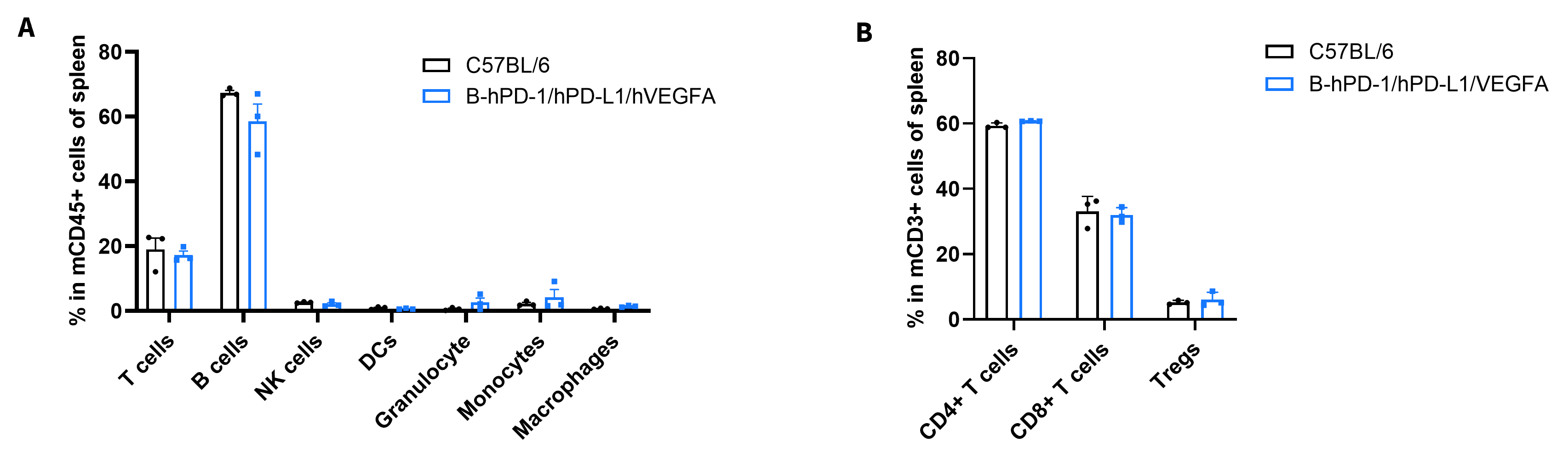
Frequency of leukocyte subpopulations in spleen by flow cytometry. Splenocytes were isolated from wild-type C57BL/6 mice and homozygous B-hPD-1/hPD-L1/hVEGFA mice (n=3, 10-week-old). A. Flow cytometry analysis of the splenocytes was performed to assess the frequency of leukocyte subpopulations. B. Frequency of T cell subpopulations. Values are expressed as mean ± SEM. Significance was determined by two-way ANOVA test. *P < 0.05, **P < 0.01, ***p < 0.001.

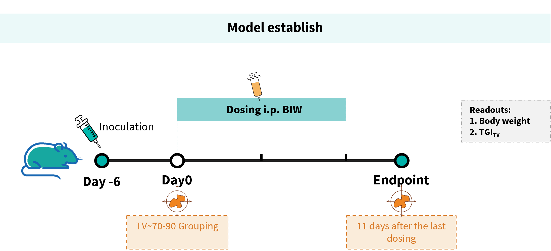
Establishment of a B-hVEGFA MC38 model and in vivo efficacy study of Ivonescimab antibody. B-hVEGFA MC38 cells were implanted subcutaneously into homozygous B-hPD-1/hPD-L1/hVEGFA mice (female, 8 weeks old, n=6). When the average tumor volume reached approximately 70~90 mm³, mice were randomized and subsequently administered the Ivonescimab analog via intraperitoneal injection.
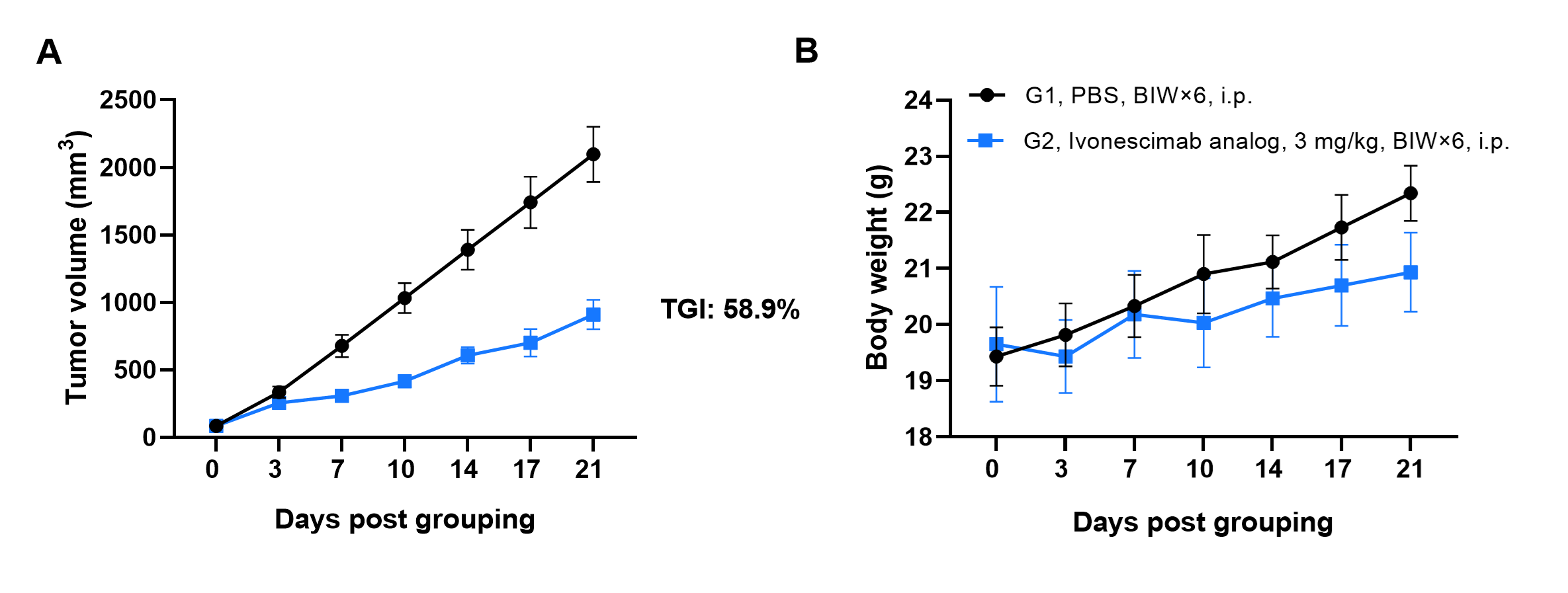
Antitumor activity of Ivonescimab analog (in-house) in B-hPD-1/hPD-L1/hVEGFA mice. (A) Tumor growth curves. (B) Body weight changes during treatment. As shown in panel A, Ivonescimab was efficacious in controlling tumor growth in B-hPD-1/hPD-L1/hVEGFA mice. Values are expressed as mean ± SEM. The overage of this tumor model is 50%.
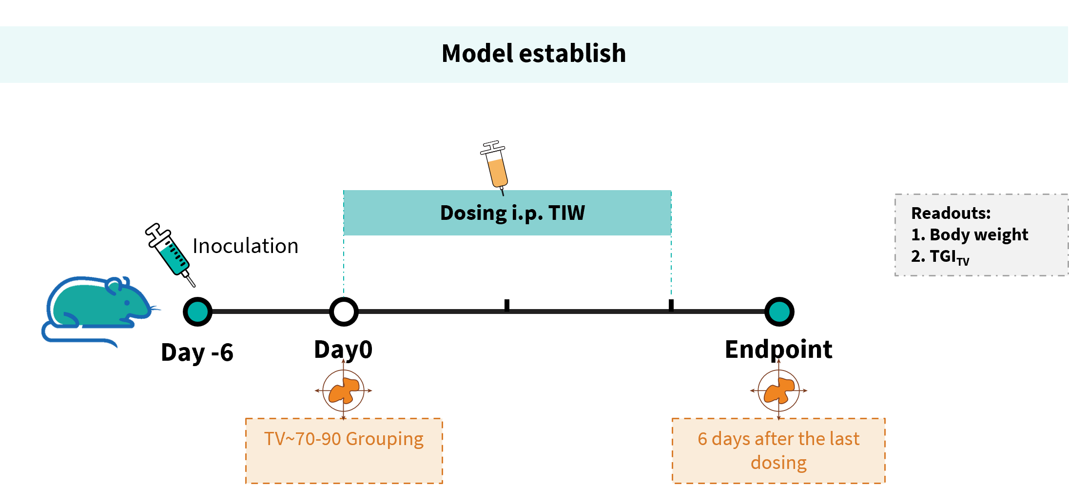
Establishment of a B-hVEGFA MC38 model and in vivo efficacy study of Bevacizumab & Keytruda combination antibody. B-hVEGFA MC38 cells were implanted subcutaneously into homozygous B-hPD-1/hPD-L1/hVEGFA mice (female, 8 weeks old, n=6). When the average tumor volume reached approximately 70~90 mm³, mice were randomized and subsequently administered with anti-PD-1 & anti-VEGFA antibody via intraperitoneal injection.
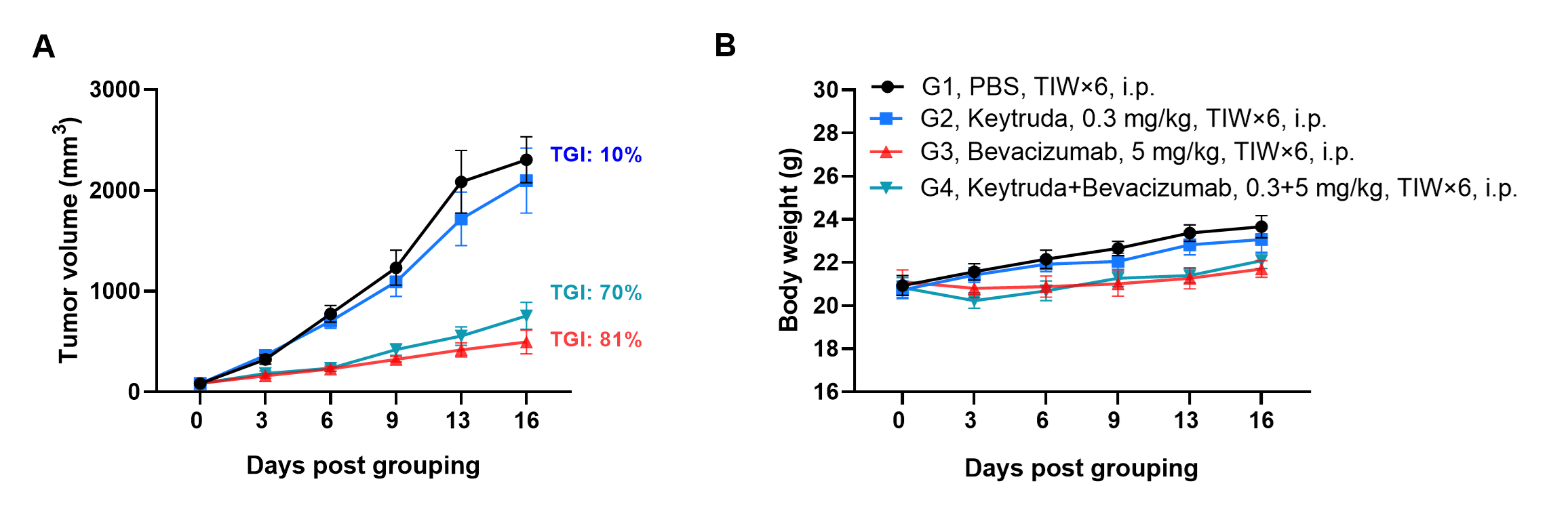
Antitumor activity of Bevacizumab & Keytruda in B-hPD-1/hPD-L1/hVEGFA mice. (A) Tumor growth curves. (B) Body weight changes during treatment. As shown in panel A, Bevacizumab was efficacious in controlling tumor growth in B-hPD-1/hPD-L1/hVEGFA mice. The combination of these two antibodies is not more effective than monotherapy. Values are expressed as mean ± SEM.
The overage of this tumor model is 40%.
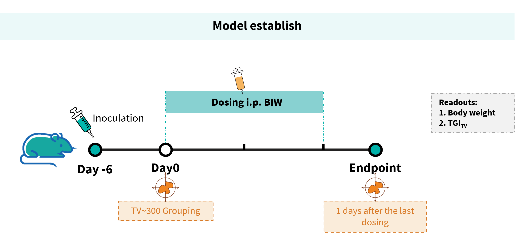
Establishment of a B-hVEGFA/hPD-L1 plus MC38 model and in vivo efficacy study of Ivonescimab antibody. B-hVEGFA/hPD-L1 plus MC38 cells were implanted subcutaneously into homozygous B-hPD-1/hPD-L1/hVEGFA mice (male, 8 weeks old, n=6). When the average tumor volume reached approximately 300 mm³, mice were randomized and subsequently administered with anti-PD-1 & anti-VEGFA antibody via intraperitoneal injection.

Antitumor activity of Ivonescimab (also known as AK112) in B-hPD-1/hPD-L1/hVEGFA mice. (A) Tumor growth curves. (B) Body weight changes during treatment. As shown in panel A, Ivonescimab was efficacious in controlling tumor growth in B-hPD-1/hPD-L1/hVEGFA mice. Values are expressed as mean ± SEM. The overage of this tumor model is 60%.
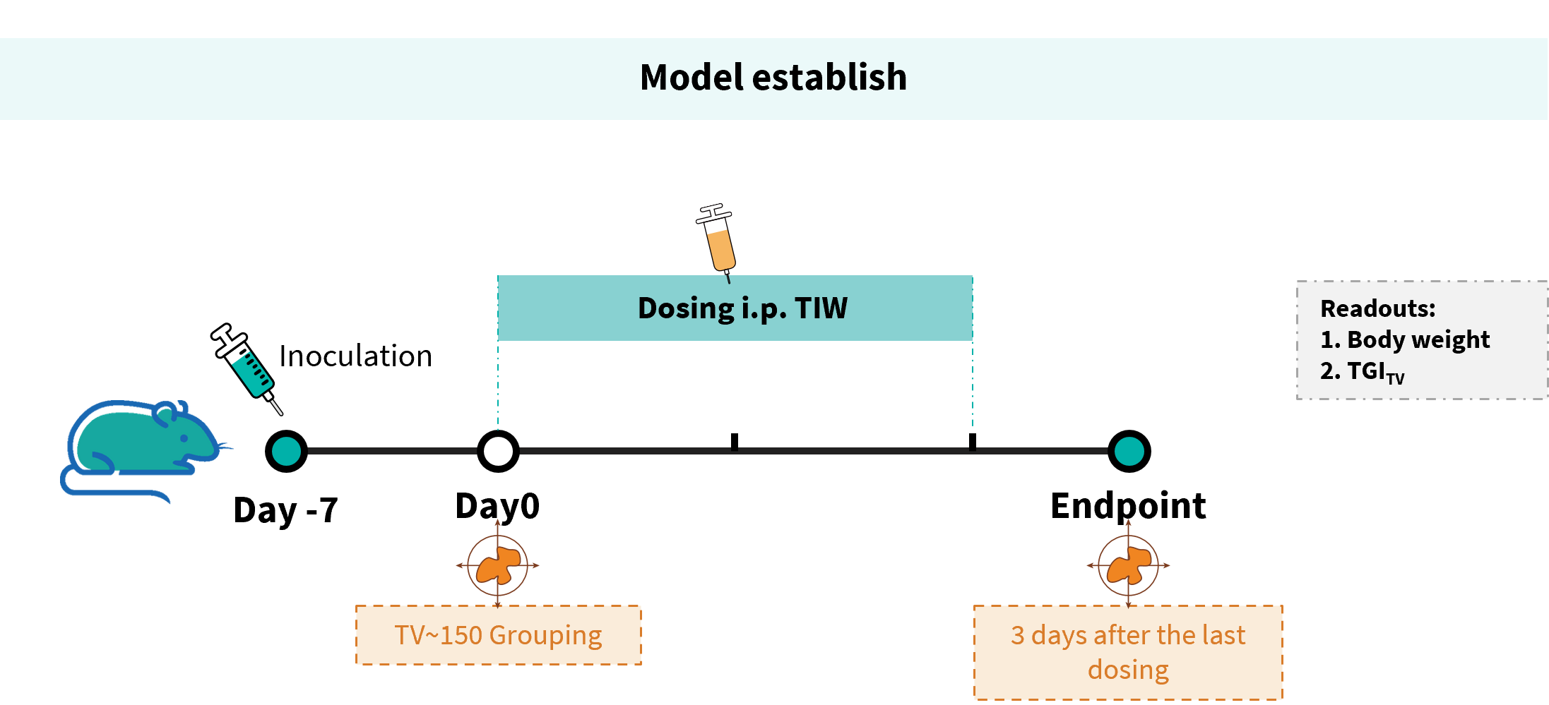
Establishment of a B-hVEGFA/hPD-L1 B16-F10 model and in vivo efficacy study of Ivonescimab antibody. B-hVEGFA/hPD-L1 B16-F10 cells were implanted subcutaneously into homozygous B-hPD-1/hPD-L1/hVEGFA mice (female, 8 weeks old, n=6). When the average tumor volume reached approximately 150 mm³, mice were randomized and subsequently administered with anti-PD-1 & anti-VEGFA antibody via intraperitoneal injection.
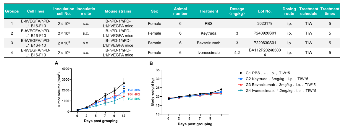
Antitumor activity of Ivonescimab (also known as AK112) in B-hPD-1/hPD-L1/hVEGFA mice. (A) Tumor growth curves. (B) Body weight changes during treatment. As shown in panel A, Ivonescimab was efficacious in controlling tumor growth in B-hPD-1/hPD-L1/hVEGFA mice. Values are expressed as mean ± SEM. The overage of this tumor model is 60%. Note: one mouse dead in G3 on day12.
