PD-1 Protein Expression Analysis in Spleen
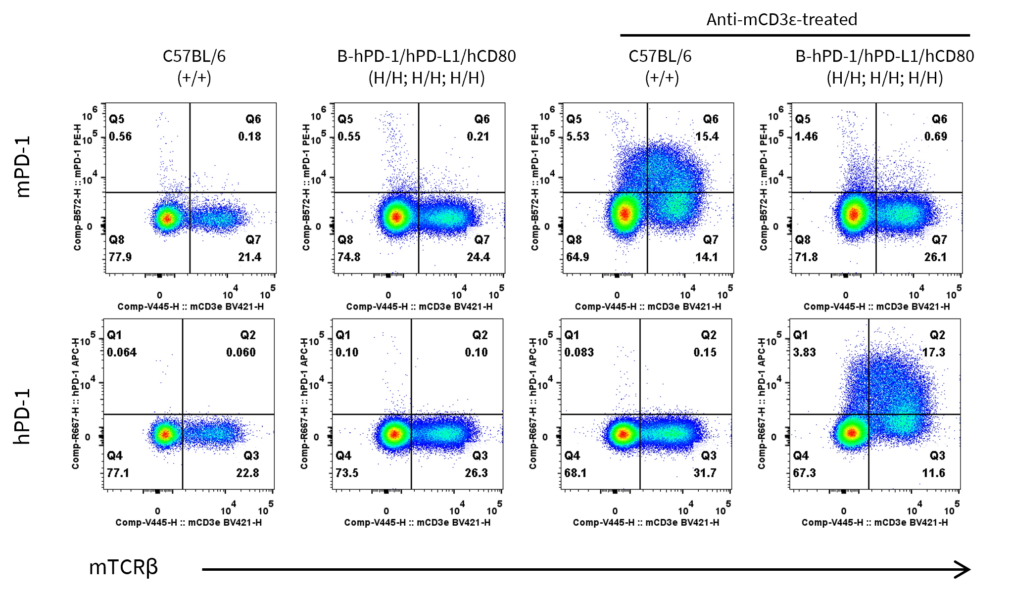
- Mouse PD-1 was detected exclusively in wild-type C57BL/6 mice
- Human PD-1 was detected in homozygous B-hPD-1/hPD-L1/hCD80 mice, but not in wild-type mice.
Strain specific PD-1 expression analysis in wild-type C57BL/6 and homozygous B-hPD-1/hPD-L1/hCD80 mice by flow cytometry. Splenocytes were collected from wild-type C57BL/6 mice (+/+) and homozygous B-hPD-1/hPD-L1/hCD80 mice (H/H, H/H, H/H) (female, 8-week-old, n=1) after stimulated with anti-mouse CD3ε antibody (7.5 μg, i.p.) in vivo for 24 hours or not. Protein expression was analyzed with anti-mouse PD-1 antibody (Biolegend, 109104) and anti-human PD-1 antibody (Biolegend, 329908) by flow cytometry.
PD-L1 Protein Expression Analysis in Spleen
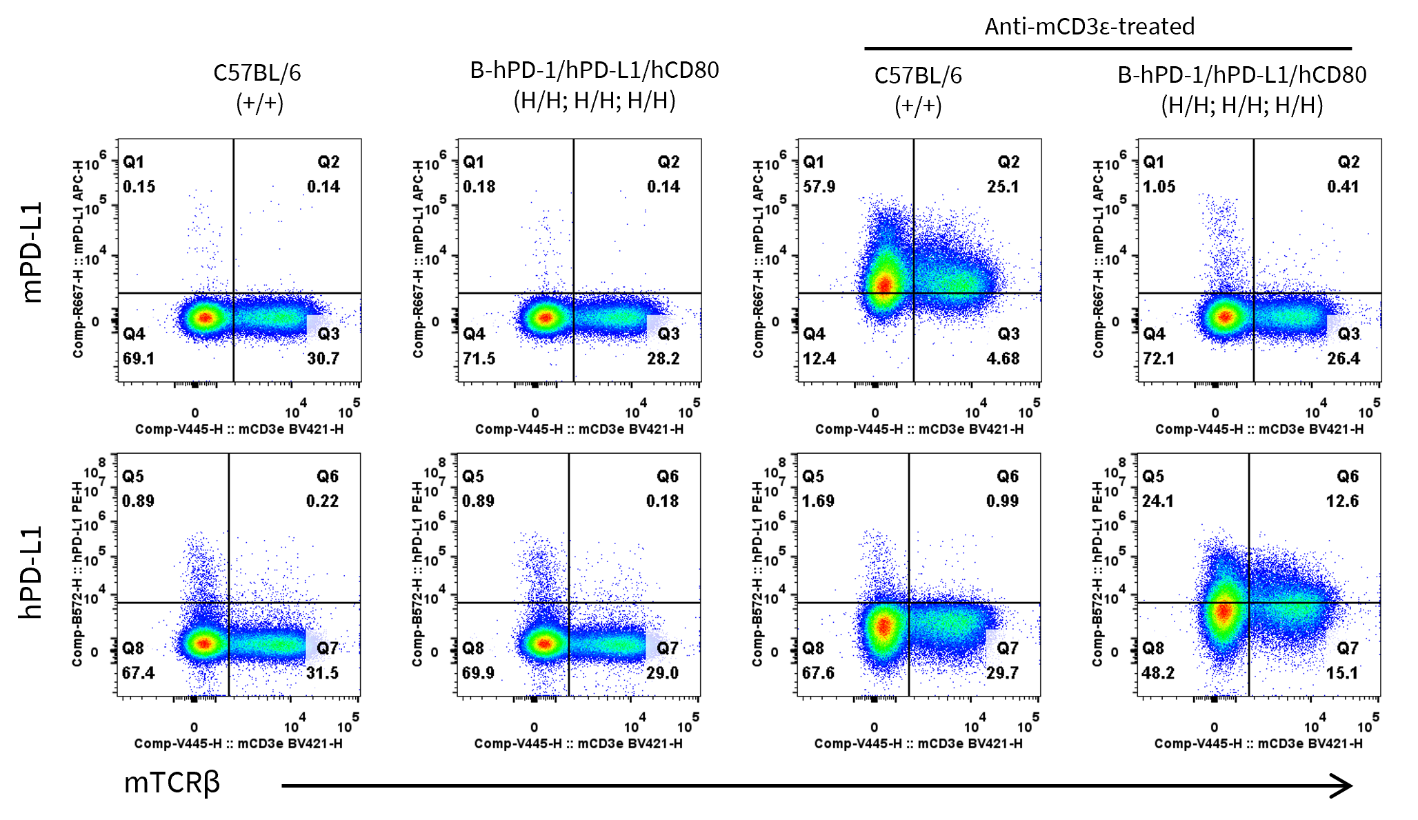
- Mouse PD-L1 was detected exclusively in wild-type C57BL/6 mice
- Human PD-L1 was detected in homozygous B-hPD-1/hPD-L1/hCD80 mice, but not in wild-type mice.
Strain specific PD-L1 expression analysis in wild-type C57BL/6 and homozygous B-hPD-1/hPD-L1/hCD80 mice by flow cytometry. Splenocytes were collected from wild-type C57BL/6 mice (+/+) and homozygous B-hPD-1/hPD-L1/hCD80 mice (H/H, H/H, H/H) (female, 8-week-old, n=1) after stimulated with anti-mouse CD3ε antibody (7.5 μg, i.p.) in vivo for 24 hours or not. Protein expression was analyzed with anti-mouse PD-L1 antibody (Biolegend, 124312) and anti-human PD-L1 antibody (Biolegend, 329706) by flow cytometry.
CD80 Protein Expression Analysis in Spleen
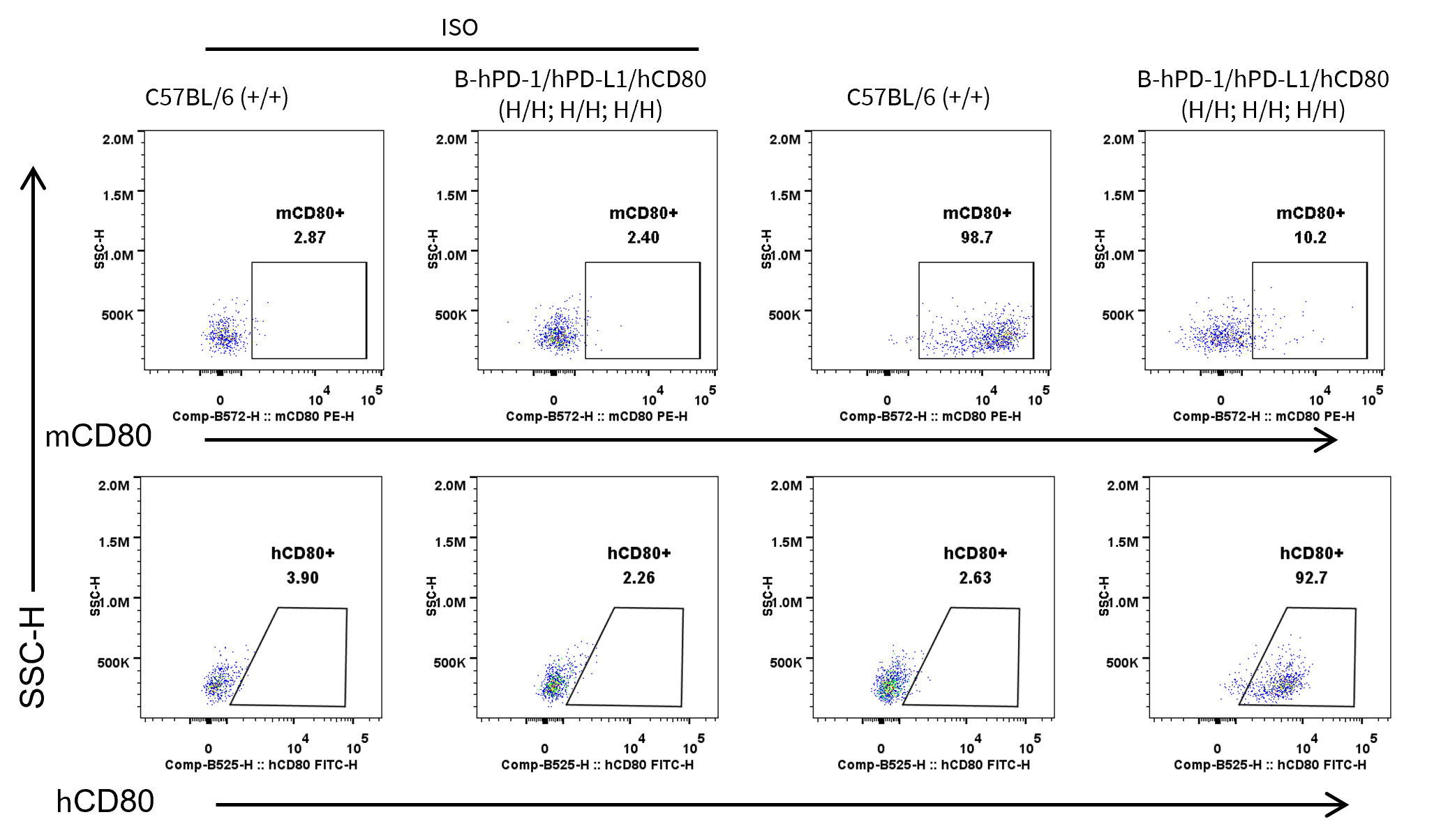
- Mouse CD80 was detected exclusively in wild-type C57BL/6 mice
- Human CD80 was detected in homozygous B-hPD-1/hPD-L1/hCD80 mice, but not in wild-type mice.
Strain specific CD80 expression analysis in wild-type C57BL/6 and homozygous B-hPD-1/hPD-L1/hCD80 mice by flow cytometry. Splenocytes were collected from wild-type C57BL/6 mice (+/+) and homozygous B-hPD-1/hPD-L1/hCD80 mice (H/H, H/H, H/H) (female, 8-week-old, n=1). Protein expression was analyzed with anti-human CD80 antibody (Biolegend, 375406) and anti-mouse CD80 antibody (Biolegend, 104708) by flow cytometry.
CD80 Protein Expression Analysis in PEMs
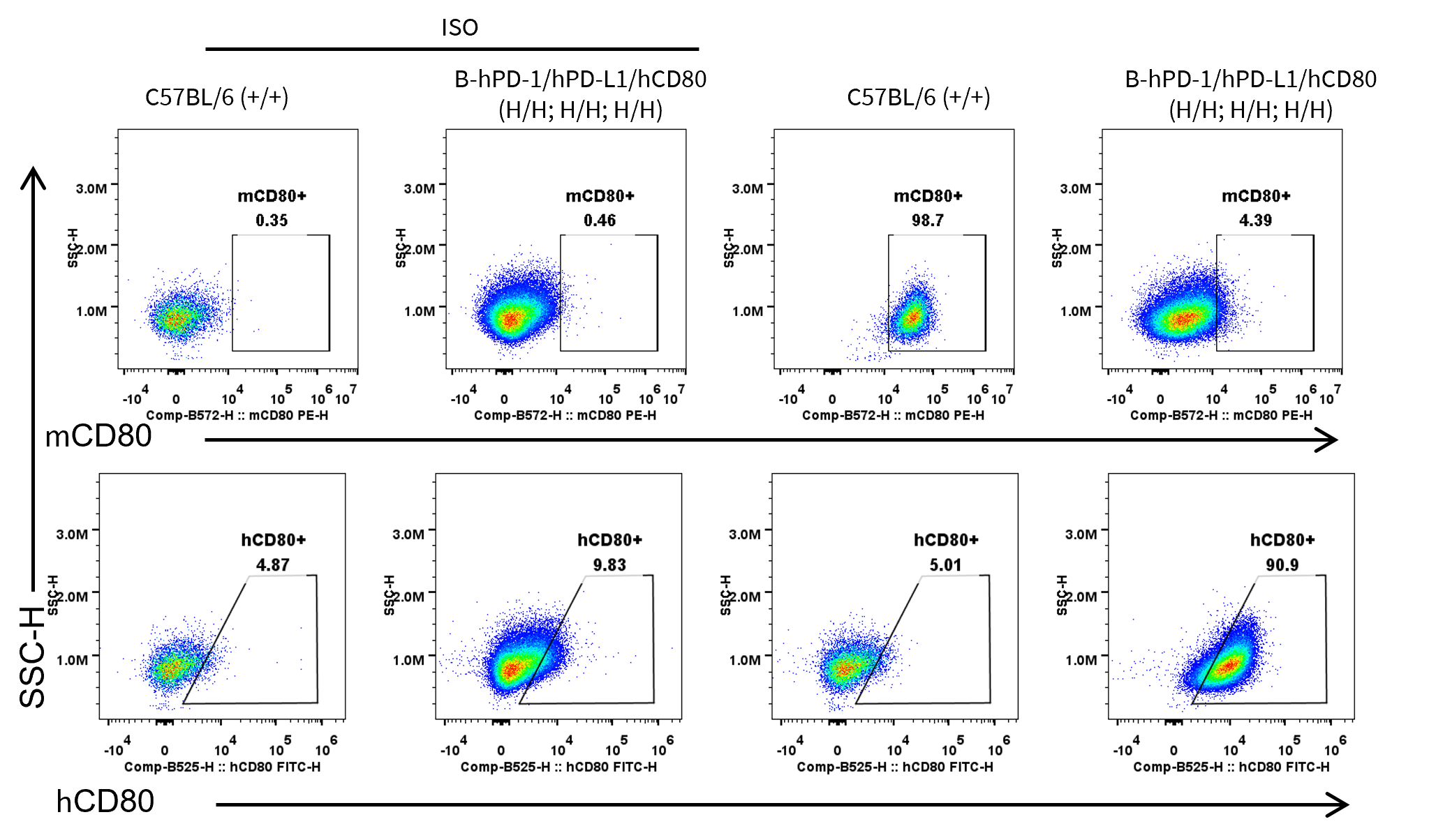
- Mouse CD80 was detected exclusively in wild-type C57BL/6 mice
- Human CD80 was detected in homozygous B-hPD-1/hPD-L1/hCD80 mice, but not in wild-type mice.
Strain specific CD80 expression analysis in wild-type C57BL/6 and homozygous B-hPD-1/hPD-L1/hCD80 mice by flow cytometry. Peritoneal exudative macrophages (PEMs) were collected from wild-type C57BL/6JNifdc mice (+/+) and homozygous B-hPD-1 plus/hPD-L1/hCD80 mice (H/H, H/H, H/H) (female, 8-week-old, n=1). Protein expression was analyzed with anti-human CD80 antibody (Biolegend, 375406) and anti-mouse CD80 antibody (Biolegend, 104708) by flow cytometry.
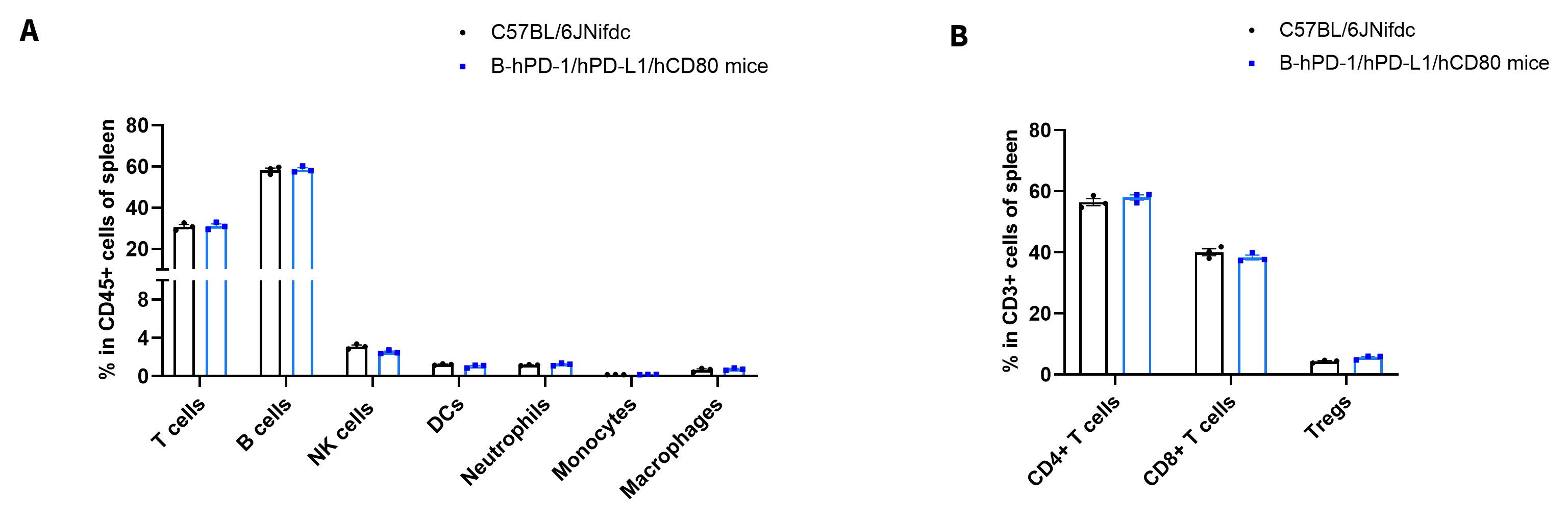
Analysis of Leukocytes Cell Subpopulation in Spleen
- The frequencies of T cells, B cells, NK cells, DCs, granulocyte, monocytes, macrophages, CD4+ T cells, CD8+ T cells and Tregs in homozygous B-hPD-1/hPD-L1/hCD80 mice were similar to those in C57BL/6 mice
- Humanization of PD-1, PD-L1 and CD80 does not change the frequency or distribution of these cell types in spleen.
Frequency of leukocyte subpopulations in spleen by flow cytometry. Splenocytes were isolated from wild-type C57BL/6 mice and homozygous B-hPD-1/hPD-L1/hCD80 mice (n=3, 8-week-old). A. Flow cytometry analysis of the splenocytes was performed to assess the frequency of leukocyte subpopulations. B. Frequency of T cell subpopulations. Values are expressed as mean ± SEM. Significance was determined by two-way ANOVA test. *P < 0.05, **P < 0.01, ***p < 0.001.
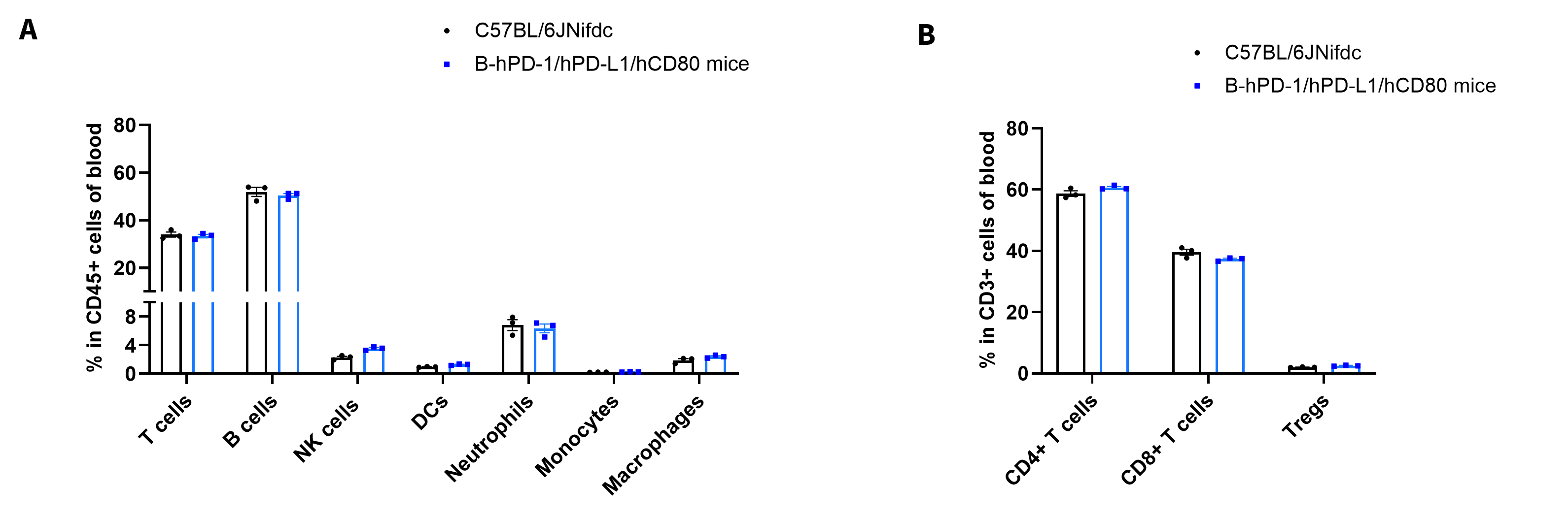
Analysis of Leukocytes Cell Subpopulation in Blood
- The frequencies of T cells, B cells, NK cells, DCs, granulocyte, monocytes, macrophages, CD4+ T cells, CD8+ T cells and Tregs in homozygous B-hPD-1/hPD-L1/hCD80 mice were similar to those in C57BL/6 mice
- Humanization of PD-1, PD-L1 and CD80 does not change the frequency or distribution of these cell types in blood.
Frequency of leukocyte subpopulations in blood by flow cytometry. Blood cells were isolated from wild-type C57BL/6 mice and homozygous B-hPD-1/hPD-L1/hCD80 mice (n=3, 8-week-old). A. Flow cytometry analysis of the blood cells were performed to assess the frequency of leukocyte subpopulations. B. Frequency of T cell subpopulations. Values are expressed as mean ± SEM. Significance was determined by two-way ANOVA test. *P < 0.05, **P < 0.01, ***p < 0.001.
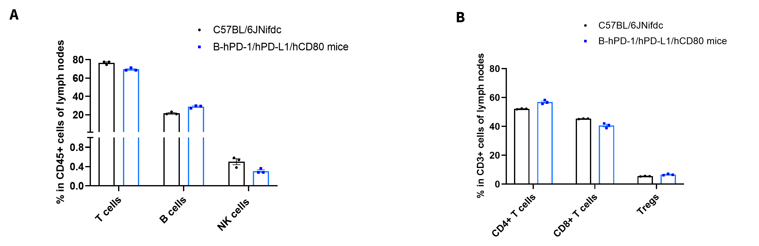
Analysis of Leukocytes Cell Subpopulation in Lymph Nodes
- The frequencies of T cells, B cells, NK cells, DCs, granulocyte, monocytes, macrophages, CD4+ T cells, CD8+ T cells and Tregs in homozygous B-hPD-1/hPD-L1/hCD80 mice were similar to those in C57BL/6 mice
- Humanization of PD-1, PD-L1 and CD80 does not change the frequency or distribution of these cell types in lymph nodes.
Frequency of leukocyte subpopulations in lymph nodes by flow cytometry. Lymph nodes were isolated from wild-type C57BL/6 mice and homozygous B-hPD-1/hPD-L1/hCD80 mice (n=3, 8-week-old). A. Flow cytometry analysis of the lymph nodes were performed to assess the frequency of leukocyte subpopulations. B. Frequency of T cell subpopulations. Values are expressed as mean ± SEM. Significance was determined by two-way ANOVA test. *P < 0.05, **P < 0.01, ***p < 0.001.
* When publishing results obtained using this animal model, please acknowledge the source as follows: The animal model [B-hPD-1 plus/hPD-L1/hCD80 mice] (Cat# 112670) was purchased from Biocytogen.