Description
TNF: A master regulator of inflammation, immune responses, and cell survival
- Gene Information: Tumor necrosis factor (TNF) is a protein-coding gene located on chromosome 6p21.33. It encodes a multifunctional proinflammatory cytokine that belongs to the tumor necrosis factor (TNF) superfamily.
- Protein Expression: TNF is primarily expressed by macrophages. TNF is synthesized as a membrane-bound precursor and cleaved into a soluble circulating protein.
- Signaling Pathway: TNF exerts its effects primarily through two receptors: TNFR1 and TNFR2.
- Therapeutic Inhibition: By blocking a central driver of the immune system's inflammatory response, adalimumab inhibits downstream inflammation and improves clinical outcomes, including reducing other inflammatory markers and reducing tissue damage in various autoimmune conditions.
IL-17A: A key pro-inflammatory cytokine in autoimmune skin and joint diseases
- Gene Information: The IL17A gene (also known as CTLA8) is a protein-coding gene located on chromosome 6p12.2. This cytokine is the founding member of the IL-17 family and primarily signals through a heterodimeric receptor complex composed of IL-17RA and IL-17RC.
- Protein Expression: IL-17A is predominantly produced by specialized CD4+ T cells Th17 cells), as well as various innate immune cells, innate lymphoid cells (ILC3s), and neutrophils. It exists primarily as a secreted, disulfide-linked homodimer (IL-17A/A) or as a heterodimer with IL-17F (IL-17A/F).
- Signaling Pathway: Upstream IL-17A production is driven by the IL-23/Th17 axis. In the downstream, IL-17A binds to the IL-17RA/IL-17RC complex on target cells (e.g., keratinocytes), recruiting Act1 to activate NF-κB and MAPKs.
- Therapeutic Inhibition: Blocking IL-17A or its interaction with its receptors dramatically reduces inflammatory responses. Therapeutic monoclonal antibodies targeting this axis are highly effective and for autoimmune conditions such as plaque psoriasis, psoriatic arthritis, and ankylosing spondylitis.
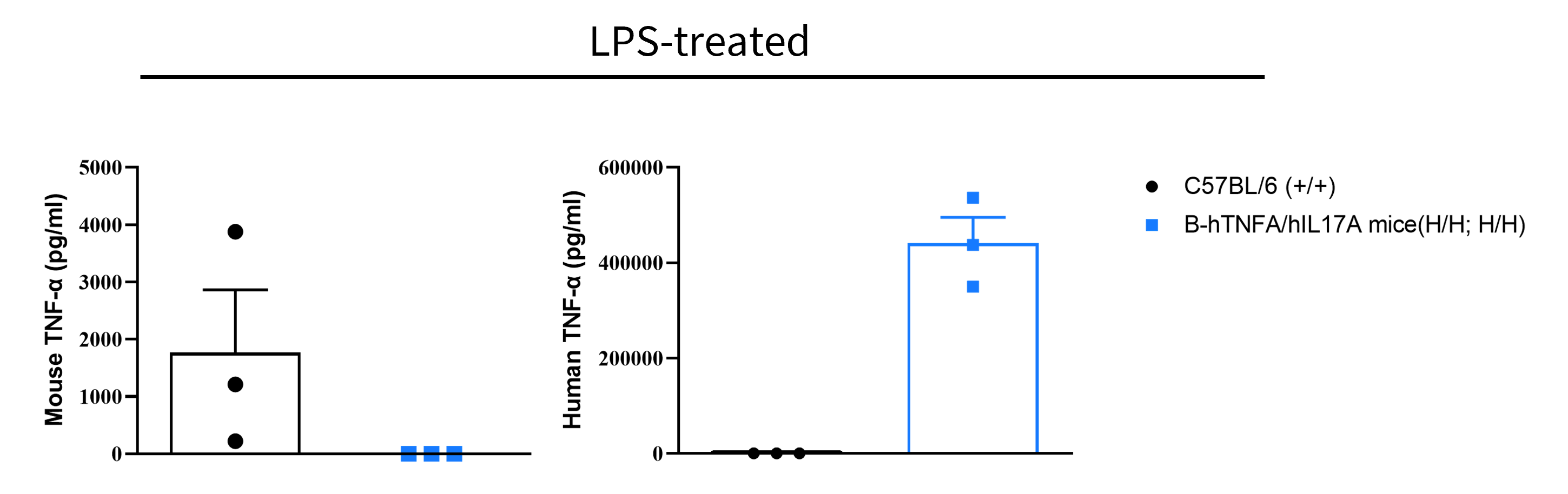
TNFA Protein Expression Analysis in Serum
- Mouse TNFA was detected exclusively in wild-type C57BL/6 mice.
- Human TNFA was detected in homozygous B-hTNFA/hIL17A mice, but not in wild-type mice.
Strain specific TNFA expression analysis in wild-type C57BL/6 mice and homozygous humanized B-hTNFA/hIL17A mice by ELISA. Serum was collected from wild-type C57BL/6 mice (+/+) and homozygous B-hTNFA/hIL17A mice (H/H;H/H) stimulated with LPS (20 μg/mice) in vivo for 1 hour (female, 8-week-old, n=3). Expression level of TNFA were analyzed by ELISA. Mouse TNFA was only detectable in wild-type C57BL/6 mice. Human TNFA was exclusively detectable in homozygous B-hTNFA/hIL17A mice. Values are expressed as mean ± SEM.
IL17A Protein Expression Analysis in Serum
- Mouse IL17A was detected exclusively in wild-type C57BL/6 mice.
- Human IL17A was detected in homozygous B-hTNFA/hIL17A mice, but not in wild-type mice.
Strain specific IL17A expression analysis in wild-type C57BL/6 mice and homozygous humanized B-hTNFA/hIL17A mice by ELISA. Serum was collected from wild-type C57BL/6 mice (+/+) and homozygous B-hTNFA/hIL17A mice (H/H;H/H) stimulated with anti-CD3ε (7.5 μg/mice, 2 hours, i.p.) and anti-CD28 (4 μg/mice, 2 hours, i.p.) in vivo, and analyzed by ELISA with species-specific IL17A ELISA kit. Mouse IL17A was only detectable in wild-type C57BL/6 mice. Human IL17A was exclusively detectable in homozygous B-hTNFA/hIL17A mice. Values are expressed as mean ± SEM.
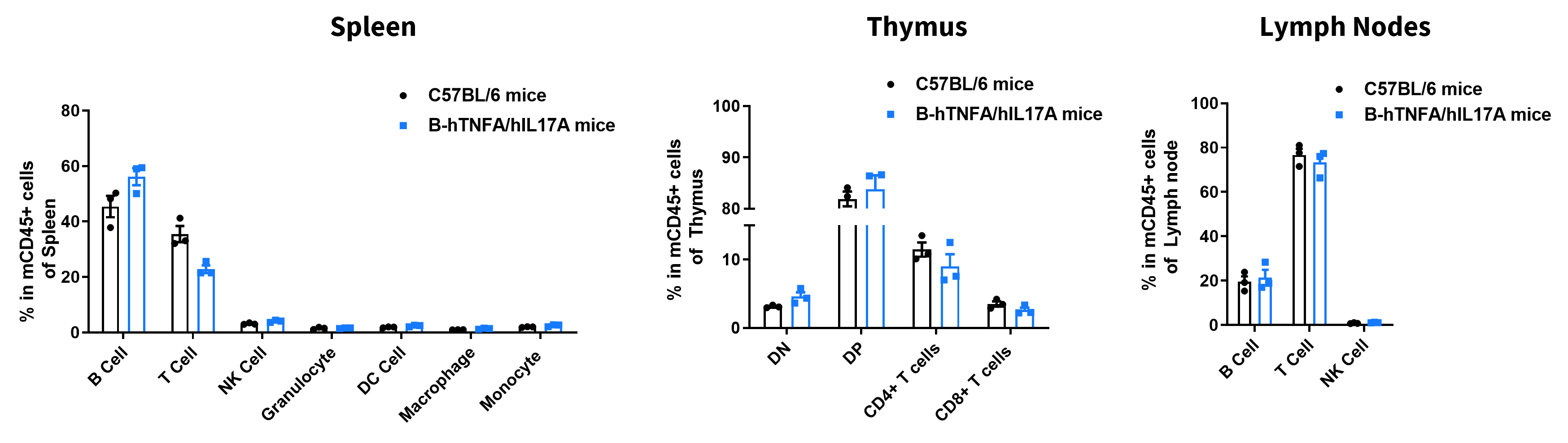
Analysis of Leukocyte Subpopulations
- The percentages of T cells, B cells, NK cells, DCs, neutrophils, monocytes, and macrophages in homozygous B-hTNFA/hIL17A mice were similar to those in C57BL/6 mice.
- Humanization of TNFA and IL17A does not affect normal immune cell development or splenic distribution.
Analysis of leukocyte subpopulations by flow cytometry in immune organs. Splenocytes, thymocytes, and lymph nodes were isolated from female C57BL/6 and B-hTNFA/hIL17A mice (female, 9-week-old, n = 3). Single live cells were gated on the CD45⁺ population and analyzed by flow cytometry as indicated. Values are expressed as mean ± SEM.
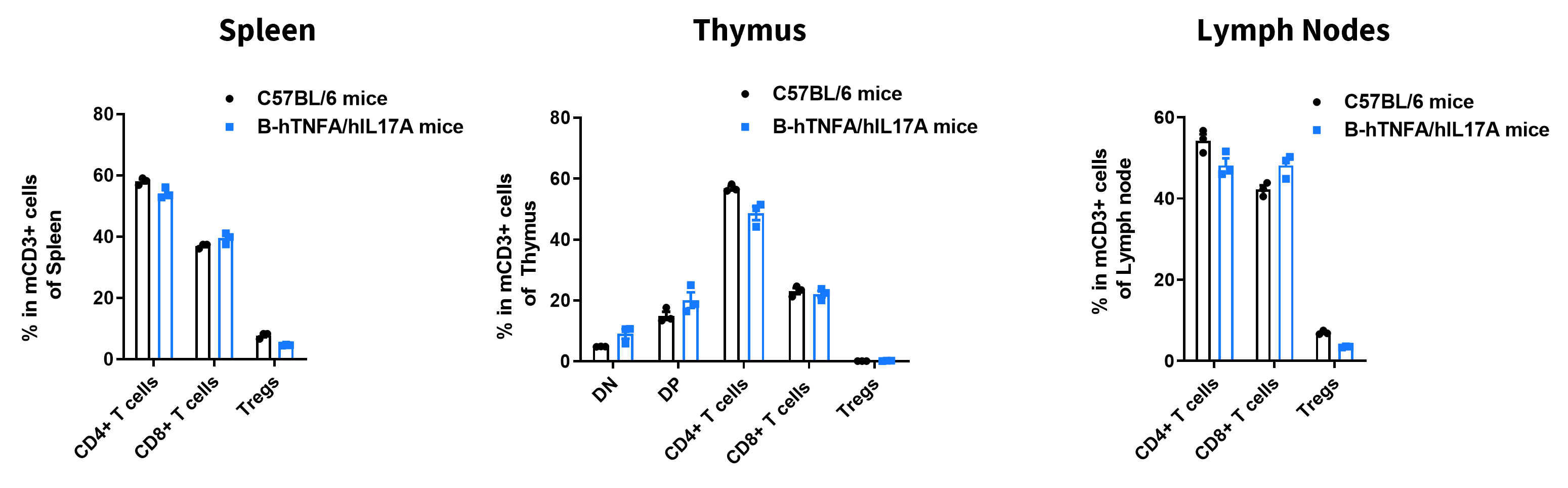
Analysis of T Cell Subpopulations
- The proportions of CD4⁺ T cells, CD8⁺ T cells, and Tregs in homozygous B-hTNFA/hIL17A mice were comparable to those in C57BL/6 mice.
- Humanization of TNFA and IL17A does not affect normal T cell development, differentiation, or splenic distribution.
Analysis of T-cell subpopulations by flow cytometry in immune organs. Splenocytes, thymocytes, and lymph nodes were isolated from female C57BL/6 and B-hTNFA/hIL17A mice (female, 9-week-old, n = 3). Single live cells were gated on the CD3⁺ T-cell population and analyzed by flow cytometry as indicated. Values are expressed as mean ± SEM.
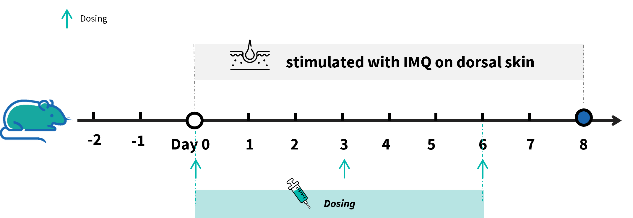
In Vivo Efficacy of Anti–Human TNFA/Human IL17A Bispecific Antibody in IMQ-induced Psoriasis Model
Experimental schedule for induction of psoriasis-like skin lesions in B-hTNFA/hIL17A mice. Mice at 10 week-old of age received a daily topical of commercially available IMQ cream on the shaved back for 6 consecutive days to induce psoriasis-like skin lesions. Severity of skin inflammation was daily scored and back skin was collected at the endpoint. Anti-hTNFA/hIL17A BsAb analog was treated on days 0, 3, and 6. IMQ: imiquimod.
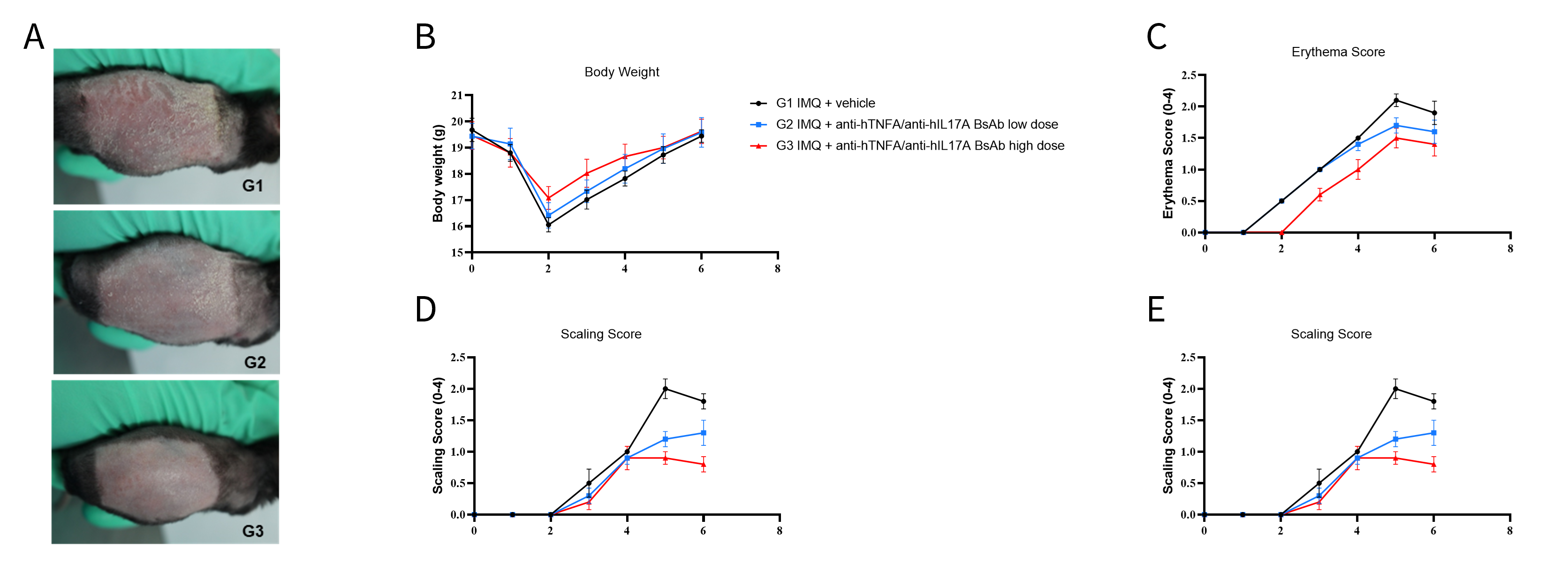
- Anti–hTNFA/hIL17A bispecific antibody treatment decreased the phenotypical presentation level and erythema and scaling score of the back compared to the modelling group.
IMQ-induced skin inflammation in B-hTNFA/hIL17A mice phenotypically resembles psoriasis. B-hTNFA/hIL17A mice (female, 10-week-old, n=5) were scored daily for up to 6 days for body weight and clinical signs of skin inflammation following treatment with imiquimod (IMQ) cream. Mice in each group were treated with different dose of antibodies produced in house. Doses are shown in legend. (A) Phenotypical presentation of mouse back skin after 6 days of treatment. (B) Body weight changes during treatment. (C-D) Erythema and scaling score of the back was scored daily on a scale from 0 to 4. Additionally, the cumulative score (erythema plus scaling) is depicted. Values are expressed as mean ± SEM.
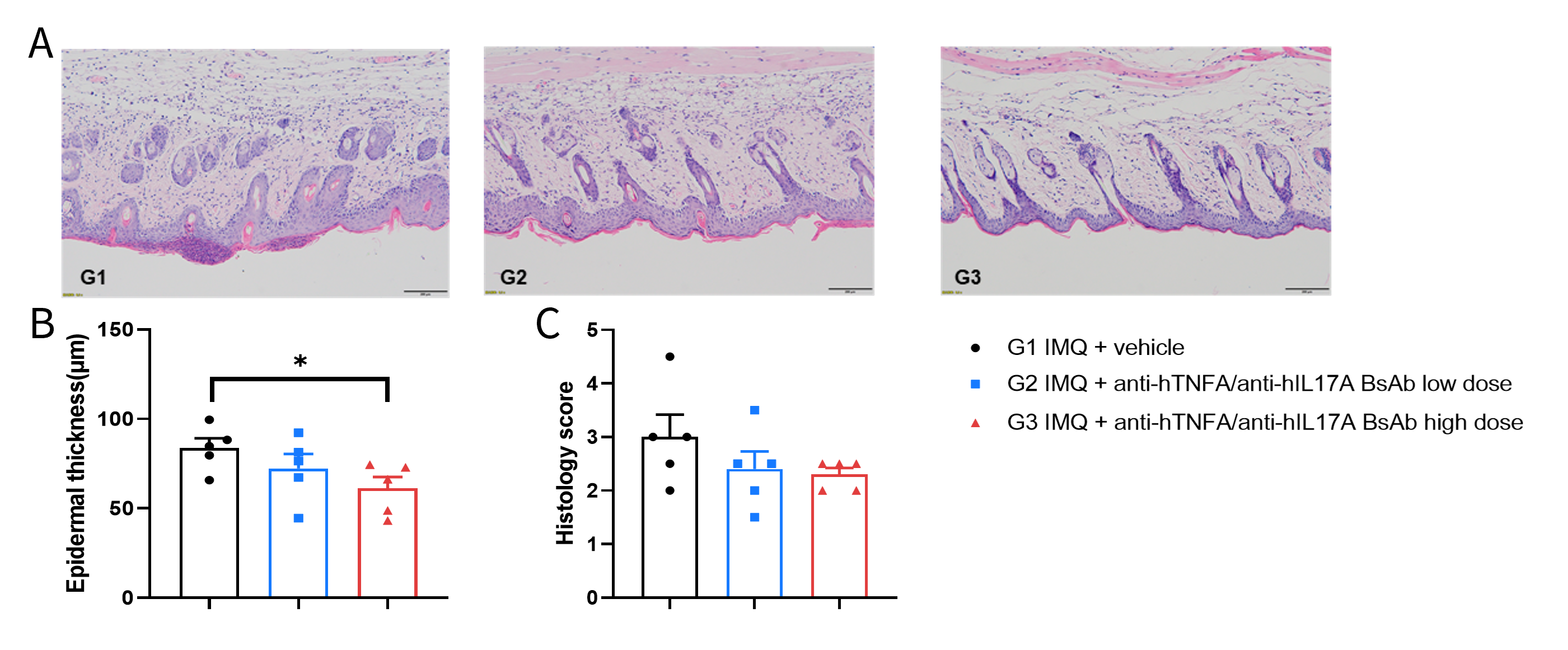
- Anti–hTNFA/hIL17A bispecific antibody treatment decreased the epidermal thickness compared to the modelling group.
Dose dependent effects of BsAbs on keratinocyte proliferation and inflammatory cell infiltration in IMQ induced psoriasis-like skin lesions in B-hTNFA/hIL17A mice. Back skin was collected at the endpoint and stained with Hematoxylin and eosin (H&E). (A) H&E staining of the back skin. (B) Epidermal thickness of the mice. (C) Histological changes were scored on a scale from 0 to 11. Results indicated that high-dose anti-hTNFA/hIL17A BsAbs analog (in house) significantly reduced psoriasis-like skin lesions in B-hTNFA/hIL17A mice, confirming that B-hTNFA/hIL17A mice provide a powerful model for in vivo evaluation of anti-human TNFA and IL17A bispecific antibodies.
* When publishing results obtained using this animal model, please acknowledge the source as follows: The animal model [B-hTNFA/hIL17A mice] (Cat# 120548) was purchased from Biocytogen.