Description
- Gene Information: PD-1 is encoded by the PDCD1 gene, while PD-L1 is encoded by the CD274 gene. Together, they form a key immune checkpoint receptor–ligand pair. ICOS is encoded by the ICOS gene, and ICOSL is encoded by the ICOSLG gene. Both belong to the CD28/B7 co-stimulatory family.
- Protein Expression: CD3: PD-1 is primarily expressed on activated T cells, B cells, and NK cells, whereas PD-L1 is expressed on antigen-presenting cells and many tumor cells, with expression often induced by IFN-γ. ICOS is induced on activated T cells, particularly T follicular helper (Tfh) cells, while ICOSL is mainly expressed on dendritic cells, B cells, and macrophages.
- Signaling Pathway: Binding of PD-L1 to PD-1 recruits SHP2 phosphatase and suppresses TCR/CD28-mediated PI3K–AKT signaling, resulting in reduced T-cell proliferation, cytokine production, and cytotoxic activity. Engagement of ICOS with ICOSL activates the PI3K–AKT–mTOR pathway, promoting T-cell survival, proliferation, cytokine production, and Tfh-cell differentiation.
- Key Therapeutic: Anti-PD-1 and anti-PD-L1 antibodies block this inhibitory pathway, restoring T-cell function and enhancing anti-tumor immune responses. ICOS agonists are being developed to enhance anti-tumor immunity, whereas ICOS/ICOSL blockade is being explored for the treatment of autoimmune diseases by reducing Tfh-mediated antibody responses.
Targeting strategy
PD-1
- A chimeric CDS that encodes human PDCD1 extracellular domain, mouse Pdcd1 transmembrane and cytoplasmic domain, followed by WPRE-pA is inserted right after mouse Pdcd1 ATG to replace the exon 1 of mouse Pdcd1 gene.
- The chimeric PDCD1 protein expression will be driven by endogenous mouse Pdcd1 promoter, while mouse Pdcd1 gene transcription and translation will be disrupted.
PD-L1
- The exon 3 of mouse Cd274 gene that encodes the IgV domain was replaced by human CD274 exon 3. Sequences of other regions still belonged to mice.
ICOS
- The exons 1-5 of mouse Icos gene that encode signal peptide, extracellular domain, transmembrane domain and cytoplasmic region were replaced by human counterparts. The promoter, 5’UTR and 3’UTR region of the mouse gene were retained.
- The human ICOS expression was driven by endogenous mouse Icos promoter, while mouse Icos gene transcription and translation will be disrupted.
ICOSL
- The exons 3-5 of mouse Icosl gene that encode extracellular domain were replaced by human counterparts. The genomic region of mouse Icosl gene that encodes signal peptide, transmembrane domain and cytoplasmic portion was retained. The promoter, 5’UTR and 3’UTR region of the mouse gene were also retained.
- The chimeric ICOSL expression was driven by endogenous mouse Icosl promoter, while mouse Icosl gene transcription and translation will be disrupted.
PD-1 and PD-L1 Protein Expression
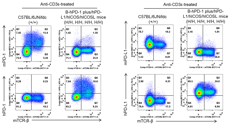
- Human PD-1 and PD-L1 were exclusively detectable in homozygous B-hPD-1 plus/hPD-L1/hICOS/hICOSL mice, but not in wild-type C57BL/6JNifdc mice.
PD-1 and PD-L1 expression analysis in wild-type C57BL/6JNifdc mice and homozygous B-hPD-1 plus/hPD-L1/hICOS/hICOSL mice by flow cytometry. Splenocytes were collected from wild-type C57BL/6JNifdc mice (+/+) and homozygous B-hPD-1 plus/hPD-L1/hICOS/hICOSL mice (H/H, H/H, H/H, H/H) (female, 8-week-old, n=1). Protein expression was analyzed with anti-human PD-1 antibody (Biolegend, 329908), anti-mouse PD-1 antibody (Biolegend, 109104), anti-human PD-L1 antibody (Biolegend, 329706) and anti-mouse PD-L1 antibody (Biolegend, 124312) by flow cytometry.
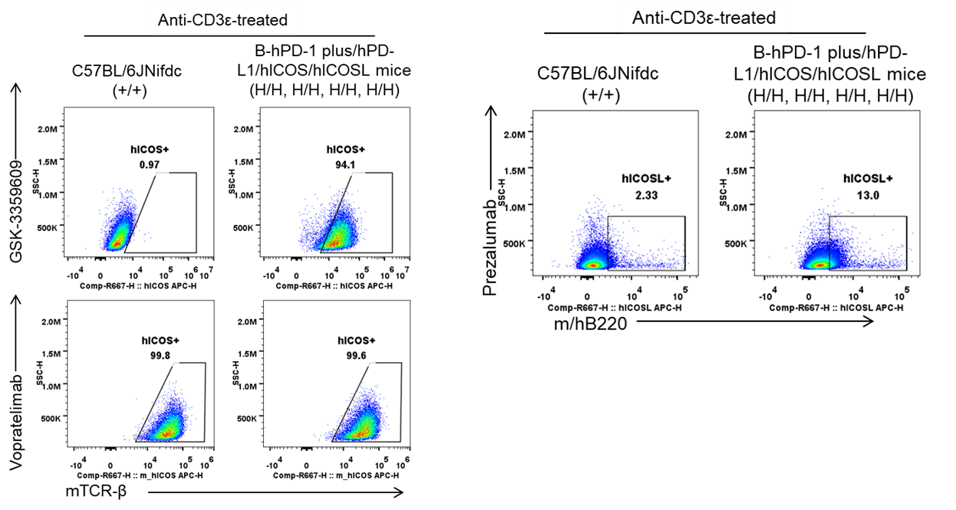
- Human ICOS and ICOSL were exclusively detectable in homozygous B-hPD-1 plus/hPD-L1/hICOS/hICOSL mice, but not in wild-type C57BL/6JNifdc mice.
ICOS and ICOSL expression analysis in wild-type C57BL/6JNifdc mice and homozygous B-hPD-1 plus/hPD-L1/hICOS/hICOSL mice by flow cytometry. Splenocytes were collected from wild-type C57BL/6JNifdc mice (+/+) and homozygous B-hPD-1 plus/hPD-L1/hICOS/hICOSL mice (H/H, H/H, H/H, H/H) stimulated in vivo with anti-CD3ε antibody (7.5 μg/mouse, i.p., for 24 hours). The splenocytes were subsequently stained for flow cytometric analysis using antibodies specific for ICOS (GSK-3359609 and Vopratelimab, in-house) and ICOSL (Prezalumab, in-house).
* When publishing results obtained using this animal model, please acknowledge the source as follows: The animal model [B-hPD-1 plus/hPD-L1/hICOS/hICOSL mice] (Cat# 114687) was purchased from Biocytogen.