Description
STING: A key innate immune sensor and therapeutic target
- Gene Information: Stimulator of interferon genes (STING, gene symbol TMEM173) is a protein-coding gene located on chromosome 5q31.2. It encodes an endoplasmic reticulum (ER)-resident transmembrane adaptor protein that plays a central role in innate immune sensing of cytosolic DNA.
- Protein Expression: STING is broadly expressed across multiple cell types, including various immune cells, endothelial cells, and epithelial cells. STING protein is localized primarily to the ER under basal conditions and undergoes activation-dependent trafficking to the Golgi apparatus.
- Signaling Pathway: Cytosolic DNA activates cGAS, leading to the production of 2′3′-cGAMP, which binds and activates STING. Activated STING traffics from the ER to the Golgi and recruits TBK1, resulting in phosphorylation and activation of IRF3 and subsequent transcription of type I interferons, particularly IFN-β. STING activation also induces NF-κB signaling and the production of inflammatory cytokines and chemokines.
- Therapeutic Intervention: STING signaling is being investigated as a therapeutic target, particularly for cancer immunotherapy. STING agonists aim to activate innate immune responses within the tumor microenvironment, promoting type I interferon production, dendritic cell activation, antigen presentation, and subsequent T-cell-mediated antitumor immunity. Conversely, STING inhibition is being explored for the treatment of inflammatory and autoimmune diseases driven by excessive STING activation.
Targeting strategy
STING
- Exons 1-8 of the mouse Sting1 gene that encode the whole molecule, including the promoter, 5' UTR, and 3' UTR, were replaced by their human counterparts in B-hSTING mice.
- Human STING expression is driven by the endogenous human STING1 promoter, while mouse Sting1 gene transcription and translation are disrupted.
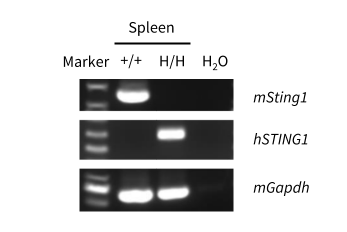
STING Expression by RT-PCR
Human STING were detectable in B-hSTING mice by RT-PCR and sequencing. Spleen tissues were isolated from wild-type C57BL/6 mice (+/+) and homozygous B-hSTING mice (H/H). Primers were designed to detect human STING1 and mouse Sting1.
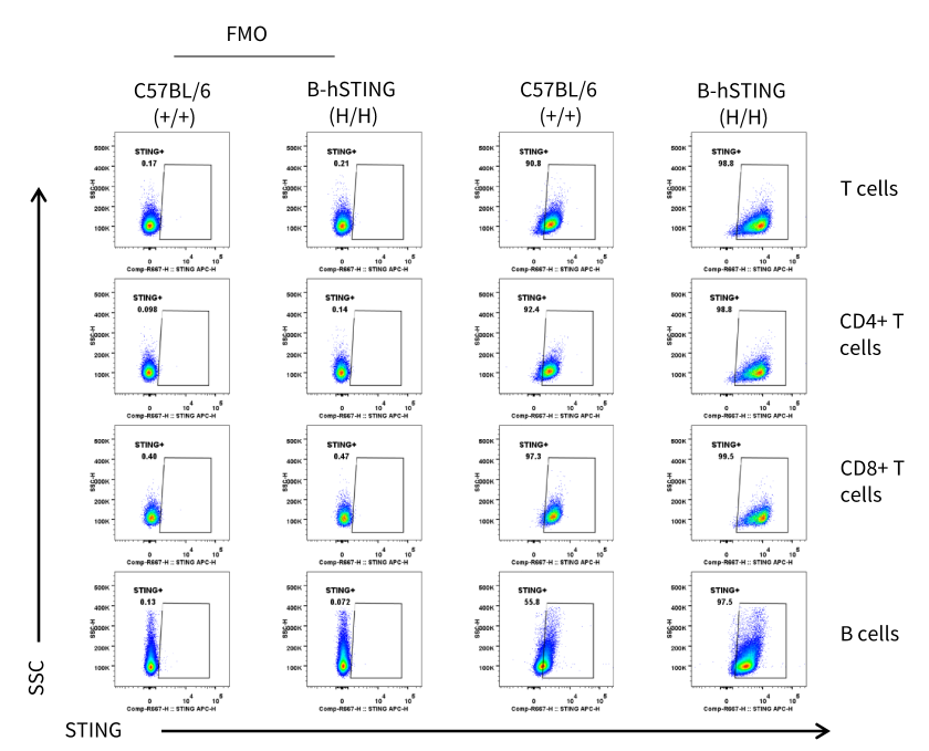
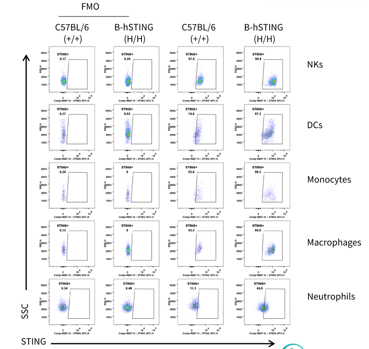
STING Protein Expression in Spleen
Mouse and human STING expression analysis in splenocytes. Splenocytes were collected from wild-type C57BL/6 mice and homozygous B-hSTING mice (male, 9-week-old, n=1). STING expression was analyzed by flow cytometry using a human/mouse cross-reactive anti-STING antibody (RD: IC7169A; internally validated).
Mouse and human STING expression analysis in splenocytes. Splenocytes were collected from wild-type C57BL/6 mice and homozygous B-hSTING mice (male, 9-week-old, n=1). STING expression was analyzed by flow cytometry using a human/mouse cross-reactive anti-STING antibody (RD: IC7169A; internally validated).
STING Protein Expression in Thymus
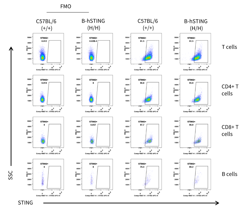
Mouse and human STING expression analysis in thymocytes. Thymocytes were collected from wild-type C57BL/6 mice and homozygous B-hSTING mice (male, 9-week-old, n=1). STING expression was analyzed by flow cytometry using a human/mouse cross-reactive anti-STING antibody (RD: IC7169A; internally validated).
STING Protein Expression in Lung
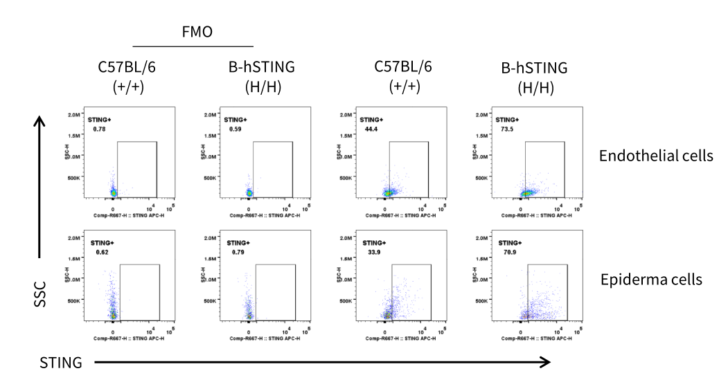
Mouse and human STING expression analysis in the lung. Pulmonary endothelial cells (top) and epithelial cells (bottom) were collected from wild-type C57BL/6 mice and homozygous B-hSTING mice (male, 9-week-old, n=1). STING expression was analyzed by flow cytometry using a human/mouse cross-reactive anti-STING antibody (RD: IC7169A; internally validated).
Functional Validation
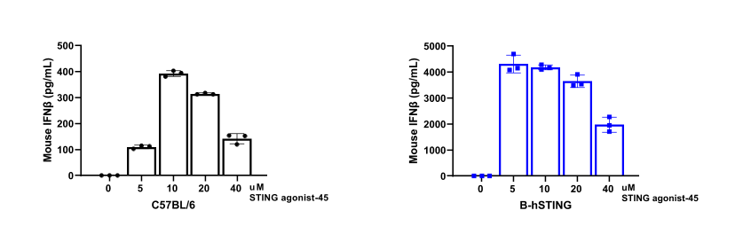
STING agonist-45 induces mIFN-β secretion in BMDMs from wild-type C57BL/6 and homozygous B-hSTING mice. Bone marrow cells were isolated from wild-type C57BL/6 and homozygous B-hSTING mice (female, 13-14-week-old, n=3) and then differentiated into BMDMs using mouse M-CSF (20 ng/mL). The resulting BMDMs were stimulated with varying concentrations of STING agonist-45 (MCE, HY-177338) for 24 hours, and the culture supernatants were collected. Mouse IFN-β in the supernatants was measured by ELISA (R&D Systems, MINFB0). STING agonist-45 induces mouse IFN-β secretion in BMDMs from wild-type C57BL/6 and homozygous B-hSTING mice.
* When publishing results obtained using this animal model, please acknowledge the source as follows: The animal model [B-hSTING mice] (Cat# 114503) was purchased from Biocytogen.