

C57BL/6JNifdc-Tg(α-MHC-hPRKAG2*R302Q)1Bcgen/Bcgen • 115060



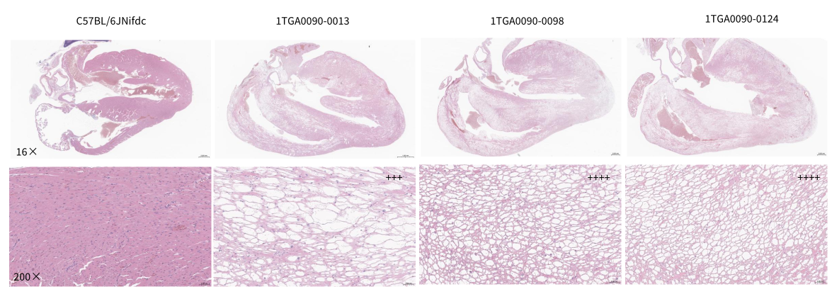
Histopathological characterization of B-Tg(hPRKAG2*R302Q) mice heart tissues via hematoxylin-eosin (HE) staining. Heart tissues were collected from wild-type control C57BL/6JNifdc mice and B-Tg(hPRKAG2*R302Q) mice (1TGA0090-0013, 1TGA0090-0098, 1TGA0090-0124) (8-11 weeks old, male). Wild-type control hearts exhibit orderly arranged cardiomyocytes with uniform cytoplasm, intact cellular structure, and no obvious vacuolation or degenerative changes in the myocardium. B-Tg(hPRKAG2*R302Q) mice display progressive, severe myocardial lesions characterized by prominent cardiomyocyte vacuolization and diffuse myocardial degeneration; with widespread cytoplasmic vacuole formation, disrupted myofibril organization, and marked disarray of myocardial tissue architecture, recapitulating the core histopathological hallmarks of human hypertrophic cardiomyopathy.
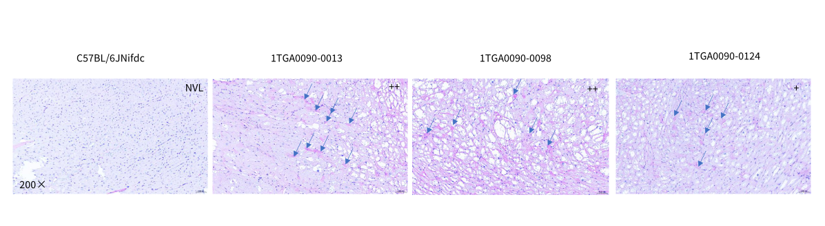
PAS staining of paraffin-embedded heart sections demonstrates glycogen accumulation in B-Tg(hPRKAG2*R302Q) mice. Heart tissues were collected from wild-type control C57BL/6JNifdc mice and B-Tg(hPRKAG2*R302Q) mice (1TGA0090-0013, 1TGA0090-0098, 1TGA0090-0124) (8-11 weeks old, male). The wild-type C57BL/6JNifdc showed no visible lesion (NVL) with minimal cytoplasmic PAS-positive glycogen. B-Tg(hPRKAG2*R302Q) mice display abundant magenta PAS-positive cytoplasmic glycogen deposits, recapitulating the core histopathological hallmarks of human hypertrophic cardiomyopathy.
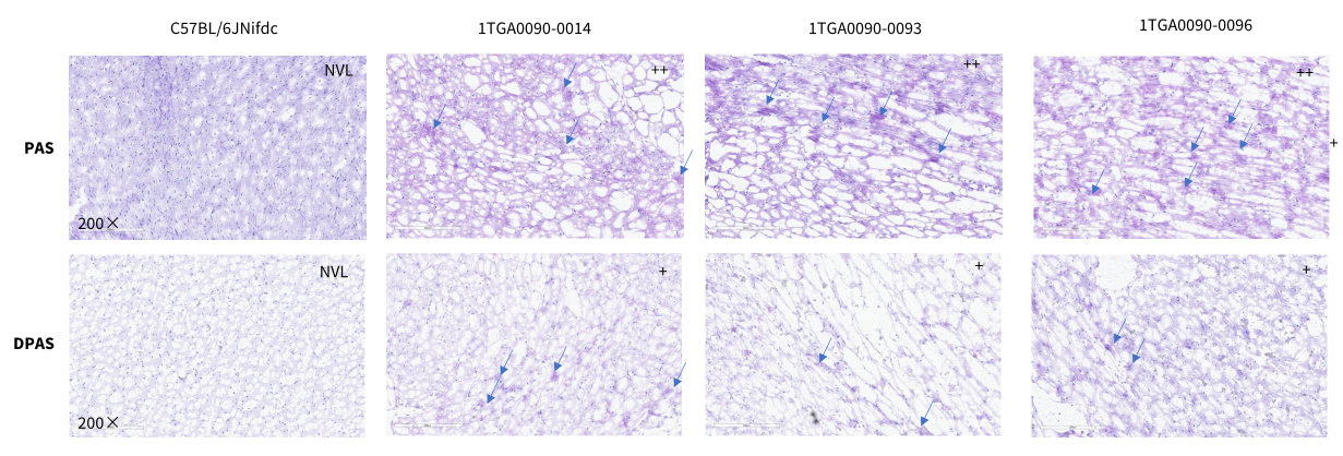
PAS staining and diastase-pretreated PAS (DPAS) staining of frozen heart sections demonstrates glycogen accumulation in B-Tg(hPRKAG2*R302Q) mice. Heart tissues were collected from wild-type control C57BL/6JNifdc mice and B-Tg(hPRKAG2*R302Q) mice (1TGA0090-0014, 1TGA0090-0093, 1TGA0090-0096) (13 weeks old, male). The wild-type C57BL/6JNifdc showed no visible lesion (NVL) with minimal cytoplasmic PAS-positive glycogen and DPAS positive signal. B-Tg(hPRKAG2*R302Q) mice display abundant magenta PAS-positive cytoplasmic glycogen deposits (upper), while DPAS staining showed marked clearing of the excessive glycogen deposition (bottom).
