SIRPA Protein Expression in Spleen
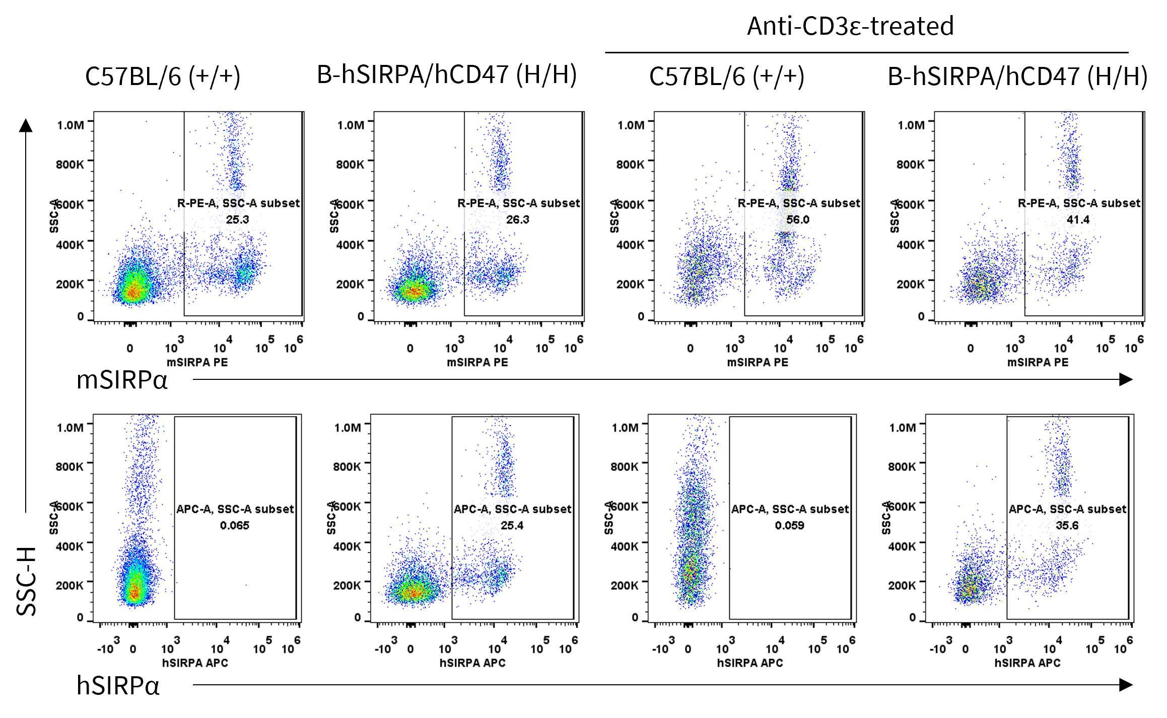
- Mouse SIRPA was detected in wild-type C57BL/6 mice. This anti-mouse SIRPα antibody also reacts crossly with human SIRPα.
- Human SIRPA was detected in homozygous B-hSIRPA/hCD47 mice, but not in wild-type C57BL/6 mice.
Mouse and human SIRPα expression analysis in Splenocytes. Splenocytes were collected from wild-type C57BL/6 mice and homozygous B-hSIRPA/hCD47 mice stimulated with or without anti-mouse CD3ε in vivo, and analyzed by flow cytometry with anti-mouse SIRPα antibody (Biolegend, 144011) and anti-human SIRPα antibody (Biolegend, 323810).
SIRPα Protein Expression on Peritoneal Lymphocytes
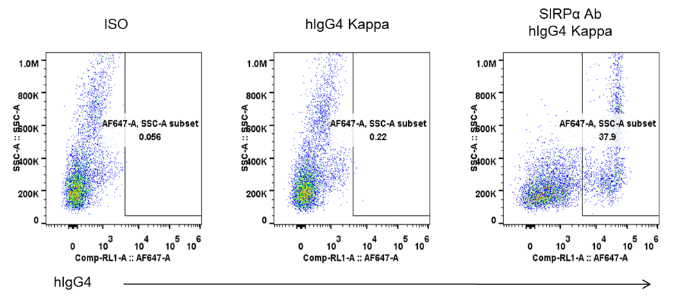
- Human SIRPα was detected on the peritoneal lymphocytes of B-hSIRPA/hCD47 mice, as evidenced by anti-human SIRPα antibody binding vs. isotype control.
Human SIRPα expression analysis on peritoneal lymphocytes. Peritoneal lymphocytes were isolated from female B-hSIRPA/hCD47 mice (n=2, 8-week-old). Flow cytometry analysis of the peritoneal lymphocytes was performed to assess the expression of human SIRPα. Single live cells were gated for the CD45 population and used for further analysis as indicated here.
CD47 Protein Expression in Spleen
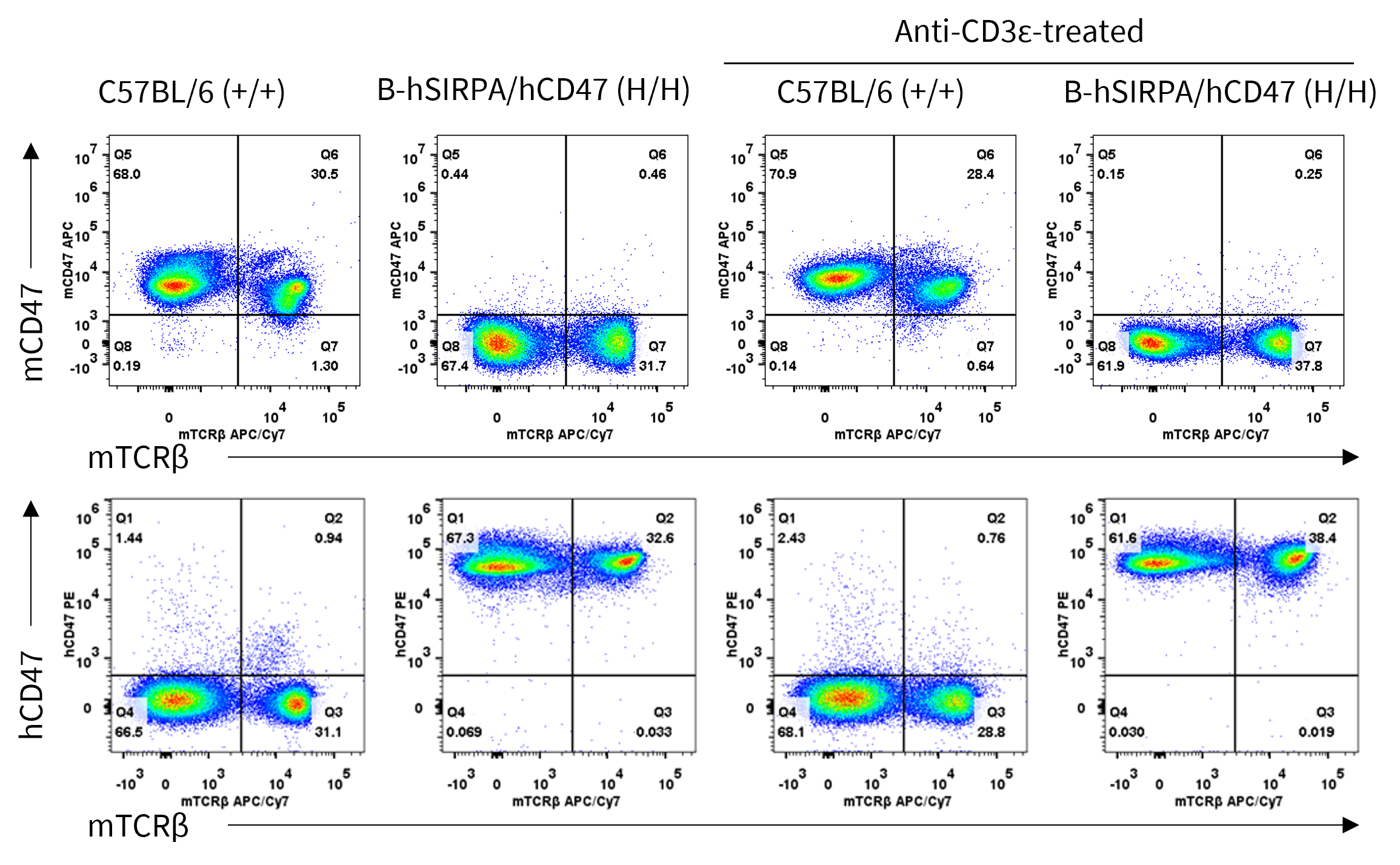
- Mouse CD47 was exclusively detected in wild-type C57BL/6 mice.
- Human CD47 was detected in homozygous B-hSIRPA/hCD47 mice, but not in wild-type C57BL/6 mice.
Mouse and human CD47 expression analysis in Splenocytes. Splenocytes were collected from wild-type C57BL/6 mice and homozygous B-hSIRPA/hCD47 mice stimulated with or without anti-mouse CD3ε in vivo, and analyzed by flow cytometry with anti-mouse CD47 antibody (Biolegend, 127514) and anti-human antibody (Biolegend, 323108).
Analysis of Leukocyte Subpopulations
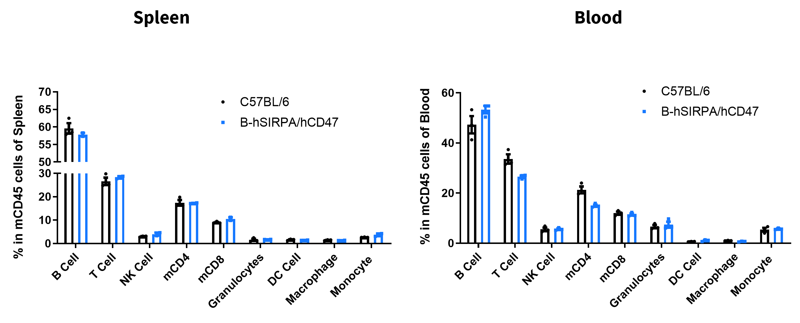
- Frequencies of B cell, T cell, NK cell, CD4+ T cell, CD8+ T cell, granulocyte, DC, macrophage and monocyte in homozygous B-hSIRPA/hCD47 mice were comparable to those in C57BL/6 mice, demonstrating that introduction of human SIRPA and CD47 in place of their mouse counterparts does not change the overall development, differentiation or distribution of these T cell subtypes in spleen or blood.
Analysis of leukocyte subpopulations by flow cytometry in Spleen. Splenocytes were collected from wild-type C57BL/6 mice and homozygous B-hSIRPA/hCD47 mice (female, 6-week-old, n = 3). Single live cells were gated on the CD45⁺ population and analyzed by flow cytometry as indicated.
Analysis of T Cell in Subpopulations
- Frequencies of CD4+ T, CD8+ T, and Tregs in homozygous B-hSIRPA/hCD47 mice were comparable to those in C57BL/6 mice, demonstrating that introduction of human SIRPA and CD47 in place of their mouse counterparts does not change the overall development, differentiation or distribution of these T cell subtypes in spleen or blood.
Analysis of T-cell subpopulations by flow cytometry in Spleen and blood. Splenocytes and blood were collected from wild-type C57BL/6 mice and homozygous B-hSIRPA/hCD47 mice (female, 6-week-old, n = 3). Single live cells were gated on the CD3⁺ T-cell population and analyzed by flow cytometry as indicated.
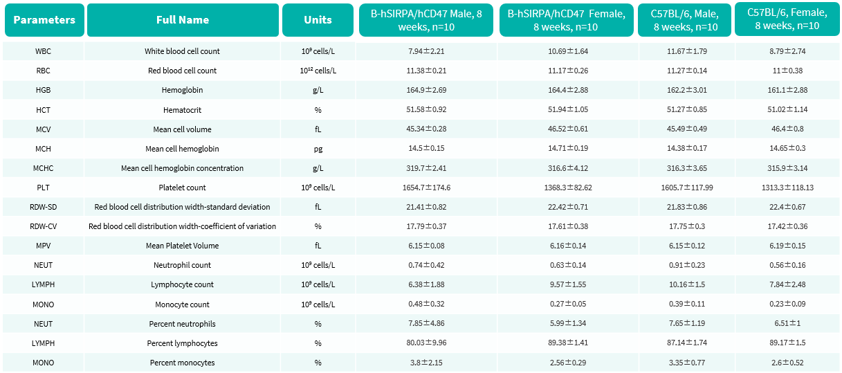
Complete Blood Count
- No significant differences were observed compared with wild-type mice.
Complete blood count (CBC) of B-hSIRPA/hCD47 mice. Values are expressed as mean ± SD.
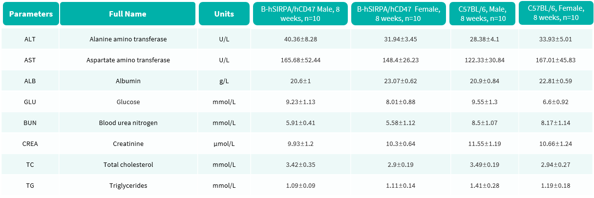
Biochemistry Analysis
- No significant differences were observed compared with wild-type mice.
Blood biochemical parameters of B-hSIRPA/hCD47 mice are shown. Values are expressed as mean ± SD.
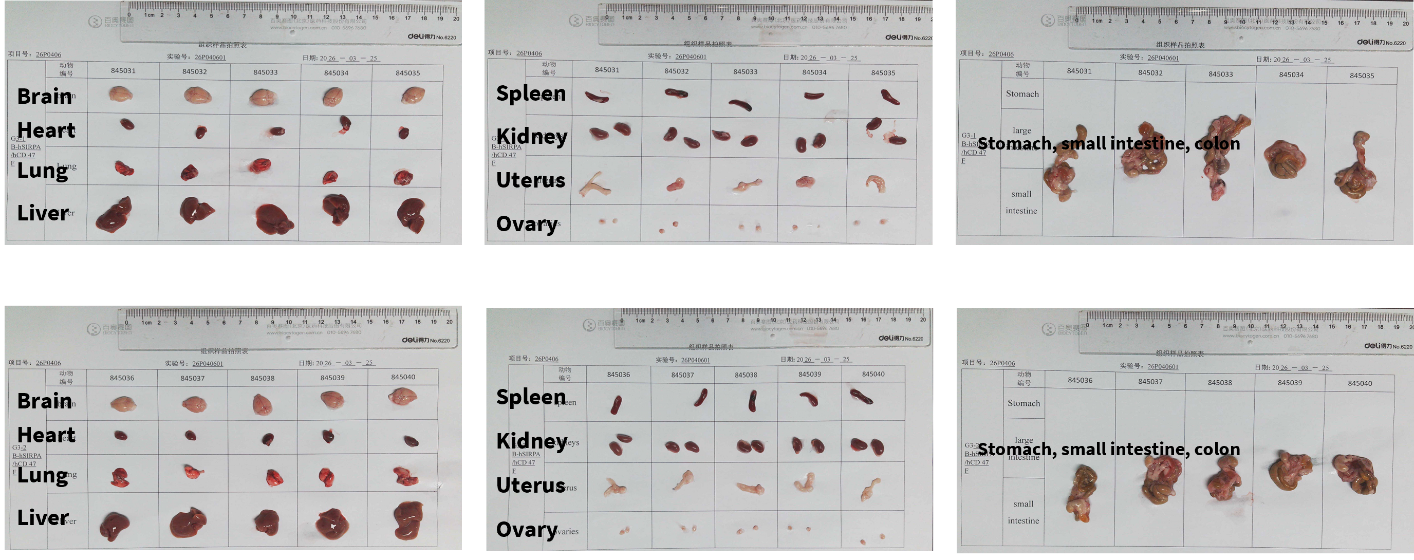
Gross Organ Anatomy (Female)
- No abnormalities were observed.
Organs of female B-hSIRPA/hCD47 mice(8-week-old, n = 10).
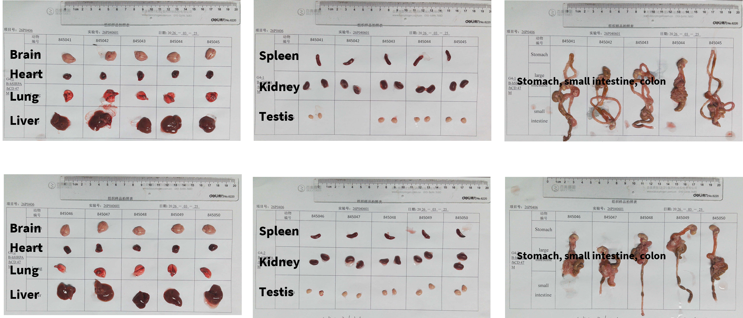
Gross Organ Anatomy (Male)
- No abnormalities were observed.
Organs of Male B-hSIRPA/hCD47 mice(8-week-old, n = 10).
Histopathological Analysis
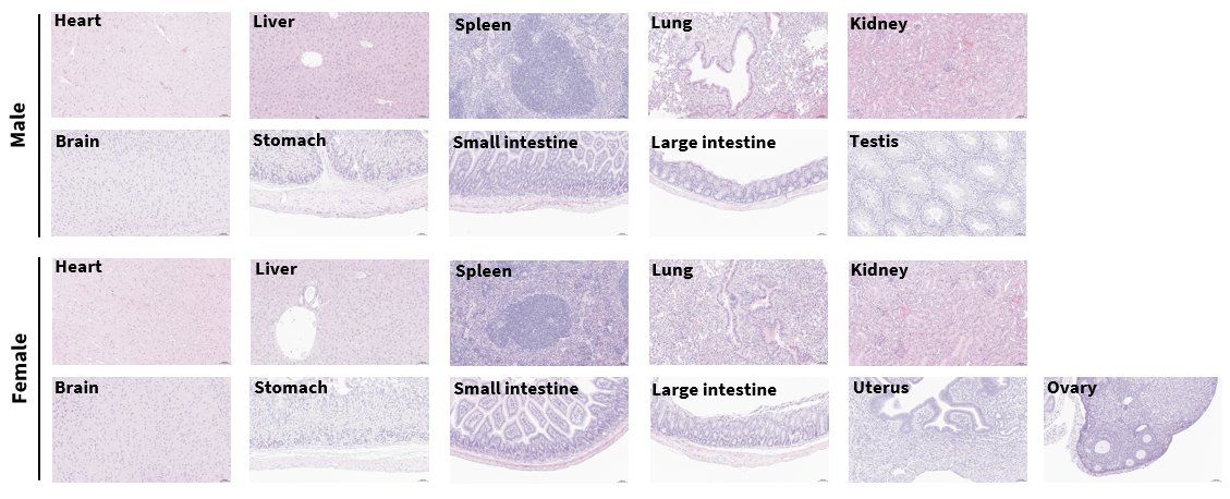
- No obvious abnormalities were observed in any organs examined (heart, liver, spleen, lung, kidney, brain, stomach, small intestine, large intestine, testis, uterus, ovary).
Histopathological analysis of organs in B-hSIRPA/hCD47 mice. Major organs from B-hSIRPA/hCD47 mice were collected at 8 weeks of age and analyzed by H&E staining (male, n = 10; female, n = 10).
Efficacy Evaluation of CD47 Specific Antibodies in the Treatment of the Subcutaneous B-hCD47 MC38 Model in B-hSIRPA/hCD47 Mice
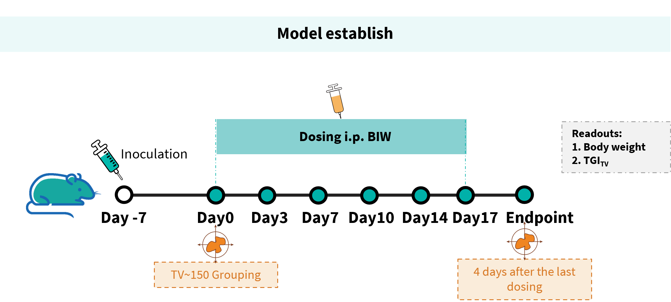
Establishment of a B-hCD47 MC38 model and in vivo efficacy study of anti-human CD47 antibodies. B-hCD47 MC38 melanoma cells were implanted subcutaneously into homozygous B-hSIRPA/hCD47mice (female, 6-8 weeks old, n=5). When the average tumor volume reached approximately 150 mm³, mice were randomized and subsequently administered the anti-human CD47 antibody via intraperitoneal injection.
Efficacy of CD47 Antibodies in B-hSIRPA/hCD47 mice. (A) Tumor growth curves. (B) Body weight changes during treatment. As shown in panel A, four anti-human CD47 antibodies differently inhibited tumor growth in B-hSIRPA/hCD47 mice, demonstrating that the B-hSIRPA/hCD47 mice provide a powerful preclinical model for in vivo evaluation of anti-human CD47 antibodies. Values are expressed as mean ± SEM.
Efficacy Evaluation of SIRPα Specific Antibody in the Treatment of the Subcutaneous B-hCD47 MC38 Model in B-hSIRPA/hCD47 Mice
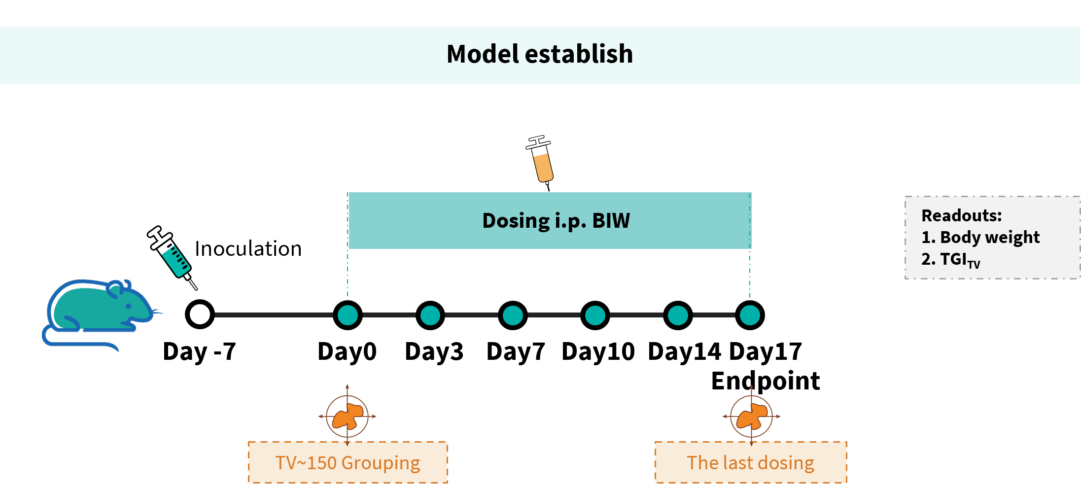
Establishment of a B-hCD47 MC38 model and in vivo efficacy study of anti-human SIRPα antibodies. B-hCD47 MC38 melanoma cells were implanted subcutaneously into homozygous B-hSIRPA/hCD47 mice (female, 6-8 weeks old, n=5). When the average tumor volume reached approximately 150 mm³, mice were randomized and subsequently administered the anti-human CD47 antibody via intraperitoneal injection.
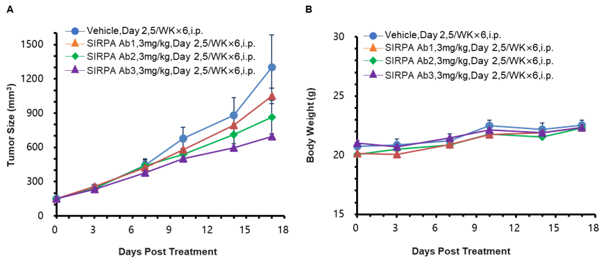
Efficacy of SIRPα Specific Antibodies in B-hSIRPA/hCD47 mice. (A) Tumor growth curves. (B) Body weight changes during treatment. As shown in panel A, three human SIRPα antibodies differently inhibited tumor growth in B-hSIRPA/hCD47 mice, demonstrating that the B-hSIRPA/hCD47 mice provide a powerful preclinical model for in vivo evaluation of anti-human SIRPα antibodies. Values are expressed as mean ± SEM.
* When publishing results obtained using this animal model, please acknowledge the source as follows: The animal model [B-hSIRPA/hCD47 mice] (Cat# 120525) was purchased from Biocytogen.