

C57BL/6N-Igf1rtm5(IGF1R)Bcgen/Bcgen • 111974
on this page

IGF1R: A pivotal receptor kinase in proliferation and its targeted therapeutic blockade

Gene targeting strategy for B-hIGF1R mice plus. A chimeric CDS that encodes mouse Igf1r signal peptide and human extracellular domain, human transmembrane and mouse cytoplasmic domain, as well as mouse 3’UTR region were inserted right after mouse Igf1r exon 2 to replace the exon 2 of mouse Igf1r gene. The chimeric IGF1R protein expression will be driven by endogenous mouse Igf1r promoter, while mouse Igf1r gene transcription and translation will be disrupted.

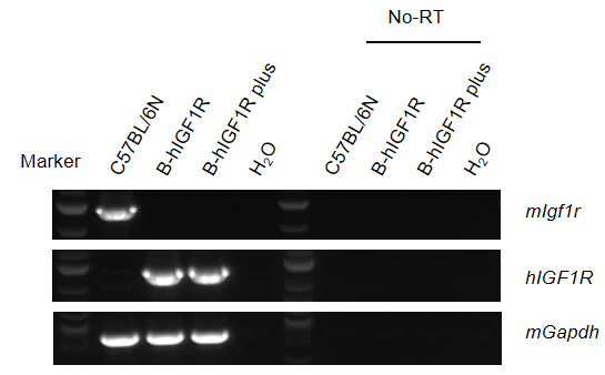
Strain specific analysis of IGF1R mRNA expression in wild-type C57BL/6N mice and B-hIGF1R mice by RT-PCR. Kidney RNA were isolated from wild-type C57BL/6N mice (+/+) (n=1, 14-week-old, male) and homozygous B-hIGF1R mice (H/H) (n=1, 8-week-old, male) and homozygous B-hIGF1R mice plus (H/H) (n=1, 14-week-old, male), then cDNA libraries were synthesized by reverse transcription, followed by PCR with IGF1R primers. No-RT: no reverse-transcripted.
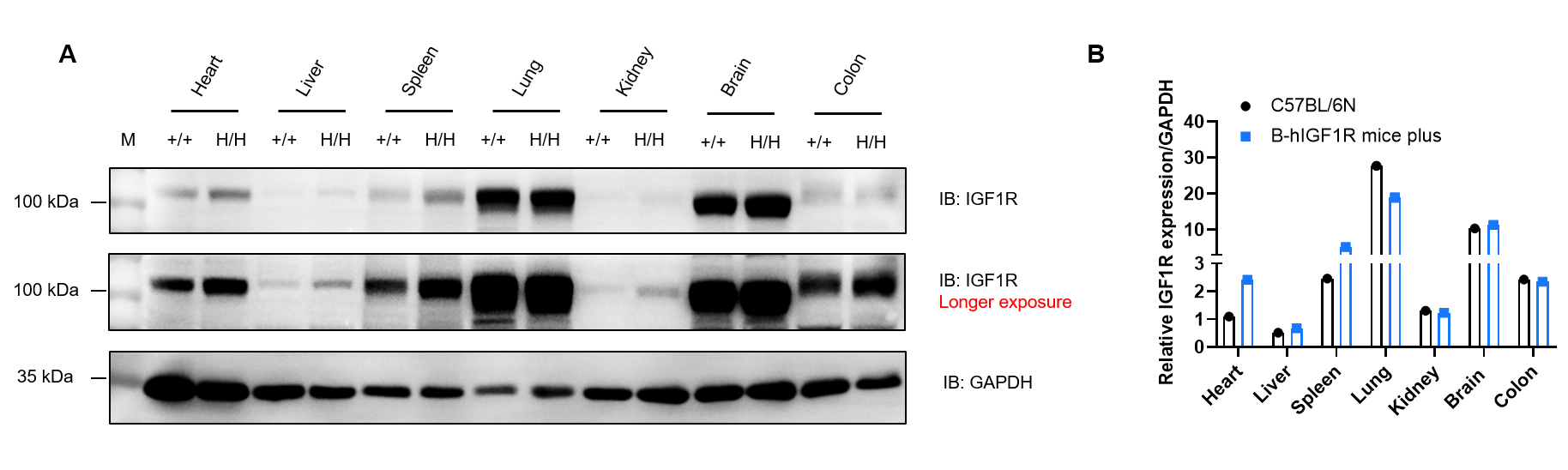
Western blot analysis of IGF1R protein expression in wild-type C57BL/6N mice and homozygous B-hIGF1R mice plus by WB. Various tissues were collected from wild-type C57BL/6N mice (+/+) and homozygous B-hIGF1R mice plus (H/H), and then analyzed by western blot with anti-IGF1R antibody (CST, 3027S). 40 μg total proteins were loaded for western blotting analysis. GAPDH were detected as internal control. M, marker.
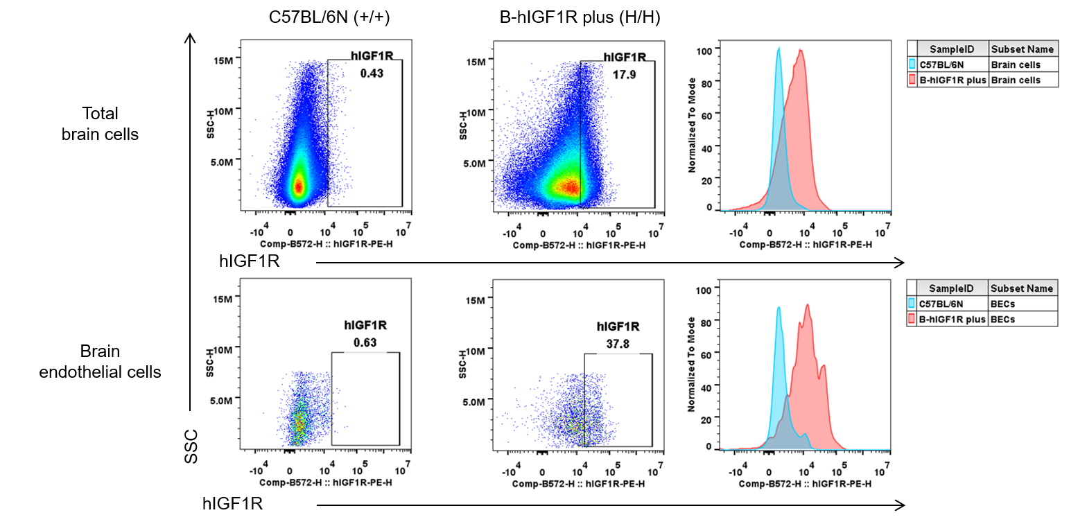
Strain specific IGF1R expression in wild-type C57BL/6N and homozygous B-hIGF1R mice plus by flow cytometry. Brain cells were collected from wild-type C57BL/6N (+/+) and homozygous B-hIGF1R mice plus (H/H) (male, 7-week-old, n=1) and analyzed by flow cytometry with anti-human IGF1R antibody (BioLegend, 351805).
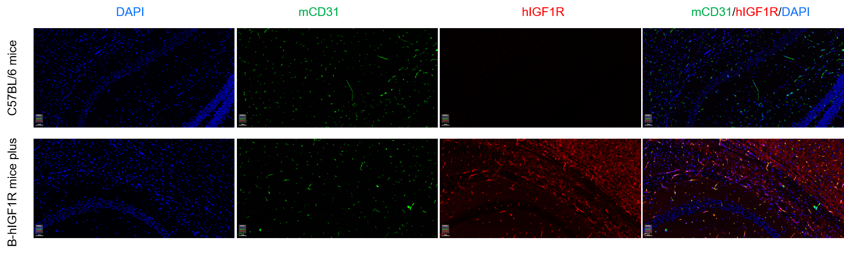
Immunofluorescence analysis of brain micro-vessels from the wild-type C57BL/6 mice and homozygous B-hIGF1R mice plus. Brain were collected from wild-type C57BL/6 mice (+/+) and homozygous B-hIGF1R mice plus (H/H), and analyzed by immunofluorescence with anti-mCD31 (abcam, ab182981) and anti-hIGF1R antibody (abcam, ab263903).
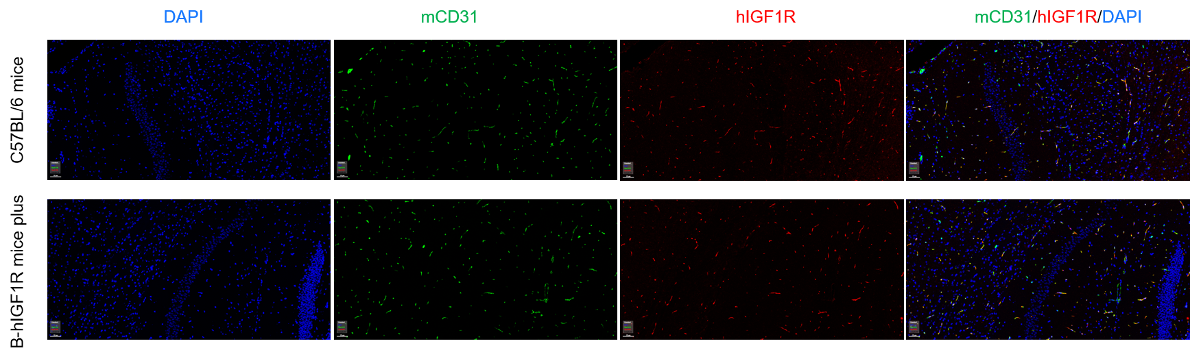
Immunofluorescence analysis of brain micro-vessels from the wild-type C57BL/6 mice and homozygous B-hIGF1R mice plus. Brain were collected from wild-type C57BL/6 mice (+/+) and homozygous B-hIGF1R mice plus (H/H), and analyzed by immunofluorescence with anti-mCD31 (abcam, ab182981) and anti-IGF1R antibody (CST, 3027S).
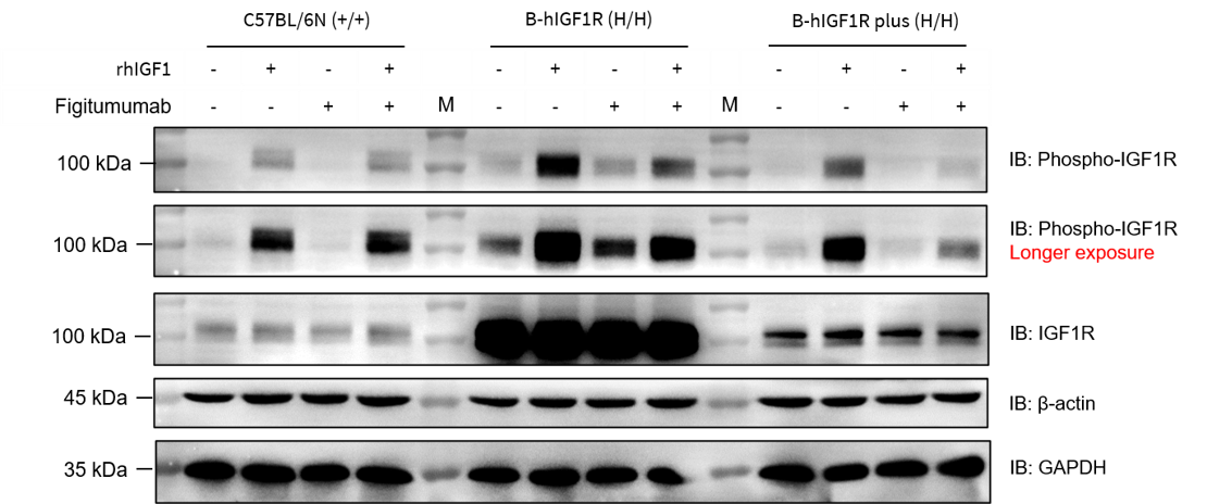
Western blot analysis of phospho-IGF1R/AKT protein expression in wild-type C57BL/6N mice and homozygous B-hIGF1R mice plus. Kidney cells were collected from wild-type C57BL/6N mice (+/+), homozygous B-hIGF1R mice (H/H) and homozygous B-hIGF1R mice plus (H/H), and then cultured with Figitumumab (1 μg/mL) or/and recombinant human IGF1 protein (200 ng/mL). Finally, these cells were analyzed by western blot with anti-IGF1R antibody (CST, 3027S), anti-phospho-IGF1R antibody (CST, 3024S) and anti-phospho-AKT antibody (CST, 9271S). M, marker.
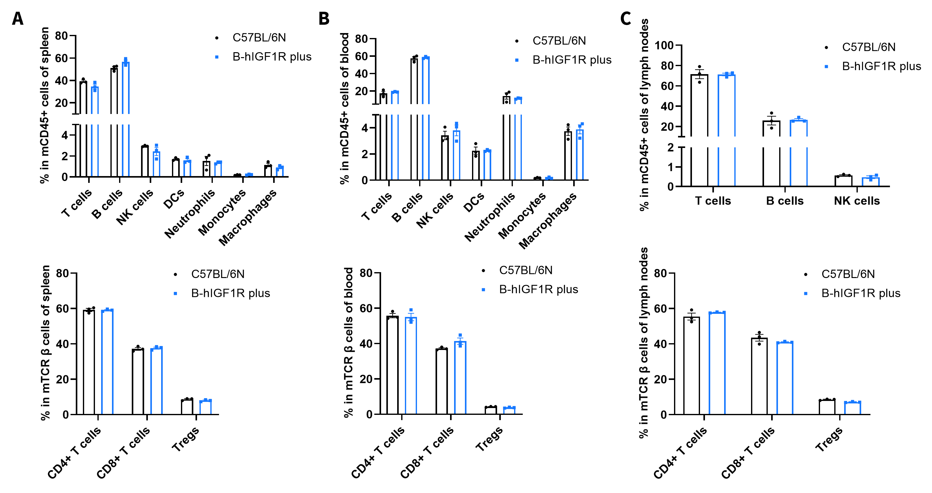
Frequency of leukocyte subpopulations by flow cytometry. Splenocytes were isolated from wild-type C57BL/6N mice (male, n=3, 7-week-old) and homozygous B-hIGF1R mice plus (male, n=3, 7-week-old). Top: Flow cytometry analysis of the splenocytes was performed to assess the frequency of leukocyte subpopulations. Bottom: Frequency of T cell subpopulations. (A) spleen. (B) blood. (C) lymph nodes. Values are expressed as mean ± SEM. Significance was determined by two-way ANOVA test. *p < 0.05, **p < 0.01, ***p < 0.001.

Growth curve of B-hIGF1R mice plus. 3-week-old mice were grouped by sex (10 males and 10 females). Body weight was measured weekly for 3 week-old to 15 week-old on the same day.
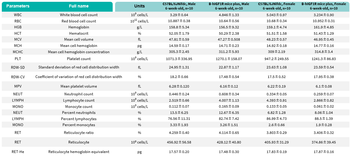
Values are expressed as mean ± SD.
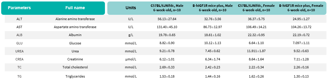
Blood chemistry tests of B-hIGF1R mice plus. Serum from male and female C57BL/6JNifdc and B-hIGF1R mice plus (n=10, 6 week-old) were collected for biochemistry analysis. There was no differences among any measurement between male C57BL/6JNifdc and male B-hIGF1R mice plus. Values are expressed as mean ± SEM.

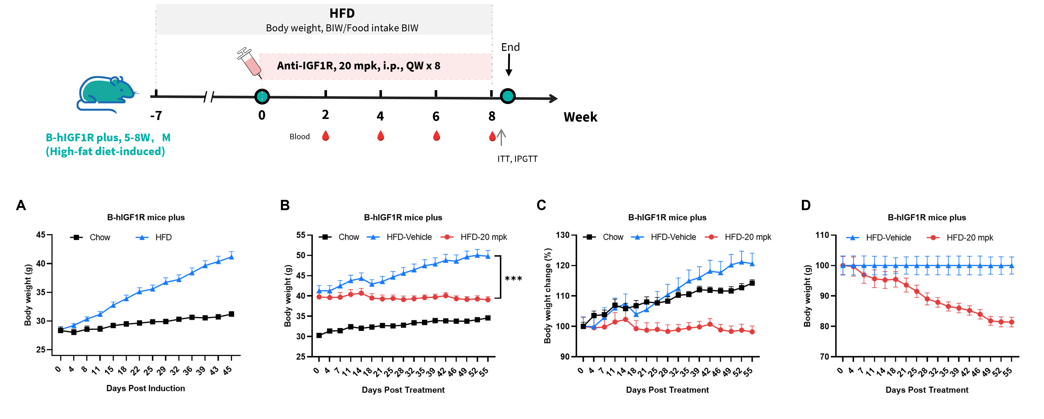
Efficacy study of anti-IGF1R antibody in HFD induced B-hIGF1R mice plus. B-hIGF1R mice plus were fed with a high-fat diet to induce obesity. (A) Body weight change after HFD induction. (B-D) Body weight change after anti-IGF1R antibody (provided by a client) treatment. ***p < 0.001. Values are expressed as mean ± SEM.
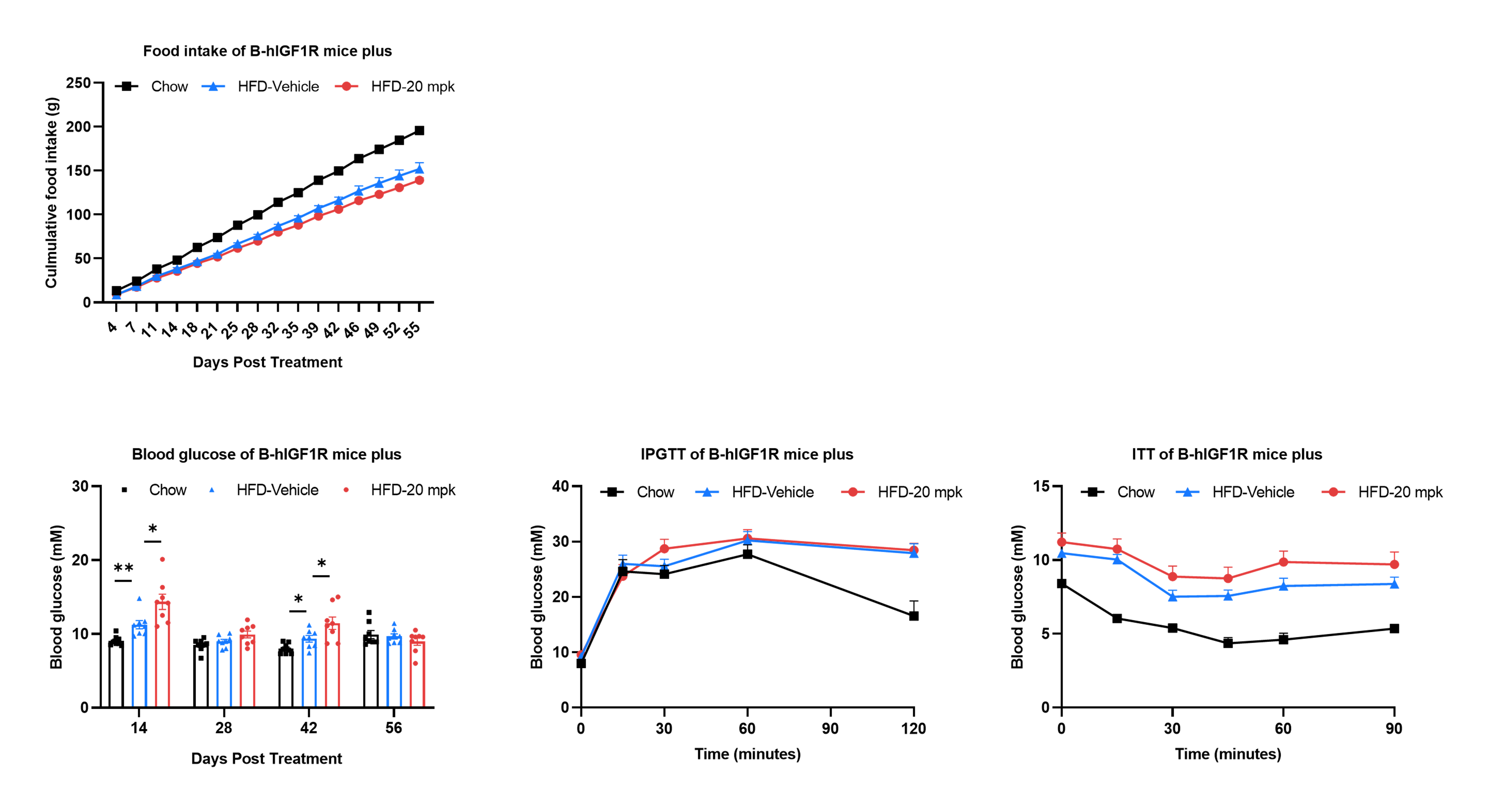
Efficacy study of anti-IGF1R antibody in HFD induced B-hIGF1R mice plus. B-hIGF1R mice plus were fed with a high-fat diet to induce obesity. (A) Effect of anti-IGF1R antibody (provided by a client) on food intake. (B) Blood glucose change after anti-IGF1R antibody treatment. (C) The glucose tolerance test for 15% D-Glucose is administered by intraperitoneal injection (IPGTT). (D) The insulin tolerance test for 0.5 U/kg is administered by intraperitoneal injection (ITT). Values are expressed as mean ± SEM. Significance was determined by the Ordinary one-way ANOVA. *p<0.05, **p<0.01, ***p<0.001.

Q1: What are B-hIGF1R mice plus?
B-hIGF1R mice plus are target-humanized mice expressing a chimeric IGF1R protein with human IGF1R extracellular and transmembrane regions, enabling evaluation of human IGF1R-targeted therapeutics in vivo.
Q2: Why is IGF1R an important therapeutic target?
IGF1R is a receptor tyrosine kinase involved in IGF signaling, cell growth, survival, metabolism, and transformation, making IGF1R relevant to oncology, metabolic disease, obesity, diabetes, and CNS delivery research.
Q3: How was IGF1R expression validated in B-hIGF1R mice plus?
Human IGF1R mRNA was validated by RT-PCR, IGF1R protein was detected by western blot across multiple tissues, and human IGF1R expression was confirmed in brain endothelial cells and brain micro-vessels.
Q4: Can B-hIGF1R mice plus be used for functional antibody studies?
Yes. Figitumumab-analog suppressed IGF1-induced activation of humanized IGF1R in B-hIGF1R mice plus, supporting functional evaluation of human IGF1R-targeted antibodies.
Q5: What are the main applications of B-hIGF1R mice plus?
Applications include anti-IGF1R antibody efficacy studies, IGF1R pathway pharmacology, obesity and metabolic disease research, oncology studies, brain endothelial IGF1R research, and preclinical safety evaluation.
