VEGFA (vascular endothelial growth factor A) is a potent angiogenic factor that regulates vascular development and pathological neovascularization. It is widely expressed in endothelial cells, smooth muscle cells, and stromal cells, and plays a critical role in tissue growth, repair, and tumor progression. Overexpression of VEGFA contributes to uncontrolled angiogenesis in cancers, diabetic retinopathy, and age-related macular degeneration (AMD), making VEGFA a central target in anti-angiogenic therapy.
In VEGFA humanized mice, the murine Vegfa gene exons 1–8 are replaced by the corresponding human VEGFA sequences, enabling the physiological expression of human VEGFA under native regulation. Importantly, these mice exhibit normal vascular morphology and angiogenesis, ensuring that human VEGFA expression does not disrupt baseline physiology.
VEGFA humanized mice express human VEGFA under endogenous regulation, supporting anti-VEGFA drug validation, angiogenesis studies, and preclinical ophthalmology and oncology research.
Key Advantages
- Fully humanized VEGFA locus ensures physiological expression of human VEGFA while preserving normal gene regulation.
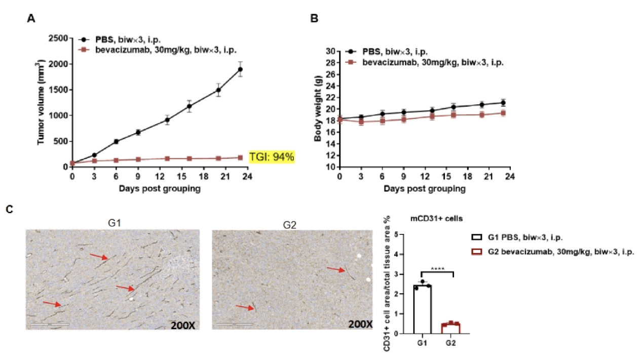
- No baseline angiogenesis disruption, as confirmed by mCD31 staining—ideal for studying pathological angiogenesis without developmental artifacts.
- Retinal morphology preserved, supporting ophthalmic studies without structural confounds.
- Robust platform for anti-VEGFA therapeutics, validated by bevacizumab efficacy and angiogenesis inhibition.
- Versatile preclinical tool, suitable for efficacy testing of anti-VEGFA and bispecific therapeutics, including combination regimens.
Validation
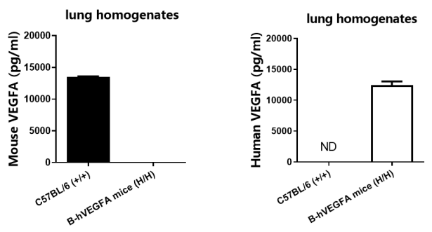
- Lung homogenates from wild-type and homozygous B-hVEGFA (H/H) mice were tested using species-specific ELISA kits. Mouse VEGFA was detected only in WT mice, whereas human VEGFA was exclusively present in B-hVEGFA mice.
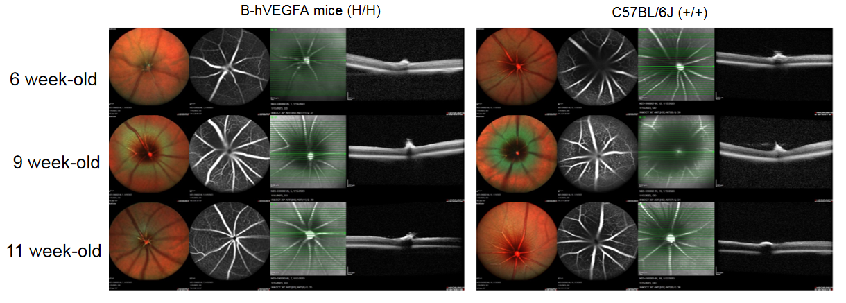
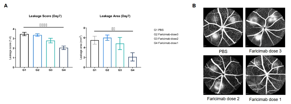
- Fundus photography of B-hVEGFA mice (male, n=3) shows no significant differences compared with WT mice, indicating preserved ocular structure.
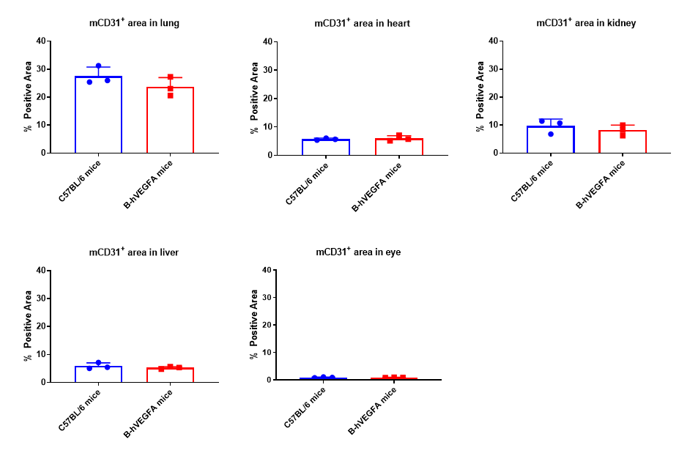
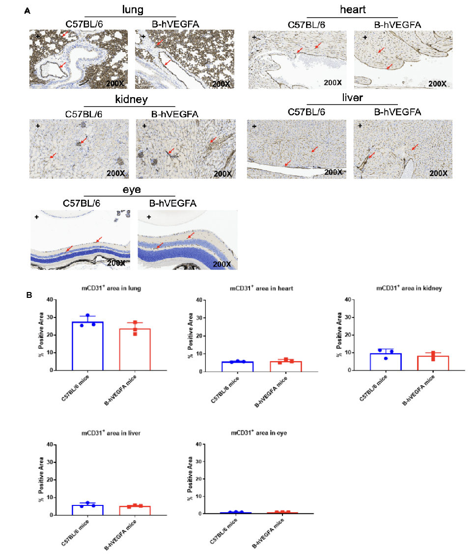
- mCD31 immunohistochemistry in heart, liver, lung, kidney, and eye tissues of B-hVEGFA and C57BL/6 mice (female, 8 weeks old, n=3) revealed no significant differences in microvascular density, suggesting humanization has no impact on baseline angiogenesis.
- Bevacizumab inhibited tumor growth in B-hVEGFA mice bearing B-hVEGFA MC38 tumors (female, 6–7 weeks old, n=6). Additionally, CD31+ area in tumor tissues was significantly reduced, demonstrating suppressed angiogenesis and confirming model suitability for anti-VEGFA therapeutic evaluation.
Application
Preclinical oncology research
- Evaluation of anti-VEGFA therapies (e.g., bevacizumab) in solid tumor models.
- Studies on tumor angiogenesis, vascular normalization, and resistance mechanisms.
Ophthalmology models
- Preclinical testing of anti-VEGFA drugs for age-related macular degeneration (AMD).
- Evaluation of therapeutic strategies for diabetic retinopathy and retinal neovascularization.
Vascular biology and angiogenesis studies
- Mechanistic studies of human VEGFA signaling in endothelial cells and stromal cells.
- Analysis of angiogenesis in physiological and pathological contexts.
Antibody and biologics validation
- In vivo efficacy testing of anti-VEGFA antibodies and biosimilars.
- Preclinical evaluation of VEGFA-targeted fusion proteins and bispecific antibodies.
Combination therapy studies
- Assessment of anti-VEGFA agents combined with chemotherapy, radiotherapy, or immunotherapy.
- Investigation of synergistic effects in tumor regression and vascular modulation.
Biomarker discovery
- Identification of VEGFA-related biomarkers for response prediction and resistance profiling.
- Development of translational endpoints for clinical trial design.