Basic Information
TNFRSF4(Tumor necrosis factor receptor superfamily, member 4, also known as OX40);
TNFSF4 ( tumor necrosis factor(TNF) superfamily member 4, also known as OX40L)
-
Gene Targeting Strategy

-
Gene targeting strategy for B-hOX40/hOX40L mice. The exons 1-5 of mouse OX40 gene that encode the extracellular domain were replaced by human OX40 exons 1-5 in B-hOX40/hOX40L mice. The exons 2-3 of mouse Ox40l gene that encode the extracellular region were replaced by human OX40L exons 2-3 in B-hOX40/hOX40L mice .
-
mRNA Expression Analysis

-

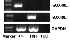
Strain specific analysis of OX40L gene expression in WT and B-hOX40/hOX40L mice by RT-PCR. (B)Mouse Ox40l mRNA was detectable in DC cell of wild-type (+/+) . Human OX40L mRNA was detectable only in B-hOX40/hOX40L (H/H) but not in +/+ mice.
-
Protein Expression Analysis

-

Species–specific OX40 expression analysis in double humanized B-hOX40/hOX40L mice. Following anti-CD3ε stimulation in vivo, splenocytes were collected from wild-type C57BL/6 (+/+) and homozygous B-hOX40/hOX40L (H/H) mice and analyzed by flow cytometry using species-specific anti-OX40 antibodies. Human OX40 was exclusively detected in B-hOX40/hOX40L mice compared to wild-type mice.
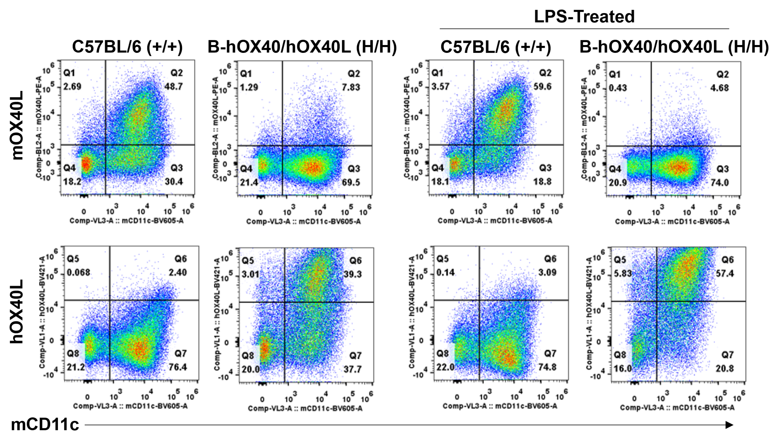
 Species–specific OX40L expression analysis in double humanized B-hOX40/hOX40L mice. Bone marrow cells or bone marrow-derived DCs stimulated with LPS were collected from wild-type C57BL/6 (+/+) and homozygous B-hOX40/hOX40L (H/H) mice and analyzed by flow cytometry using species-specific anti-OX40L antibodies. Human OX40L was exclusively detected in B-hOX40/hOX40L mice compared to wild-type mice.
 -
Analysis of Immune Cells

-
Analysis of spleen leukocytes cell subpopulations in B-hOX40/hOX40L mice


Analysis of spleen leukocyte subpopulations by FACS. Splenocytes were isolated from female C57BL/6 and B-hOX40/hOX40L mice (n=3, 6-week-old). Flow cytometry analysis of the splenocytes was performed to assess leukocyte subpopulations. (A) Representative FACS plots. Single live cells were gated for the CD45+ population and used for further analysis as indicated here. (B) Results of FACS analysis. Percent of T cells, B cells, NK cells, dendritic cells, granulocytes, monocytes and macrophages in homozygous B-hOX40/hOX40L mice were similar to those in the C57BL/6 mice, demonstrating that introduction of hOX40/hOX40L in place of its mouse counterpart does not change the overall development, differentiation or distribution of these cell types in spleen. Values are expressed as mean ± SEM.
Analysis of spleen T cell subpopulations in B-hOX40/hOX40L mice

Analysis of spleen T cell subpopulations by FACS. Splenocytes were isolated from female C57BL/6 and B-hOX40/hOX40L mice (n=3, 6-week-old). Flow cytometry analysis of the splenocytes was performed to assess leukocyte subpopulations. (A) Representative FACS plots. Single live CD45+ cells were gated for TCRβ+ T cell population and used for further analysis as indicated here. (B) Results of FACS analysis. The percent of CD8+ T cells, CD4+ T cells, and Tregs in homozygous B-hOX40/hOX40L mice were similar to those in the C57BL/6 mice, demonstrating that introduction of hOX40/hOX40L in place of its mouse counterpart does not change the overall development, differentiation or distribution of these T cell subtypes in spleen. Values are expressed as mean ± SEM.
Analysis of blood leukocytes cell subpopulations in B-hOX40/hOX40L mice


Analysis of blood leukocyte subpopulations by FACS. Blood cells were isolated from female C57BL/6 and B-hOX40/hOX40L mice (n=5, 6-week-old). Flow cytometry analysis of the blood cell was performed to assess leukocyte subpopulations. (A) Representative FACS plots. Single live cells were gated for the CD45+ population and used for further analysis as indicated here. (B) Results of FACS analysis. Percent of T cells, B cells, NK cells, dendritic cells, granulocytes, monocytes and macrophages in homozygous B-hOX40/hOX40L mice were similar to those in the C57BL/6 mice, demonstrating that introduction of hOX40/hOX40L in place of its mouse counterpart does not change the overall development, differentiation or distribution of these cell types in blood. Values are expressed as mean ± SEM.
Analysis of blood leukocytes cell subpopulations in B-hOX40/hOX40L mice

Analysis of blood T cell subpopulations by FACS. Blood cells were isolated from female C57BL/6 and B-hOX40/hOX40L mice (n=3, 6-week-old). Flow cytometry analysis of the blood was performed to assess leukocyte subpopulations. (A) Representative FACS plots. Single live CD45+ cells were gated for TCRβ+ T cell population and used for further analysis as indicated here. (B) Results of FACS analysis. The percent of CD8+ T cells, CD4+ T cells, and Tregs in homozygous B-hOX40/hOX40L mice were similar to those in the C57BL/6 mice, demonstrating that introduction of hOX40/hOX40L in place of its mouse counterpart does not change the overall development, differentiation or distribution of these T cell subtypes in blood. Values are expressed as mean ± SEM.
Analysis of lymph node leukocytes cell subpopulations in B-hOX40/hOX40L mice

Analysis of lymph node leukocyte subpopulations by FACS. Lymph nodes were isolated from female C57BL/6 and B-hOX40/hOX40L mice (n=3, 6-week-old). Flow cytometry analysis of the leukocytes was performed to assess leukocyte subpopulations. (A) Representative FACS plots. Single live cells were gated for the CD45+ population and used for further analysis as indicated here. (B) Results of FACS analysis. Percent of T cells, B cells, NK cells in homozygous B-hOX40/hOX40L mice were similar to those in the C57BL/6 mice, demonstrating that introduction of hOX40/hOX40L in place of its mouse counterpart does not change the overall development, differentiation or distribution of these cell types in lymph node. Values are expressed as mean ± SEM.
Analysis of lymph node T cell subpopulations in B-hOX40/hOX40L mice

Analysis of lymph node T cell subpopulations by FACS. Lymph nodes were isolated from female C57BL/6 and B-hOX40/hOX40L mice (n=3, 6-week-old). Flow cytometry analysis of the leukocytes was performed to assess leukocyte subpopulations. (A) Representative FACS plots. Single live CD45+ cells were gated for TCRβ+ T cell population and used for further analysis as indicated here. (B) Results of FACS analysis. The percent of CD8+ T cells, CD4+ T cells, and Tregs in homozygous B-hOX40/hOX40L mice were similar to those in the C57BL/6 mice, demonstrating that introduction of hOX40/hOX40L in place of its mouse counterpart does not change the overall development, differentiation or distribution of these T cell subtypes in lymph node. Values are expressed as mean ± SEM.
-
Experimental Schedule for Induction of AD-Like Skin Lesions and In Vivo Efficacy of Anti-Human OX40L Antibody

-

Experimental schedule for Induction of AD-like skin lesions and in vivo efficacy of anti-human OX40L antibody. OXZ was applied to dorsal and ear skin of mice on day 0, and then challenge to the same site of skin nine times from days 7 to 25. Anti-human OX40L antibody KY1005 (in house) was administered by intraperitoneal injection twice a week on days 6 to 23. Serum was collected at the endpoint on day 26. AD: atopic dermatitis; OXZ: oxazolone.
-
In Vivo Efficacy of Anti-Human OX40L Antibody with AD Model

-

Efficacy of anti-human OX40L antibody in B-hOX40/hOX40L mice. Mice in each group were treated with different dose of KY1005 produced in house. Doses are shown in legend. Total IgE levels were measured by ELISA on day 26. The concentrations of total serum IgE were negative related with the doses of antibody. (n = 5).
-
Summary

-
Protein expression analysis:
- Human OX40 was exclusively detectable in T cells of the homozygous B-hOX40/hOX40L mice but not in wild-type mice.
- Human OX40L was exclusively detectable in DCs of the homozygous B-hOX40/hOX40L mice but not in wild-type mice.
Leukocytes subpopulation analysis:
- OX40 and OX40L humanized does not change the overall development, differentiation or distribution of immune cell types in spleen, bone marrow, and blood.
In vivo efficacy:
- Anti-human OX40L antibody was efficacious in controlling atopic dermatitis like symptoms in B-hOX40/hOX40L mice, the concentrations of total serum IgE was decreased after treat with Anti-human OX40L antibody.
-
Poster

-
AACR 2023: Humanized OX40/OX40L Mice as a Tool for Evaluating Novel Therapeutics


