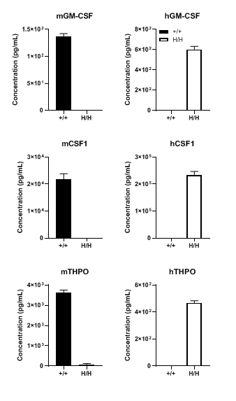
Protein expression analysis
Strain specific GM-CSF, CSF1 and THPO expression analysis in wild-type B-NDG mice and homozygous B-NDG MGMT3 mice by ELISA. Serum was collected from the two mice stimulated with LPS in vivo and analyzed by ELISA (n=3). Mouse GM-CSF, CSF1 and THPO were only detectable in B-NDG mice (+/+) but not in B-NDG MGMT3 mice (H/H). Human GM-CSF, CSF1 and THPO were only detectable in B-NDG MGMT3 mice. As IL3 is mainly expressed in activated T cells and there is no mature T cells in B-NDG background mice, mouse or human IL3 was not detectable in both of the two mice.
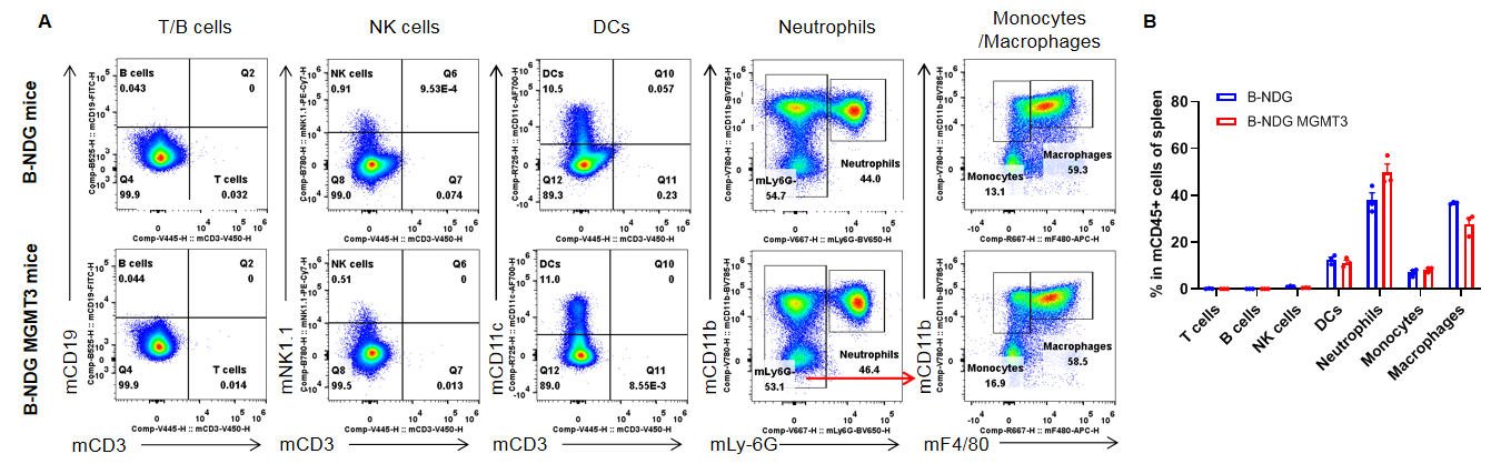
Analysis of leukocyte subpopulation in spleen
Analysis of leukocyte subpopulations in spleen by flow cytometry. Splenocytes were isolated from male B-NDG and B-NDG MGMT3 mice (n=3, 7-8-week-old). Flow cytometry analysis of the splenocytes was performed to assess leukocyte subpopulations. A. Representative FACS plots. Single live cells were gated for the CD45+ population and used for further analysis as indicated here. B. Results of FACS analysis. Percentages of T cells, B cells, NK cells, dendritic cells, neutrophils, monocytes and macrophages in homozygous B-NDG MGMT3 mice were similar to those in the B-NDG mice, demonstrating that IL3, GM-CSF, CSF1 and THPO humanized does not change the overall development, differentiation or distribution of these cell types in spleen. Values are expressed as mean ± SEM.
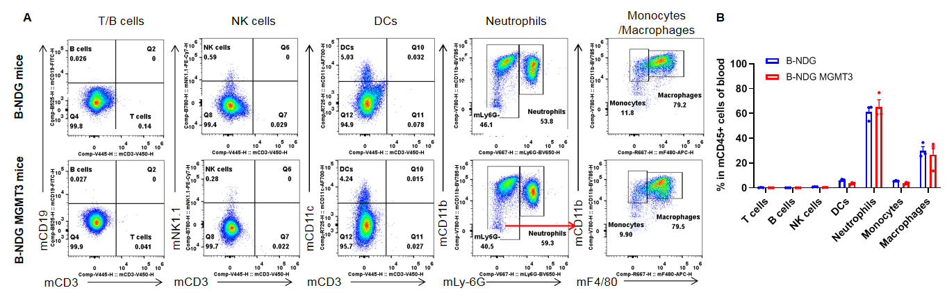
Analysis of leukocyte subpopulation in blood
Analysis of leukocyte subpopulations in blood by flow cytometry. Blood was isolated from male B-NDG and B-NDG MGMT3 mice (n=3, 7-8-week-old). Flow cytometry analysis of the blood cells was performed to assess leukocyte subpopulations. A. Representative FACS plots. Single live cells were gated for the CD45+ population and used for further analysis as indicated here. B. Results of FACS analysis. Percentages of T cells, B cells, NK cells, dendritic cells, neutrophils, monocytes and macrophages in homozygous B-NDG MGMT3 mice were similar to those in the B-NDG mice, demonstrating that IL3, GM-CSF, CSF1 and THPO humanized does not change the overall development, differentiation or distribution of these cell types in blood. Values are expressed as mean ± SEM.
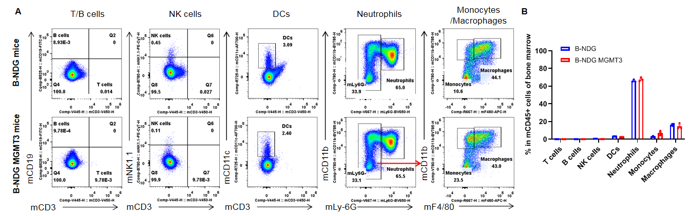
Analysis of leukocyte subpopulation in bone marrow
Analysis of leukocyte subpopulations in bone marrow by flow cytometry. Bone marrow was isolated from male B-NDG and B-NDG MGMT3 mice (n=3, 7-8-week-old). Flow cytometry analysis of the cells from bone marrow was performed to assess leukocyte subpopulations. A. Representative FACS plots. Single live cells were gated for the CD45+ population and used for further analysis as indicated here. B. Results of FACS analysis. Percent of T cells, B cells, NK cells, dendritic cells, neutrophils, monocytes and macrophages in homozygous B-NDG MGMT3 mice were similar to those in the B-NDG mice, demonstrating that IL3, GM-CSF, CSF1 and THPO humanized does not change the overall development, differentiation or distribution of these cell types in bone marrow. Values are expressed as mean ± SEM.
Human CD34+ HSCs engraftment for human immune system reconstitution
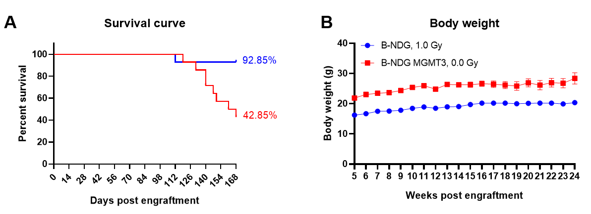
Human CD34+ HSCs (3E4) were intravenous (temporal vein) engrafted into wild-type B-NDG mice and homozygous B-NDG MGMT3 mice (both sex, 24-72 hr after birth, n=15). B-NDG mice were treated with 1.0 Gy-irradiation. B-NDG MGMT3 mice were not irradiated. A. Survival rates of the mice were analyzed with Kaplan Meier survival curves. B. Body weight. Results showed that the survival rate of B-NDG MGMT3 mice was similar to that of B-NDG mice until 18 weeks after human CD34+ HSCs engraftment and then decreased to 42.85% at 24 weeks post engraftment. But the body weight of B-NDG MGMT3 mice was significantly higher than that of B-NDG mice and increased steadily during the whole reconstitution. Values are expressed as mean ± SEM. HSCs: hematopoietic stem cells.
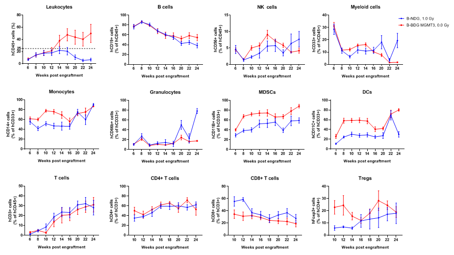
Human CD34+ HSCs (3E4) were intravenous (temporal vein) engrafted into wild-type B-NDG mice and homozygous B-NDG MGMT3 mice (both sex, 24-72 hr after birth, n=15). B-NDG mice were treated with 1.0 Gy-irradiation. B-NDG MGMT3 mice were not irradiated. Peripheral blood lymphocytes from the two mice after engraftment with human CD34+ HSCs were analyzed with flow cytometry. Results showed that the proportion of CD45+ cells in B-NDG MGMT3 mice reached 25% starting from 12 weeks after engraftment and continued to rise, significantly higher than that in B-NDG mice. The proportions of monocytes, MDSCs, DCs and Tregs in B-NDG MGMT3 mice were higher than that in B-NDG mice. Values are expressed as mean ± SEM.
Human CD34+ HSC engraftment for human immune system reconstitution
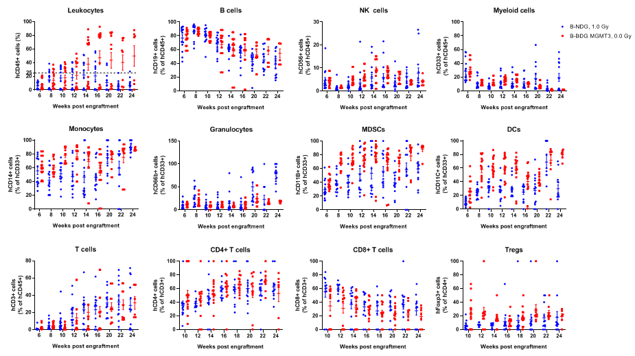
Human CD34+ HSCs (3E4) were intravenous (temporal vein) engrafted into wild-type B-NDG mice and homozygous B-NDG MGMT3 mice (both sex, 24-72 hr after birth, n=15). B-NDG mice were treated with 1.0 Gy-irradiation. B-NDG MGMT3 mice were not irradiated. Peripheral blood lymphocytes from the two mice after engraftment with human CD34+ HSCs were analyzed with flow cytometry. Results showed that the proportion of CD45+ cells in B-NDG MGMT3 mice reached 25% starting from 12 weeks after engraftment and continued to rise, significantly higher than that in B-NDG mice. The proportions of monocytes, MDSCs, DCs and Tregs in B-NDG MGMT3 mice were higher than that in B-NDG mice. Values are expressed as mean ± SEM.
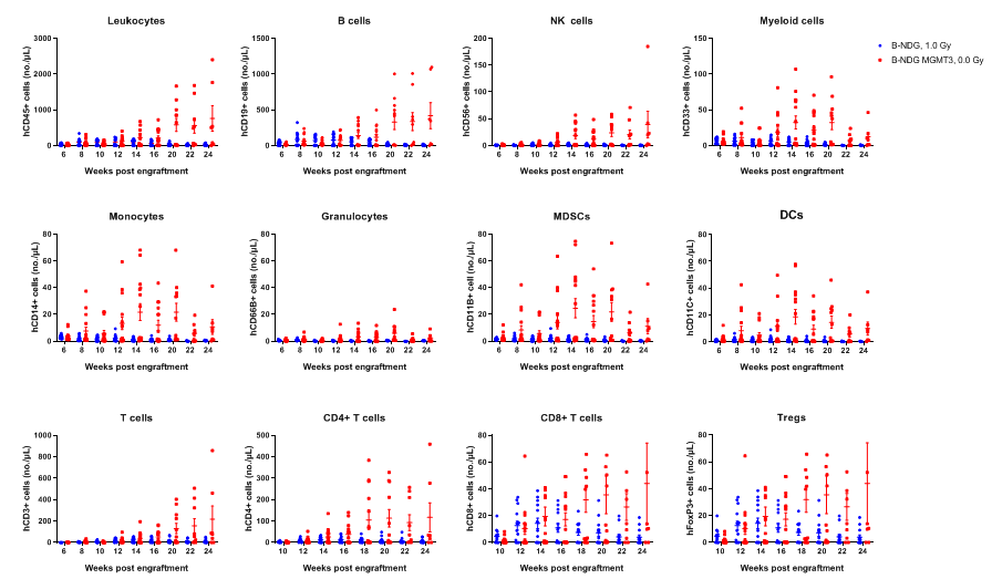
Human CD34+ HSCs (3E4) were intravenous (temporal vein) engrafted into wild-type B-NDG mice and homozygous B-NDG MGMT3 mice (both sex, 24-72 hr after birth, n=15). B-NDG mice were treated with 1.0 Gy-irradiation. B-NDG MGMT3 mice were not irradiated. Peripheral blood lymphocytes from the two mice after engraftment with human CD34+ HSCs were analyzed with flow cytometry. Results showed that the cell numbers of all the cells analyzed from 12 weeks after engraftment in B-NDG MGMT3 mice were higher than that in B-NDG mice. Values are expressed as mean ± SEM. Values are expressed as mean ± SEM.
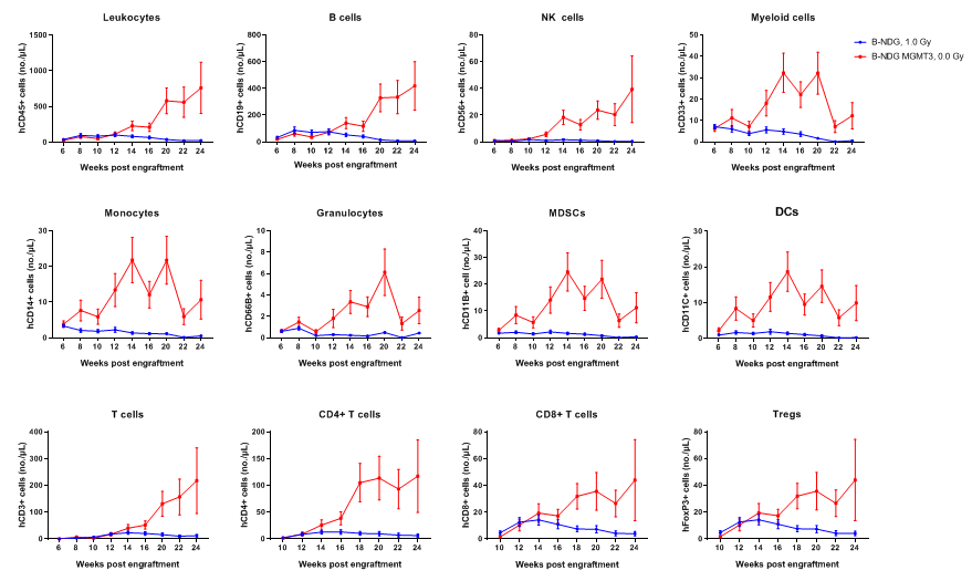
Human CD34+ HSCs (3E4) were intravenous (temporal vein) engrafted into wild-type B-NDG mice and homozygous B-NDG MGMT3 mice (both sex, 24-72 hr after birth, n=15). B-NDG mice were treated with 1.0 Gy-irradiation. B-NDG MGMT3 mice were not irradiated. Peripheral blood lymphocytes from the two mice after engraftment with human CD34+ HSCs were analyzed with flow cytometry. Results showed that the cell numbers of all the cells analyzed from 12 weeks after engraftment in B-NDG MGMT3 mice were higher than that in B-NDG mice. Values are expressed as mean ± SEM. Values are expressed as mean ± SEM.
* When publishing results obtained using this animal model, please acknowledge the source as follows: The animal model [B-NDG MGMT3 mice] (Cat# 111882) was purchased from Biocytogen.