Description
The immunodeficient B-NDG mice (NOD.CB17-Prkdcscid Il2rgtm1Bcgen/Bcgen) was independently designed and generated by Biocytogen. B-NDG mice were generated by deleting the Il2rg gene from NOD scid mice with severe immunodeficient phenotype. This mouse model lacks mature T cells, B cells and functional NK cells. It is internationally recognized as an immunodeficient mouse model well suited for human-derived tissue or cell engraftment.
- NOD (non-obese diabetes) genetic background: Spontaneous type I diabetes; defective function of T cells, NK cells, macrophages, and dendritic cells and lack hemolytic function of complement.
- Prkdc (protein kinase, DNA activated, catalytic polypeptide) gene mutation: no mature T cells and B cells, severe combined immunodeficiency (scid) of cellular and humoral immunity.
- Il2rg gene knockout: the gamma chain of IL2 receptor (IL2Rγc, CD132) is located on the X chromosome of mouse, and is the common receptor subunit of cytokines IL2, IL4, IL7, IL9, IL15 and IL21 with important immune functions. The immune function of Il2rg knockout mice was severely reduced, especially the activity of NK cells was almost lost.
B-NDG mice: Combined NOD scid-Il2rg null background features, severe immunodeficient phenotype, absence of mature T cells, B cells, and functional NK cells, decreased function of macrophages and dendritic cells. It is very suitable for the transplantation of human hematopoietic stem cells (CD34+ HSCs) and peripheral blood mononuclear cells (PBMC) to obtain humanized mice with human immune system.
Engraftment of human CD34+ HSCs in B-NDG mice to reconstitute human immune system (adult mice)
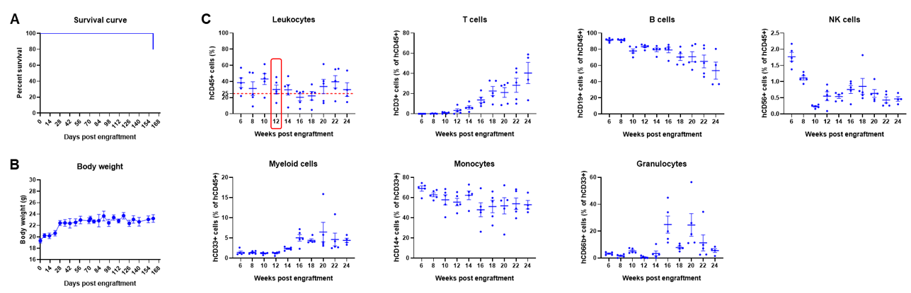
Engraftment of human CD34+ HSCs in adult B-NDG mice successfully reconstituted human T and B cells, a small number of NK and myeloid cells. Human CD34+ HSCs (1E5) were engrafted via the tail veins of B-NDG mice (female, 6-week-old, n=5) within 4-12 hours after being irradiated with 1.0 Gy of X-ray. The reconstitution level of human immune cells in peripheral blood was analyzed by flow cytometry. (A) Survival curve of human CD34+ HSCs engrafted mice; (B) Body weight; (C) Percentages of reconstituted human immune cells. The results showed that no mice died before 22 weeks of human CD34+ HSCs engraftment. Only one mouse died after 160 days of reconstitution. All the mice continued to gain body weight. The percentage of human CD45+ cells exceeded 25% at 6 weeks of reconstitution and can still be maintained at about 25% after 24 weeks of reconstitution. The percentage of human T cells begins to increase persistently at 10 weeks of reconstitution. The percentage of human B cells reached more than 90% at 6 weeks of reconstitution and continued to decline after 8 weeks. A small percentage of human NK cells, total myeloid cells, monocytes and granulocytes can also be detected throughout the reconstitution process.
Engraftment of human CD34+ HSCs in B-NDG mice to reconstitute human immune system (neonatal mice)
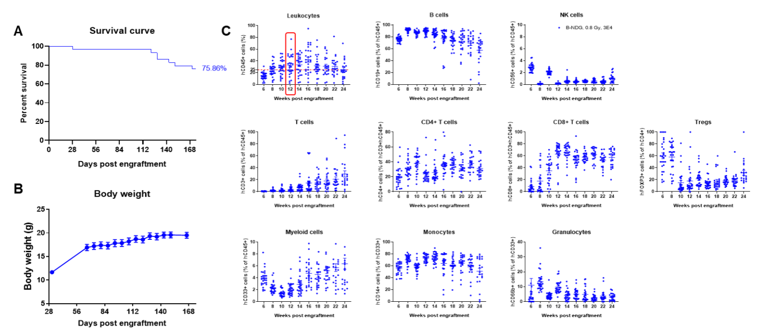
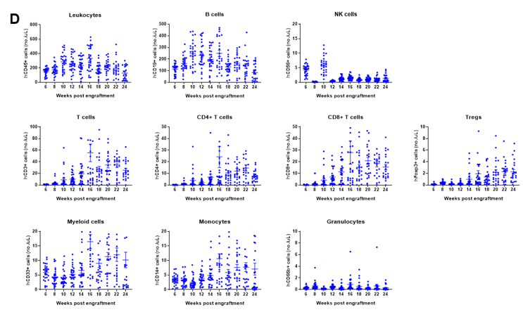
Engraftment of human CD34+ HSCs in neonatal B-NDG mice successfully reconstituted human T and B cells, a small number of NK and myeloid cells. Human CD34+ HSCs (1.5E5) were engrafted via the facial vein of B-NDG mice (male and female, 24-8 hour-old, n=28) within 4-12 hours after being irradiated with 0.8 Gy of X-ray. The reconstitution level of human immune cells in peripheral blood was analyzed by flow cytometry. (A) Survival curve of human CD34+ HSCs engrafted mice; (B) Body weight; (C) Percentages of reconstituted human immune cells; (D) Number of human immune cells. The results showed that the mice began to die from 17 weeks of reconstitution. But the lived mice continued to gain body weight. The percentage of human CD45+ cells exceeded 25% from 8 weeks of reconstitution. But there are obvious individual differences. The percentage of human T cells began to increase continuously at 10 weeks of reconstitution. Human CD4+ T cells, CD8+ T cells and Tregs could be detected at a relatively high level. The percentage of human B cells reached more than 70% at 6 weeks of reconstitution and continued to decline slowly after reaching the highest value at 8 weeks. A small percentage of human NK cells, total myeloid cells, monocytes and granulocytes can also be detected throughout the reconstitution process.
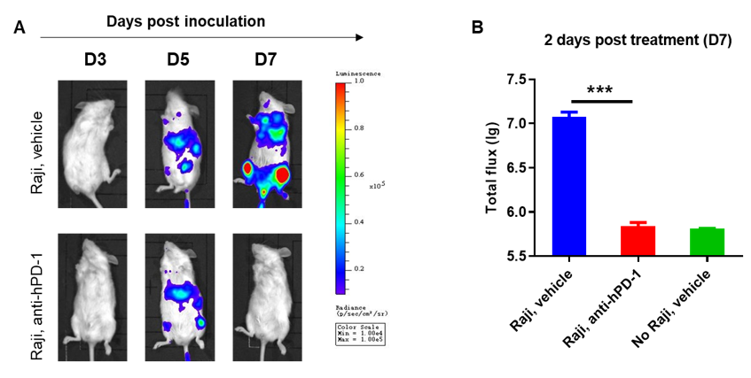
Engraftment of human CD34+ HSCs in B-NDG mice to reconstitute human immune system and evaluate the efficacy of anti-human PD-1 antibody
B-NDG mice reconstituted with CD34+ HSCs were used for drug efficacy evaluation. Human lymphoma cell line B-luciferase-GFP Raji cells (5E5) were inoculated via tail veins of adult B-NDG mice reconstituted with human CD34+ HSCs. Mice were treated with anti-human PD-1 antibody 5 days after tumor cell implantation. A dramatic inhibitory effect of the anti-human PD-1 antibody on tumor cell growth was observed at day 7. The results indicate that establishing a CDX tumor model in B-NDG mice with reconstituted HSCs provide a powerful preclinical model for in vivo evaluation of antibodies. Values are expressed as mean ± SEM.
* When publishing results obtained using this animal model, please acknowledge the source as follows: The animal model [huHSC-B-NDG mice] (Cat# 112470) was purchased from Biocytogen.