Basic Information
-
Targeting strategy

-
Gene targeting strategy for B-HLA-A11.1 mice. The B2M gene (Exon1 to Exon3) of mouse were replaced by the sequence encompassing the human B2M CDS and HLA-A*1101 gene that included leader sequence, α1 and α2 domains ligated to a fragment of the murine H-2Db gene containing the α3, transmembrane and cytoplasmic domains.
-
Protein expression analysis

-

Strain specific B2M and HLA-A expression analysis in homozygous B-HLA-A11.1 mice by flow cytometry. Splenocytes were collected from wild-type C57BL/6 mice (+/+) and homozygous B-HLA-A11.1 mice (H/H) and analyzed by flow cytometry with species-specific anti-B2M and anti-HLA-A antibodies. Mouse B2M and H-2Db were detectable in wild-type C57BL/6 mice. Human B2M and HLA-A11.1 were exclusively detectable in homozygous B-HLA-A11.1 mice but not in wild-type mice.
-
Analysis of leukocytes cell subpopulation in spleen

-
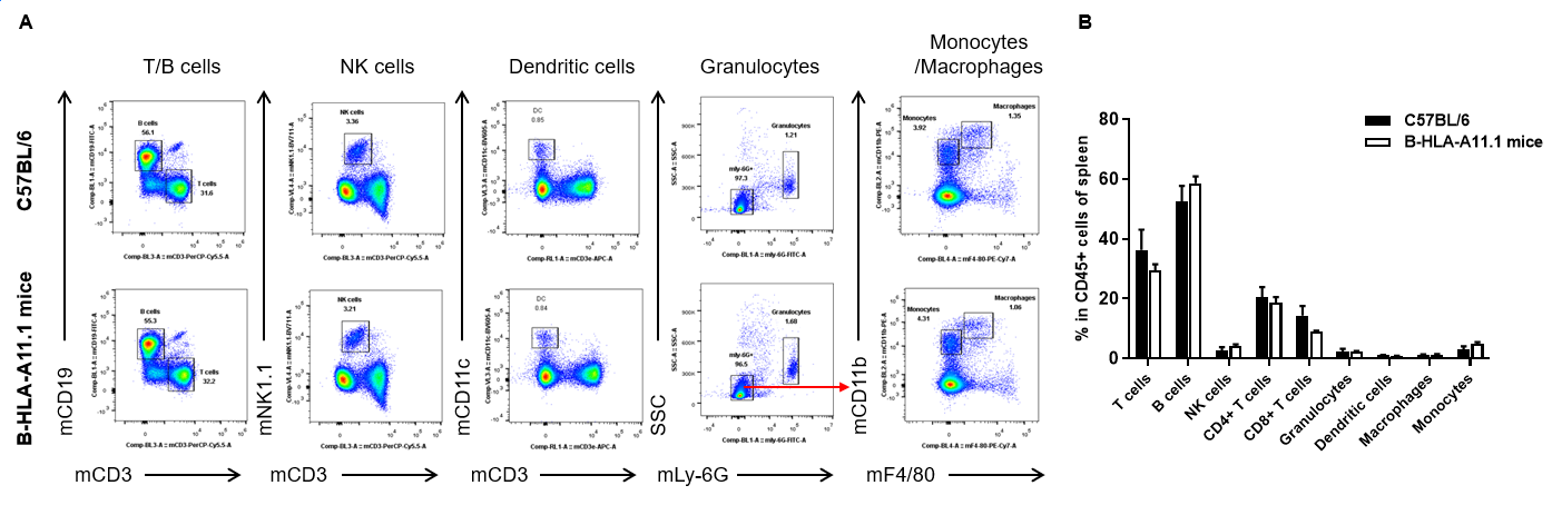
Analysis of spleen leukocyte subpopulations by FACS. Splenocytes were isolated from female C57BL/6 and B-HLA-A11.1 mice (n=3, 8-week-old). Flow cytometry analysis of the splenocytes was performed to assess leukocyte subpopulations. A. Representative FACS plots. Single live cells were gated for the CD45+ population and used for further analysis as indicated here. B. Results of FACS analysis. Percent of total T cells, B cells, NK cells, dendritic cells, granulocytes, monocytes and macrophages in homozygous B-HLA-A11.1 mice were similar to those in the C57BL/6 mice. Percent of CD8+ T cells were significantly decreased, demonstrating that introduction of hB2M-HLA-A11.1-H-2D in place of mouse B2M may affected the development of CD8 + T cells, which in turn affected the proportion of T cell subtypes in the spleen. Values are expressed as mean ± SEM.
-
Analysis of T cell subpopulation in spleen

-

Analysis of spleen T cell subpopulations by FACS. Splenocytes were isolated from female C57BL/6 and B-HLA-A11.1 mice (n=3, 8-week-old). Flow cytometry analysis of the splenocytes was performed to assess leukocyte subpopulations. A. Representative FACS plots. Single live CD45+ cells were gated for TCRβ+ T cell population and used for further analysis as indicated here. B. Results of FACS analysis. The percent of Tregs in homozygous B-HLA-A11.1 mice were similar to those in the C57BL/6 mice. Percent of CD8+ T cells were significantly decreased while percent of CD4+ T cells were significantly increased, demonstrating that introduction of hB2M-HLA-A11.1-H-2D in place of mouse B2M may affected the development of CD8 + T cells, which in turn affected the proportion of T cell subtypes in the spleen. Values are expressed as mean ± SEM.
-
Analysis of leukocytes cell subpopulation in lymph node

-

Analysis of lymph node leukocyte subpopulations by FACS. Lymph nodes were isolated from female C57BL/6 and B-HLA-A11.1 mice (n=3, 8-week-old). Flow cytometry analysis of the splenocytes was performed to assess leukocyte subpopulations. A. Representative FACS plots. Single live cells were gated for the CD45+ population and used for further analysis as indicated here. B. Results of FACS analysis. Percent of total T cells, B cells, NK cells, dendritic cells, granulocytes, monocytes and macrophages in homozygous B-HLA-A11.1 mice were similar to those in the C57BL/6 mice. Percent of CD8+ T cells were significantly decreased, demonstrating that introduction of hB2M-HLA-A11.1-H-2D in place of mouse B2M may affected the development of CD8 + T cells, which in turn affected the proportion of T cell subtypes in the lymph nodes. Values are expressed as mean ± SEM.
-
Analysis of T cell subpopulation in lymph node

-
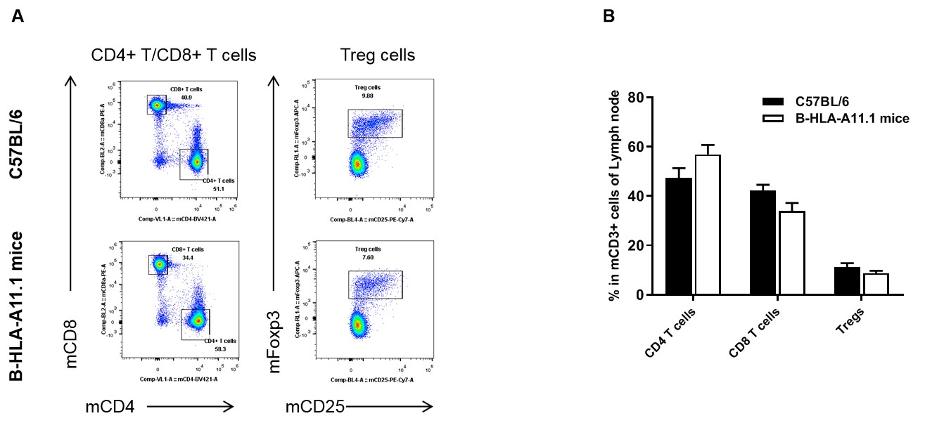
Analysis of lymph node T cell subpopulations by FACS. Lymph nodes were isolated from female C57BL/6 and B-HLA-A11.1 mice (n=3, 8-week-old). Flow cytometry analysis of the splenocytes was performed to assess leukocyte subpopulations. A. Representative FACS plots. Single live CD45+ cells were gated for TCRβ+ T cell population and used for further analysis as indicated here. B. Results of FACS analysis. The percent of Tregs in homozygous B-HLA-A11.1 mice were similar to those in the C57BL/6 mice. Percent of CD8+ T cells were significantly decreased while percent of CD4+ T cells were significantly increased, demonstrating that introduction of hB2M-HLA-A11.1-H-2D in place of mouse B2M may affected the development of CD8 + T cells, which in turn affected the proportion of T cell subtypes in the lymph nodes. Values are expressed as mean ± SEM.
-
Analysis of leukocytes cell subpopulation in blood

-

Analysis of blood leukocyte subpopulations by FACS. Blood cells were isolated from female C57BL/6 and B-HLA-A11.1 mice (n=3, 8-week-old). Flow cytometry analysis of the splenocytes was performed to assess leukocyte subpopulations. A. Representative FACS plots. Single live cells were gated for the CD45+ population and used for further analysis as indicated here. B. Results of FACS analysis. Percent of total T cells, B cells, NK cells, dendritic cells, granulocytes, monocytes and macrophages in homozygous B-HLA-A11.1 mice were similar to those in the C57BL/6 mice. Percent of CD8+ T cells were significantly decreased, demonstrating that introduction of hB2M-HLA-A11.1-H-2D in place of mouse B2M may affected the development of CD8 + T cells, which in turn affected the proportion of T cell subtypes in the blood. Values are expressed as mean ± SEM.
-
Analysis of T cell subpopulation in blood

-
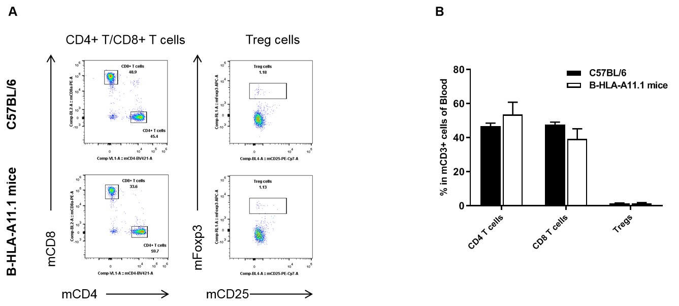
Analysis of blood T cell subpopulations by FACS. Blood cells were isolated from female C57BL/6 and B-HLA-A11.1 mice (n=3, 8-week-old). Flow cytometry analysis of the splenocytes was performed to assess leukocyte subpopulations. A. Representative FACS plots. Single live CD45+ cells were gated for TCRβ+ T cell population and used for further analysis as indicated here. B. Results of FACS analysis. The percent of Tregs in homozygous B-HLA-A11.1 mice were similar to those in the C57BL/6 mice. Percent of CD8+ T cells were significantly decreased while percent of CD4+ T cells were significantly increased, demonstrating that introduction of hB2M-HLA-A11.1-H-2D in place of mouse B2M may affected the development of CD8 + T cells, which in turn affected the proportion of T cell subtypes in the blood. Values are expressed as mean ± SEM.
-
Peptide vaccines induced immune responses in B-HLA-A11.1 mice

-
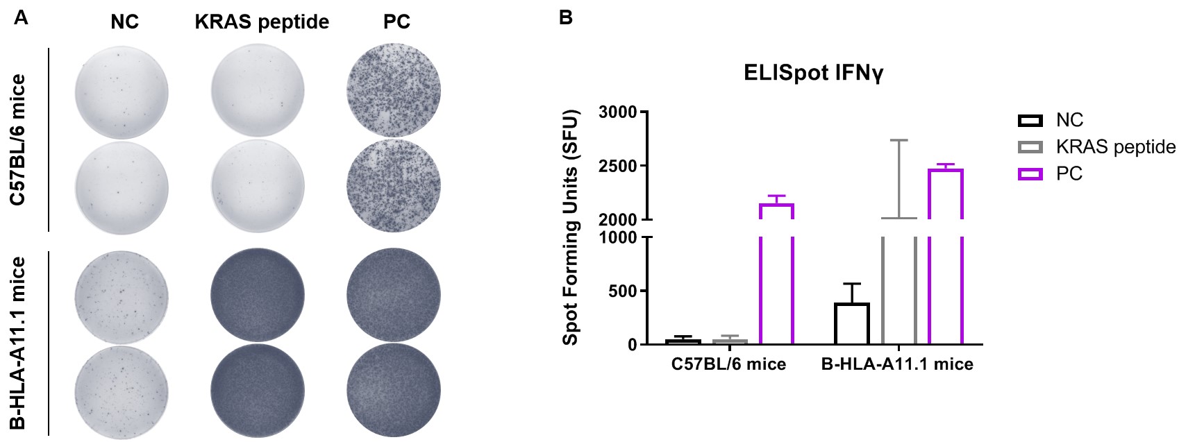
Detection of vaccine-induced immune responses in B-HLA-A11.1 mice by IFN-γ ELISpot assay. Female C57BL/6 mice and B-HLA-A11.1 mice at the age of 7–8 weeks were divided into PBS group and KRAS peptide group (n=2), and then inoculated PBS or vaccines at the inside muscle of both legs. Three weeks after the last immunization, mice were sacrificed. The splenocytes were extracted, stimulated with individual peptide or target-unrelated polypeptide as negative control (NC) or anti-CD3 as positive control(PC), and then measured for IFN-γ secretion. No significant difference in body weight among groups (Data was not shown). (A) Representative results showing stimulation of splenocytes harvested from immunized mice with negative control, or peptide vaccines, or positive control in duplicates. (B) Summary of results. The results demonstrate that B-HLA-11.1 mice provide a powerful preclinical model for in vivo evaluation of vaccines. NC: negative control, PC: positive control.
-
Identification of target peptide specific TCRs from B-HLA-A11.1 mice

-
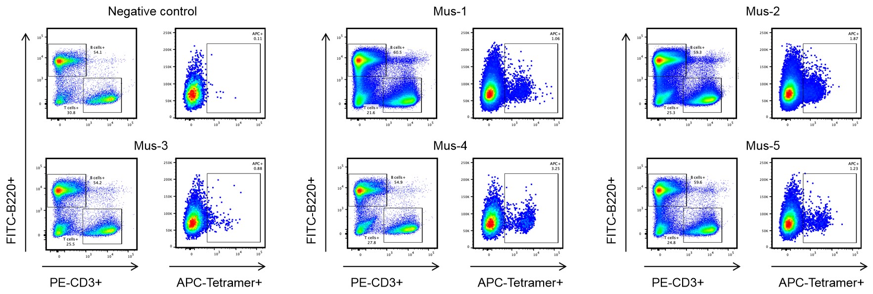
Single-cell isolation of target peptide-specific T cells from B-HLA-A11.1 mice. B-HLA-A11.1 mice were subcutaneously immunized with the target peptide and the spleen cells from five mice displayed specific responses against the target peptide. Spleen cells from five mice (Mus-1, Mus-2, Mus-3, Mus-4 and Mus-5) that showed substantial target peptide specific responses were stained with target peptide/HLA-A*11:01 tetramer, and the tetramer positive CD8+ T cells were sorted by single-cell sorting with flow Cytometry. Mouse without target peptide immunization was enrolled as the negative control. These results demonstrated that the B-HLA-A11.1 mice could be used for identifying target peptide specific TCRs and investigating mechanisms of peptide presentation and TCR recognition of cancer targets epitopes in the context of HLA-A*11:01 (All results were provided by the client.)
-
Summary

-
- Protein expression analysis:
Mouse B2M and H-2Db were detectable in wild-type C57BL/6 mice. Human B2M and HLA-A11.1 were exclusively detectable in homozygous B-HLA-A11.1 mice, while mouse B2M and/ H-2Db were not detectable in the homozygous B-HLA-A11.1 mice.
- Leukocytes cell subpopulation analysis:
Introduction of hB2M-HLA-A11.1-H-2D in place of mouse B2M may affected the development of CD8+ T cells, which in turn affected the proportion of T cell subtypes in spleen, lymph nodes and blood.


