Description
CD3EDG: Signaling in Tregs for Mechanism and Therapeutic Potential
- Gene Information: The CD3E, CD3D, and CD3G genes, located in a cluster on chromosome 11q23, encode the three invariant polypeptide chains that assemble into the CD3εδ and CD3εγ heterodimers of the T-cell receptor (TCR) complex.
- Protein Expression: These proteins are exclusively and constitutively expressed on the surface of mature T lymphocytes and thymocytes, serving as definitive lineage markers for the T-cell population.
- Signaling Pathway: Upon TCR engagement by an antigen-MHC complex, the CD3 chains transduce activation signals through their cytoplasmic Immunoreceptor Tyrosine-based Activation Motifs (ITAMs), which recruit ZAP-70 to trigger downstream MAPK, NF-κB, and calcium signaling pathways.
- Therapeutic Inhibition: Therapeutic strategies utilize monoclonal antibodies or bispecific T-cell engagers (TCEs) to either modulate TCR signaling for immunosuppression in transplantation or to bypass MHC restriction by directly linking T cells to tumor cells for targeted cytotoxicity.
4-1BB: Signaling in Tregs for Mechanism and Therapeutic Potential
- Gene Information: The TNFRSF9 gene, located on human chromosome 1p36, encodes 4-1BB (CD137), a inducible type I transmembrane glycoprotein and a prominent member of the tumor necrosis factor receptor superfamily.
- Protein Expression: While absent on resting cells, 4-1BB is transiently expressed on activated T cells, NK cells, and regulatory T cells (Tregs), as well as certain myeloid and endothelial cells within the inflammatory tumor microenvironment.
- Signaling Pathway: Ligand binding triggers TRAF1/2 recruitment to activate NF-κB, MAPK, and PI3K/Akt pathways, robustly enhancing T-cell survival, proliferation, and metabolic fitness.
- Therapeutic Inhibition: Targeted therapeutically via agonist antibodies or integrated into CAR-T co-stimulatory domains, 4-1BB serves to amplify anti-tumor immunity rather than acting as a traditional checkpoint inhibitor.
Targeting strategy
CD3E
- The exons 2-6 of mouse Cd3e gene that encode the extracellular domain were replaced by human CD3E exons 2-7 in B-hCD3E/h4-1BB mice.
4-1BB
- The exons 2-7 of mouse Tnfrsf9 gene that encode the extracellular domain were replaced by human TNFRSF9 exons 3-8 in B-hCD3E/h4-1BB mice.
CD3E Protein Expression Analysis
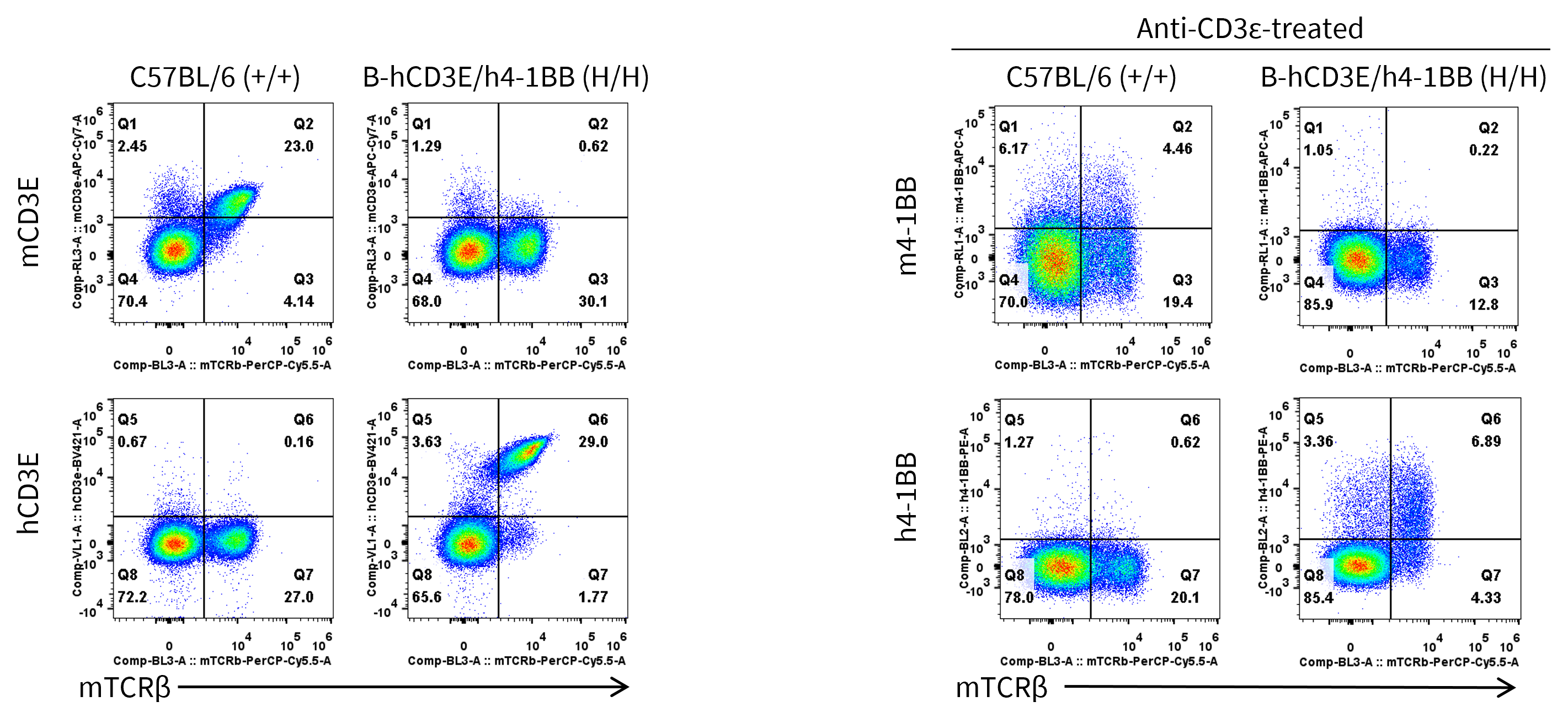
- Mouse CD3E and 4-1BB were detectable only in splenocytes of wild-type C57BL/6 mice (+/+) but not in the homozygous B-hCD3E/h4-1BB mice. Human CD3E and 4-1BB were exclusively detectable in homozygous B-hCD3E/h4-1BB mice.
Strain specific CD3E and 4-1BB expression analysis in homozygous B-hCD3E/h4-1BB mice by flow cytometry. Splenocytes were collected from wild type C57BL/6 mice (+/+) and homozygous B-hCD3E/h4-1BB mice (H/H) stimulated with or without anti-CD3ε antibody in vivo, and analyzed by flow cytometry with anti-mouse CD3E antibody (Biolegend, 100330), anti-human CD3E antibody (BD Pharmingen, 562426), anti-mouse 4-1BB antibody (Biolegend, 106110), and anti-human 4-1BB antibody (Biolegend, 309804).
Analysis of Leukocyte Subpopulations
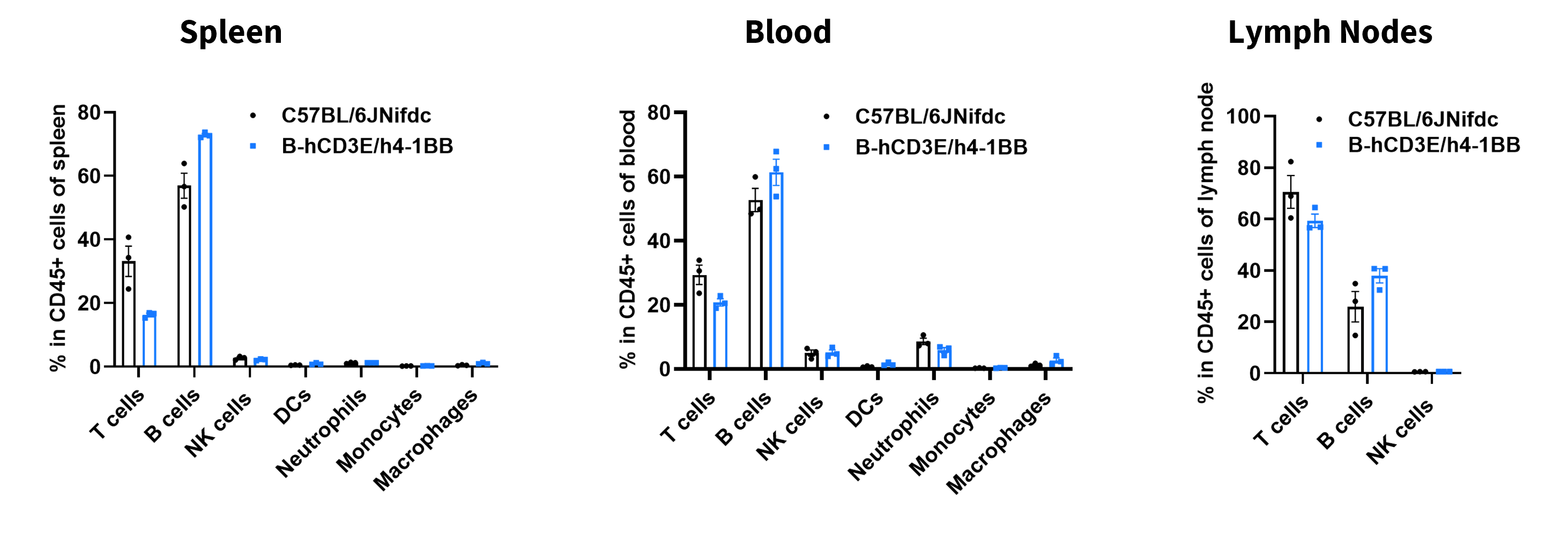
- The frequencies of T cells, B cells, NK cells, DCs, neutrophils, and monocytes, macrophages in homozygous B-hCD3E/h4-1BB mice were similar to those in C57BL/6JNifdc mice, demonstrating that humanization of CD3E and 4-1BB does not change the frequency or distribution of these cell types in spleen, peripheral blood, or lymph nodes.
Analysis of leukocyte subpopulations by flow cytometry in immune organs. Splenocytes, peripheral blood, and lymph nodes were isolated from wild-type C57BL/6JNifdc mice and B-hCD3E/h4-1BB mice (female, 8-week-old, n = 3). Single live cells were gated on the CD45⁺ population and analyzed by flow cytometry as indicated. Values are expressed as mean ± SEM.
Analysis of T Cell Subpopulations
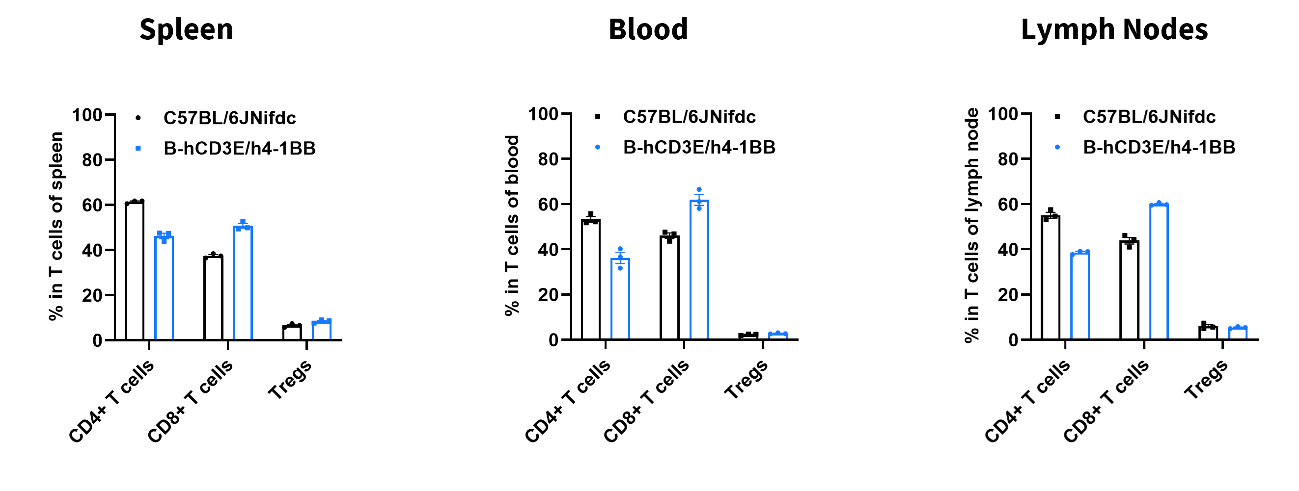
- The frequencies of CD4+ T, CD8+ T, and Tregs in homozygous B-hCD3E/h4-1BB mice were comparable to those in C57BL/6JNifdc mice, demonstrating that introduction of human CD3E and 4-1BB in place of its mouse counterpart does not change the overall development, differentiation or distribution of these T cell sub types in spleen, peripheral blood, or lymph nodes.
Analysis of T-cell subpopulations by flow cytometry in immune organs. Splenocytes, peripheral blood, and lymph nodes were isolated from wild-type C57BL/6JNifdc mice and B-hCD3E/h4-1BB mice (female, 8-week-old, n = 3). Single live cells were gated on the CD3⁺ T-cell population and analyzed by flow cytometry as indicated. Values are expressed as mean ± SEM.
Anti-tumor Effect of Anti-CD3E/4-1BB/CDCP1 against B-hCDCP1 MC38 Tumor Cells
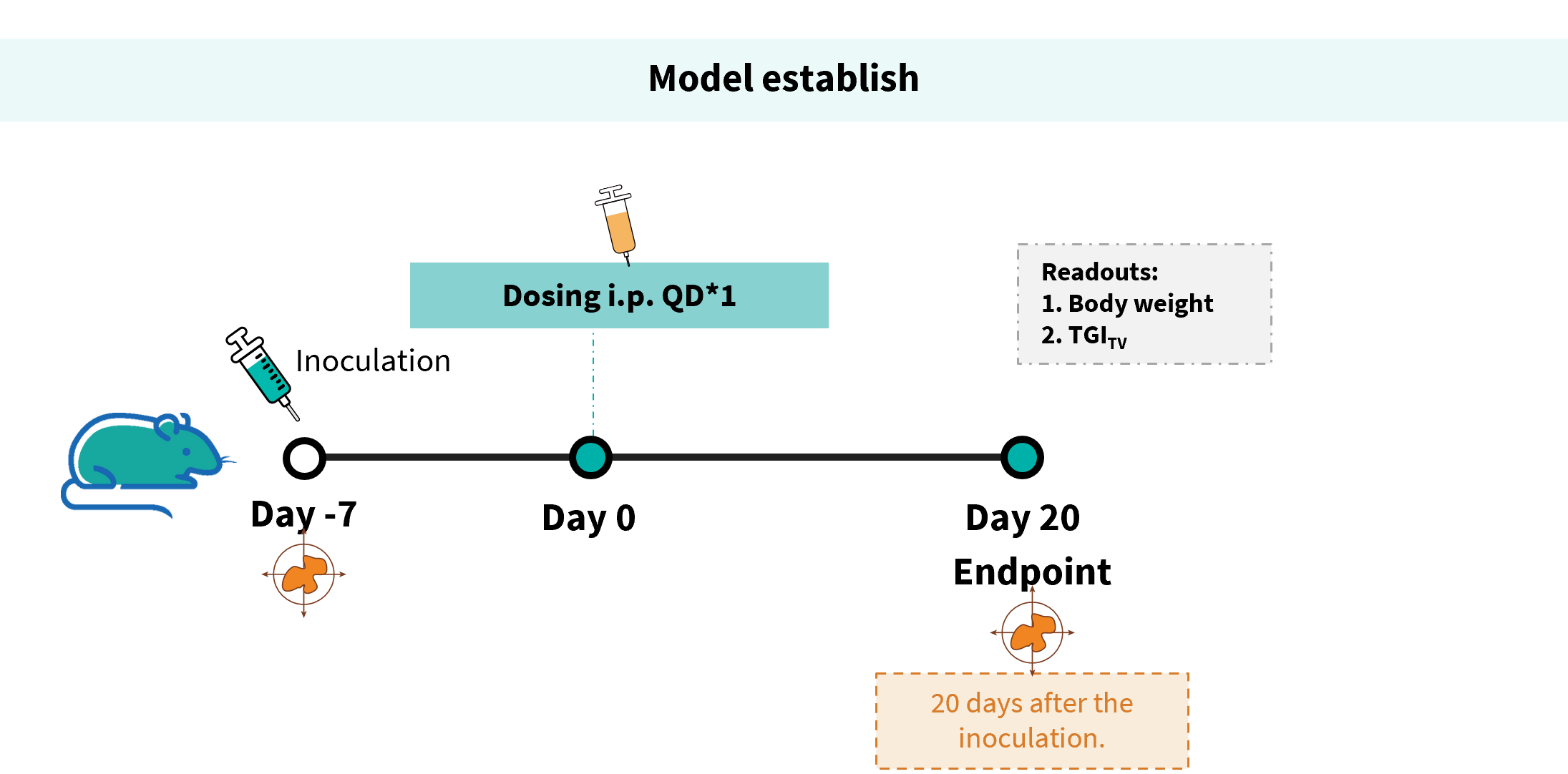
Establishment of a B-hCDCP1 MC38 model and in vivo efficacy study of an anti-human CD3E/4-1BB/CDCP1 antibody. B-hCDCP1 MC38 cells were implanted subcutaneously into homozygous B-hCD3E/h4-1BB mice (female, 7-8-week-old, n = 6).
Antitumor activity of anti-human CD3E/4-1BB/CDCP1 antibody in B-hCD3E/h4-1BB mice. (A) Tumor growth curves. (B) Body weight changes during treatment. These results demonstrate that B-hCD3E/h4-1BB mice provide a powerful preclinical model for in vivo evaluation of anti-human CD3E/4-1BB/CDCP1 antibody.
The overage of this tumor model is 40%.
Antitumor activity of anti-human CD3E/4-1BB/CDCP1 antibody in B-hCD3E/h4-1BB mice. Tumor cells growth of individual mouse. These results demonstrate that B-hCD3E/h4-1BB mice provide a powerful preclinical model for in vivo evaluation of anti-human CD3E/4-1BB/CDCP1 antibody.
* When publishing results obtained using this animal model, please acknowledge the source as follows: The animal model [B-hCD3E/h4-1BB mice] (Cat# 121281) was purchased from Biocytogen.