Expression by RT-PCR
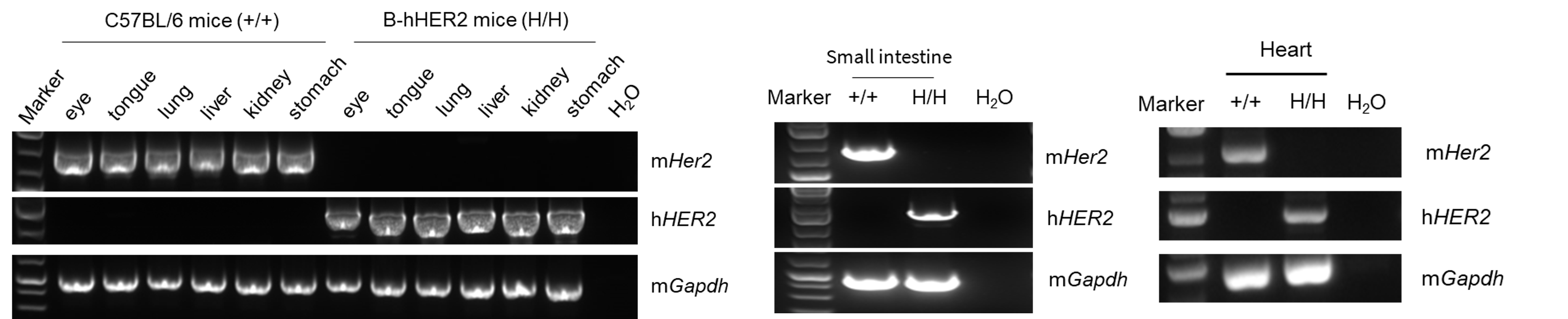
- Mouse Her2 mRNA was detectable in wild-type C57BL/6 mice.
- Human HER2 mRNA was detectable only in homozygous B-hHER2 mice but not in wild-type mice.
Strain specific analysis of HER2 mRNA expression in wild-type C57BL/6 mice and homozygous B-hHER2 mice by RT-PCR. Small intestine, heart, eye, tongue, lung, liver, kidney and stomach RNA were isolated from wild-type C57BL/6 mice (+/+) and homozygous B-hHER2 mice (H/H), then cDNA libraries were synthesized by reverse transcription, followed by PCR with mouse or human HER2 primers. Sequencing of the short-form PCR products confirmed that the amplified sequences were consistent with database reference sequences.
HER2 Protein Expression
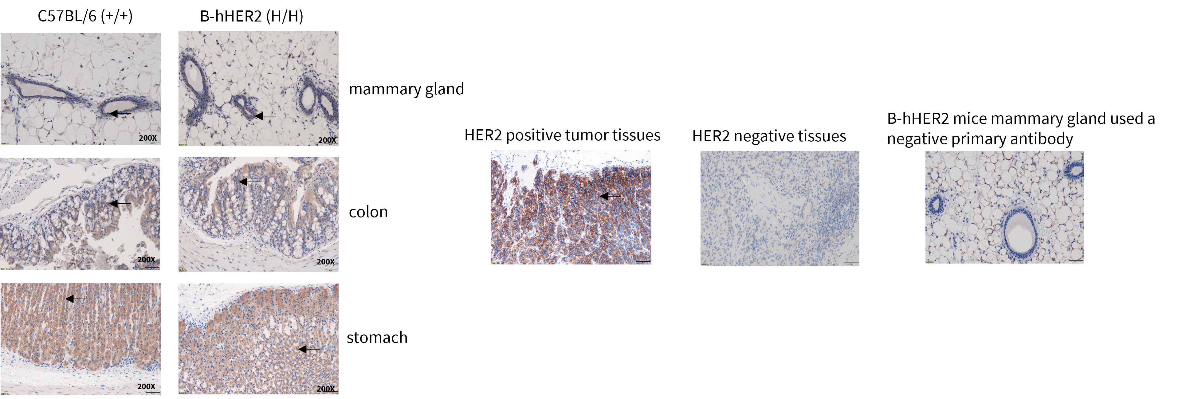
- HER2 was detectable in wild-type mice and homozygous B-hHER2 mice due to the cross-reactivity of the antibody.
Immunohistochemical (IHC) analysis of HER2 protein expression in wild-type mice and B-hHER2 mice. The mammary gland, colon and stomach tissues were collected from wild-type mice and homozygous B-hHER2 mice (H/H) and analyzed by IHC with anti-HER2 antibody (Abcam, ab214275). The arrow indicates tissue cells with positive HER2 staining (brown).
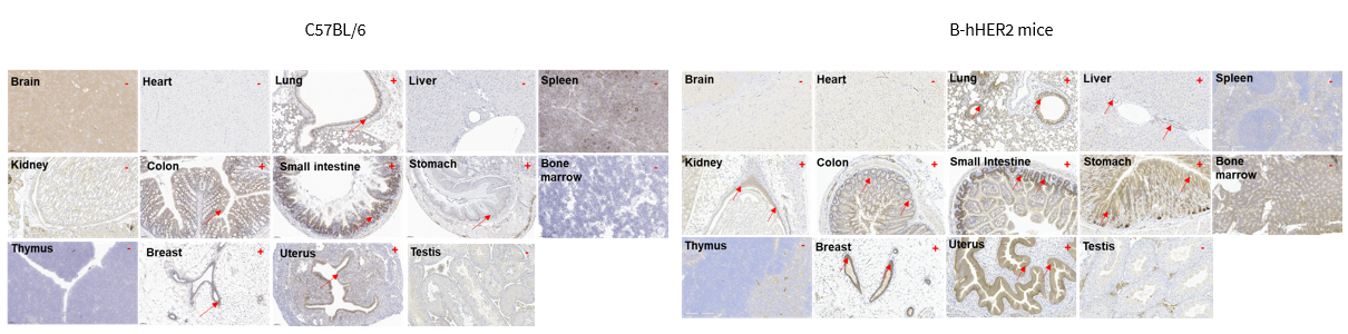
- Mouse HER2 was detected in the lung, stomach, colon, small intestine, uterus and breast of wild-type mice.
- Human HER2 was detected in the lung, stomach, colon, small intestine, uterus, breast, kidney and liver of homozygous B-hHER2 mice.
Immunohistochemical (IHC) analysis of HER2 protein expression in wild-type mice and B-hHER2 mice. Fourteen major tissues were collected from wild-type mice and homozygous B-hHER2 mice and analyzed by IHC with anti-mouse HER2 antibody (ab214275) and anti-human HER2 antibody (ab16662).
Growth Curve
Growth curve of B-hHER2 mice. Three-week-old mice were grouped by sex (10 males and 10 females). Body weight was measured weekly for 13 weeks on the same day each week. The minimum and maximum body weights shown in the table were calculated from the mean ± SD.
Hematology Analysis
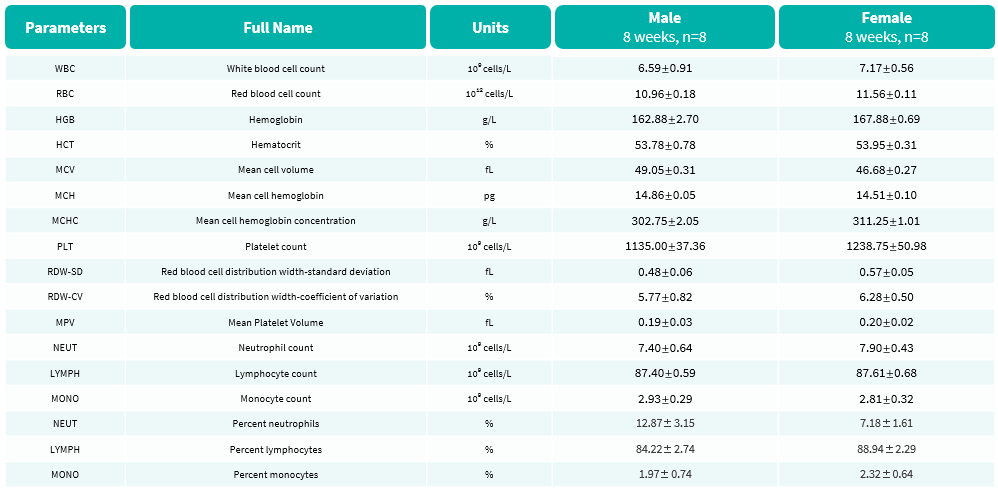
- No significant differences were observed compared with wild-type mice.
Complete blood count (CBC) of B-hHER2 mice. Values are expressed as mean ± SD.
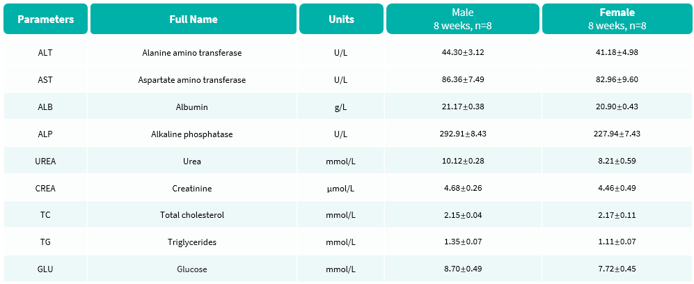
Blood Biochemical Analysis
- No significant differences were observed compared with wild-type mice.
Blood biochemical parameters of B-hHER2 mice are shown. Values are expressed as mean ± SD.
Analysis of Leukocyte Subpopulations
- The percentages of T cells, B cells, NK cells, DCs, granulocytes, monocytes, and macrophages in homozygous B-hHER2 mice are similar to those in C57BL/6 mice.
- Humanization of HER2 does not affect normal immune cell development or distribution.
Analysis of leukocyte subpopulations by flow cytometry in immune organs and blood. Splenocytes, peripheral blood, and Lymph nodes were isolated from female C57BL/6 and B-hHER2 mice (female, 8-week-old, n = 3). Single live cells were gated on the CD45⁺ population and analyzed by flow cytometry as indicated. Values are expressed as mean ± SEM.
Analysis of T Cell Subpopulations
- The proportions of CD4⁺ T cells, CD8⁺ T cells, and Tregs in homozygous B-hHER2 mice are comparable to those in C57BL/6 mice.
- Humanization of HER2 does not affect normal T cell development, differentiation, or distribution.
Analysis of T-cell subpopulations by flow cytometry in immune organs and blood. Splenocytes, peripheral blood, and Lymph nodes were isolated from female C57BL/6 and B-hHER2 mice (female, 8-week-old, n=3). Single live cells were gated on the TCRβ⁺ T-cell population and analyzed by flow cytometry as indicated. Values are expressed as mean ± SEM.
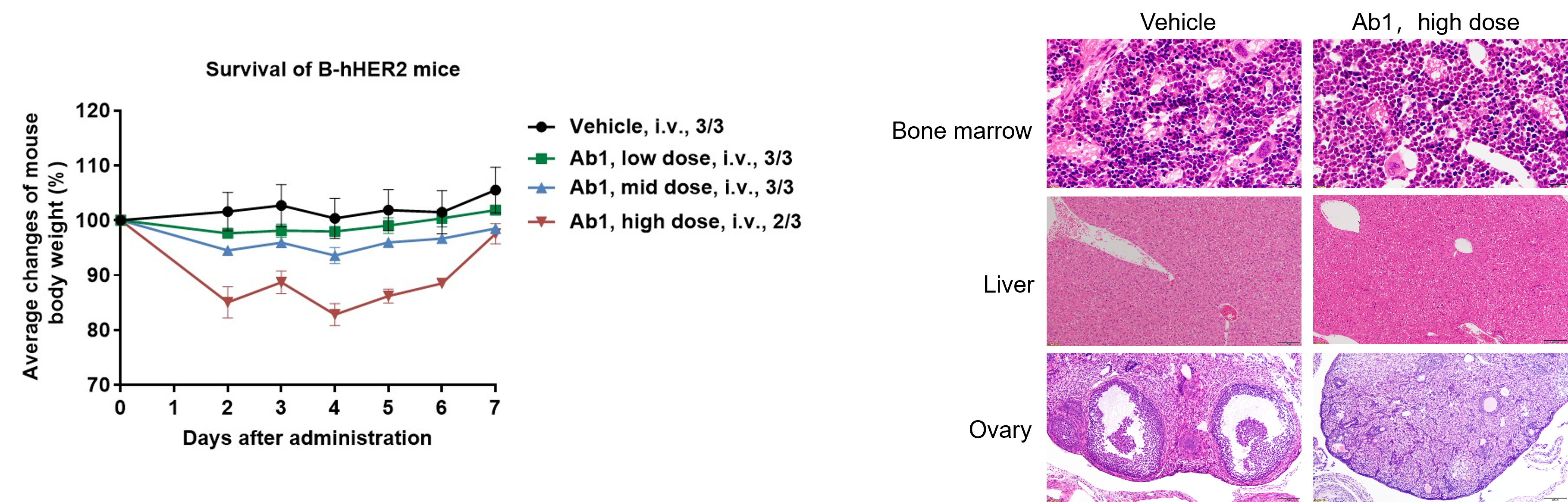
Toxicity Test in B-hHER2 mice-Case 1
- Histopathological analysis revealed no significant abnormal changes in liver, but no immature follicles were seen in ovaries, and the myeloid cells in the bone marrow increased and the red blood cell count decreased.
In vivo toxicity test of HER2 antibody (by the clients). Different doses of anti-human HER2 antibody Ab1 were injected into B-hHER2 mice via a single intravenous injection. The body weight of B-hHER2 mice decreased in a dose-dependent manner. In the high-dose group, one mouse died. Histopathological analysis revealed no significant abnormal changes in liver, but no immature follicles were seen in ovaries, and the myeloid cells in the bone marrow increased and the red blood cell count decreased. This suggests that B-hHER2 mice can be used to assess the toxicity of anti-human HER2 antibodies. Data is obtained from a partner. Values are expressed as mean ± SEM.
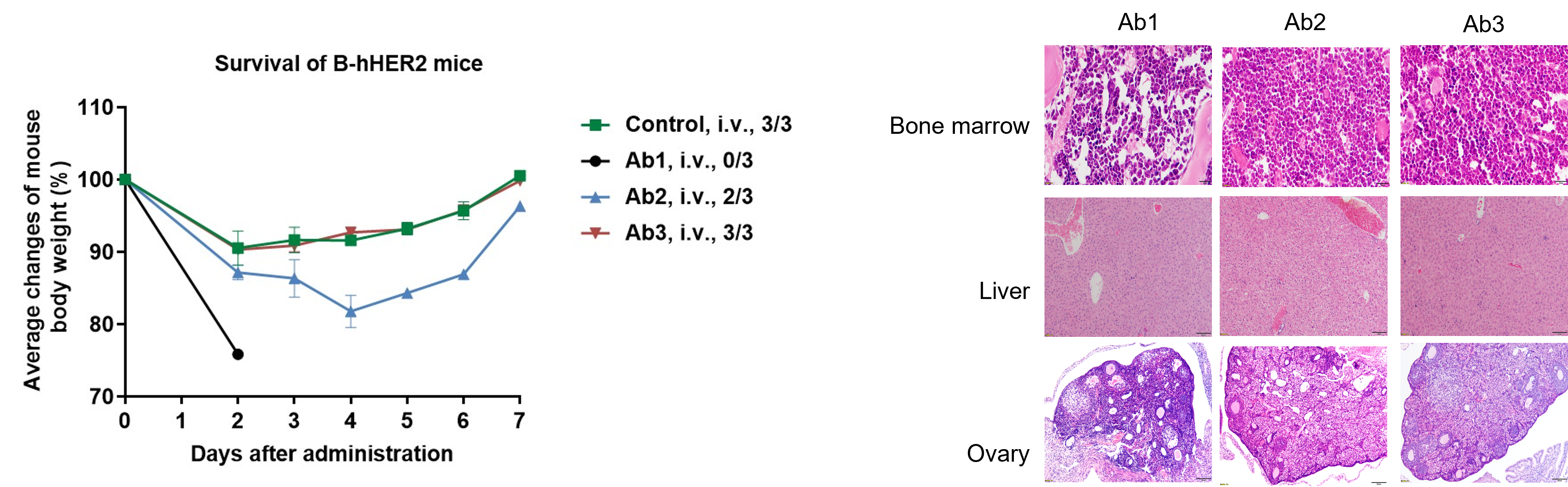
Toxicity Test in B-hHER2 mice-Case 2
- Mild toxicity was observed in the bone marrow and ovaries through histopathological analysis.
In vivo toxicity test of HER2 antibody (by the clients). Different anti-human HER2 antibodies were injected into B-hHER2 mice through a single intravenous injection. The mice in different antibody treatment groups had varying rates of weight loss. In antibody Ab1 treatment group, all mice were euthanized due to rapid weight loss. Mild toxicity was observed in the bone marrow and ovaries through histopathological analysis. This suggests that B-hHER2 mice can be used to assess the toxicity of anti-human HER2 antibodies. Data is obtained from a partner. Values are expressed as mean ± SEM.
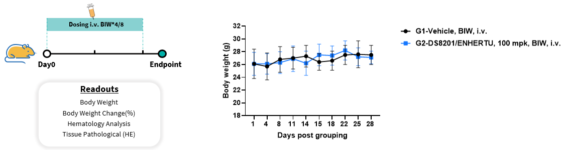
In vivo Toxicity Test of DS8201/ENHERTU (ADC)-Case-3
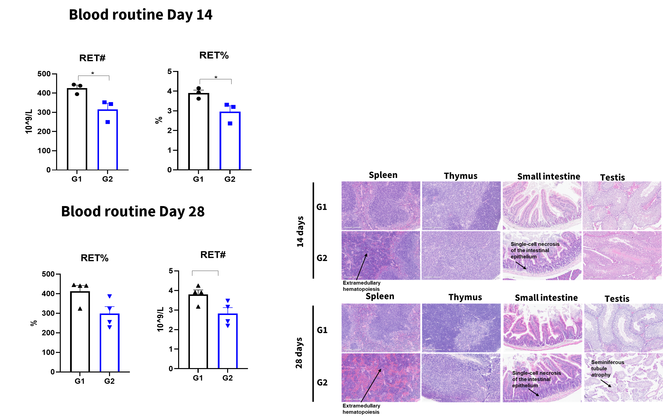
In vivo toxicity test of DS8201/ENHERTU. Anti-human HER2 ADC DS8201/ENHERTU (Commercialized) were intravenously injected into B-hHER2 mice (Male, 8 weeks old, n=3). Mice were weighed every two days, and their condition was observed daily. At the end of the experiment, blood samples were collected for complete blood count test. Additionally, tissue samples were collected from spleen, thymus, small intestine and testis and then subjected to pathological analysis. Values are expressed as mean ± SEM.
- RET reduction reflects potential bone marrow erythroid suppression caused by off-target effects of Enhertu’s DXd payload on rapidly dividing hematopoietic stem cells.
- In the G2 group, some pathological changes were observed in the spleen, thymus, small intestine, and testis (see the chart above for details), whereas no changes were found in the control group.
In vivo toxicity test of DS8201/ENHERTU. At the end of the experiment, blood samples were collected for complete blood count test. Additionally, tissue samples were collected from spleen, thymus, small intestine and testis and then subjected to pathological analysis.
In vivo Efficacy of anti-human HER2 antibody-drug conjugate (ADC)
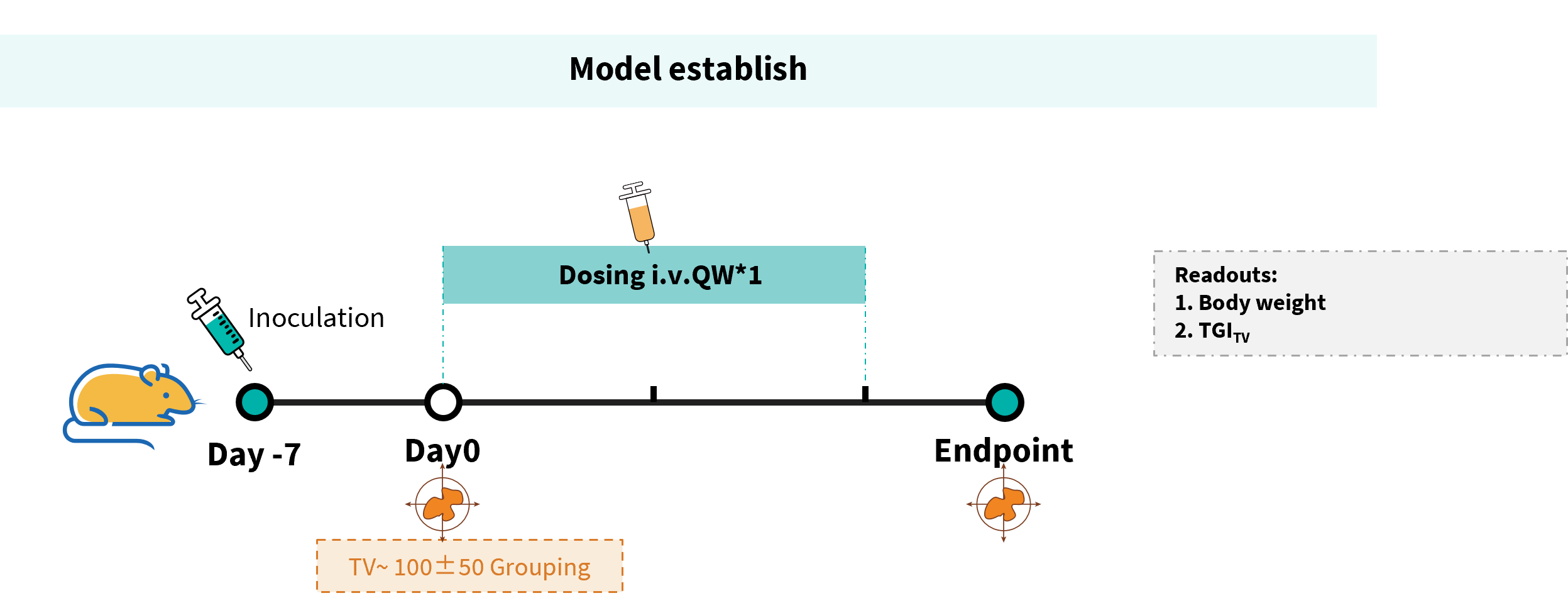
Establishment of a MC38 model in B-hHER2 mice and in vivo efficacy study of anti-HER2 antibody (purchased from AstraZeneca/Daiichi Sankyo). Murine colon cancer B-hHER2 MC38 plus cells (Cat# 322353) were subcutaneously implanted into homozygous B-hHER2 mice (female, 10 weeks-old, n=6). Mice were grouped when tumor volume reached approximately 100 mm3, at which time they were intravenously injected with anti-human HER2 ADC DS8201 (purchased from AstraZeneca/Daiichi Sankyo) indicated in panel.
- Anti-human HER2 ADC DS8201 (purchased from AstraZeneca/Daiichi Sankyo) was efficacious in controlling tumor growth in B-hHER2 mice, demonstrating that the B-hHER2 mice provide a powerful preclinical model for in vivo evaluation of anti-human HER2 ADC.
Efficacy of anti-human HER2 ADC in B-hHER2 mice. (A) Tumor growth curves. (B) Body weight changes during treatment. (C) In vivo efficacy of anti-human HER2 antibodies-individual tumor growth curves. Values are expressed as mean ± SEM.
- Anti-human HER2 ADC Trastuzumab analog-MMAE (in-house) was efficacious in controlling tumor growth in B-hHER2 mice, demonstrating that the B-hHER2 mice provide a powerful preclinical model for in vivo evaluation of anti-human HER2 ADC.
Establishment of a MC38 model in B-hHER2 mice and in vivo efficacy study of anti-HER2 antibody (Trastuzumab analog-MMAE, in-house). Murine colon cancer B-hHER2 MC38 plus cells (Cat# 322353) were subcutaneously implanted into homozygous B-hHER2 mice (female, 7-8-week-old, n=6). Mice were grouped when tumor volume reached approximately 100 mm3, at which time they were intravenously injected with anti-human HER2 ADC Trastuzumab analog-MMAE (in-house) indicated in panel. Values are expressed as mean ± SEM.
- Anti-human HER2 ADC Trastuzumab analog-MMAE (in-house) was efficacious in controlling tumor growth in B-hHER2 mice in a dose-dependent manner, demonstrating that the B-hHER2 mice provide a powerful preclinical model for in vivo evaluation of anti-human HER2 ADC.
In vivo efficacy of anti-human HER2 ADC-individual tumor growth curves.
In vivo Efficacy of HER2 ADC combined with anti-mPD-1 Antibody
- Anti-human HER2 ADC Trastuzumab analog-MMAE combined with anti-PD-1 antibody therapy showed superior anti-tumor efficacy compared with monotherapy in B-hHER2 mice, demonstrating that this model is a valuable platform for evaluating cancer immunotherapies and combination treatment strategies.
Establishment of a MC38 model in B-hHER2 mice and In vivo Efficacy of HER2 ADC combined with anti-mPD-1 Antibody. Murine colon cancer B-hHER2 MC38 plus cells (Cat# 322353) were subcutaneously implanted into homozygous B-hHER2 mice (female, 7-8-week-old, n=6). Mice were grouped when tumor volume reached approximately 100 mm3, at which time they were intravenously injected with anti-human HER2 ADC Trastuzumab analog-MMAE (in-house) or Intraperitoneal injected mPD-1 antibody (in-house).
In vivo Efficacy of HER2 ADC DS8201 combined with anti-PD-1 Antibody
- Anti-human HER2 ADC Trastuzumab analog-MMAE combined with anti-PD-1 antibody therapy showed superior anti-tumor efficacy compared with monotherapy in B-hHER2 mice, demonstrating that this model is a valuable platform for evaluating cancer immunotherapies and combination treatment strategies.
In vivo Efficacy of HER2 ADC combined with anti-mPD-1 Antibody-individual tumor growth curves.
In vivo efficacy of anti-human HER2 antibody-drug conjugate (ADC)
- Anti-human HER2 ADC DS8201 was efficacious in controlling tumor growth in B-hHER2 mice in a dose-dependent manner, demonstrating that the B-hHER2 mice provide a powerful preclinical model for in vivo evaluation of anti-human HER2 ADC.
Establishment of a MC38 model in B-hHER2 mice and in vivo efficacy study of HER2 ADC DS8201 (purchased from Daiichi Sankyo). Murine melanoma B-hHER2 B16-F10 cells (Cat# 322393) were subcutaneously implanted into homozygous B-hHER2 mice (female, 9-week-old, n=6). Mice were grouped when tumor volume reached approximately 100 mm3, at which time they were intravenously injected with anti-human HER2 ADC DS8201 (purchased from Daiichi Sankyo) indicated in panel. (A) Tumor growth curves. (B) Body weight changes during treatment In vivo. (C) Efficacy of HER2 ADC DS8201 individual tumor growth curves.
In vivo Efficacy of anti-human HER2 antibody-drug conjugate (ADC)
- Anti-human HER2 ADC DS8201 was efficacious in controlling tumor growth in B-hHER2 mice in a dose-dependent manner, demonstrating that the B-hHER2 mice provide a powerful preclinical model for in vivo evaluation of anti-human HER2 ADC.
Establishment of a MC38 model in B-hHER2 mice and in vivo efficacy study of HER2 ADC DS8201 (purchased from Daiichi Sankyo). Murine melanoma B-hHER2 B16-F10 cells (Cat# 322393) were subcutaneously implanted into homozygous B-hHER2 mice (female, 9-week-old, n=6). Mice were grouped when tumor volume reached approximately 100 mm3, at which time they were intravenously injected with anti-human HER2 ADC DS8201 (purchased from Daiichi Sankyo) indicated in panel. (A) Tumor growth curves. (B) Body weight changes during treatment In vivo. (C) Efficacy of HER2 ADC DS8201 individual tumor growth curves.
* When publishing results obtained using this animal model, please acknowledge the source as follows: The animal model [B-hHER2 mice] (Cat# 110812) was purchased from Biocytogen.