NA • 322473
In the experiment, it is necessary to ensure that the number of animals inoculated subcutaneously is at least 1.6 times the actual grouping number.
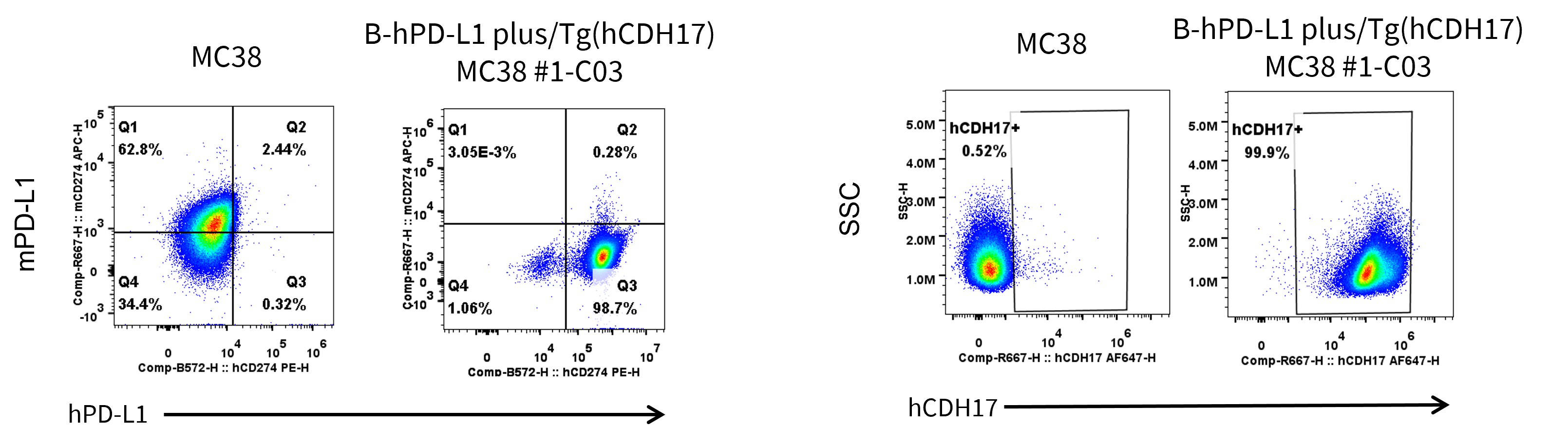
PD-L1 and CDH17 expression analysis in B-hPD-L1 plus/Tg(hCDH17) MC38 by flow cytometry. Single cell suspensions from wild-type MC38 and B-hPD-L1 plus/Tg(hCDH17) MC38 #1-C03 cultures were stained with anti-mouse PD-L1 antibody (Biolegend, 124312), anti-human PD-L1 antibody (Biolegend, 329706), and anti-human CDH17 antibody (in house) .
Subcutaneous tumor growth of B-hPD-L1 plus/Tg(hCDH17) MC38 cells. B-hPD-L1 plus/Tg(hCDH17) MC38 (1×10⁶) and wild-type MC38 cells (5×10⁵) were subcutaneously implanted into B-hCDH17 mice (female, 8-week-old, n=6). Tumor volume and body weight were measured twice a week. (A) Average tumor volume. (B) Body weight. Volume was expressed in mm3 using the formula: V=0.5 × long diameter × short diameter2. Results indicate that B-hPD-L1 plus/Tg(hCDH17) MC38 cells were able to establish tumors in vivo and can be used for efficacy studies. Values are expressed as mean ± SEM.
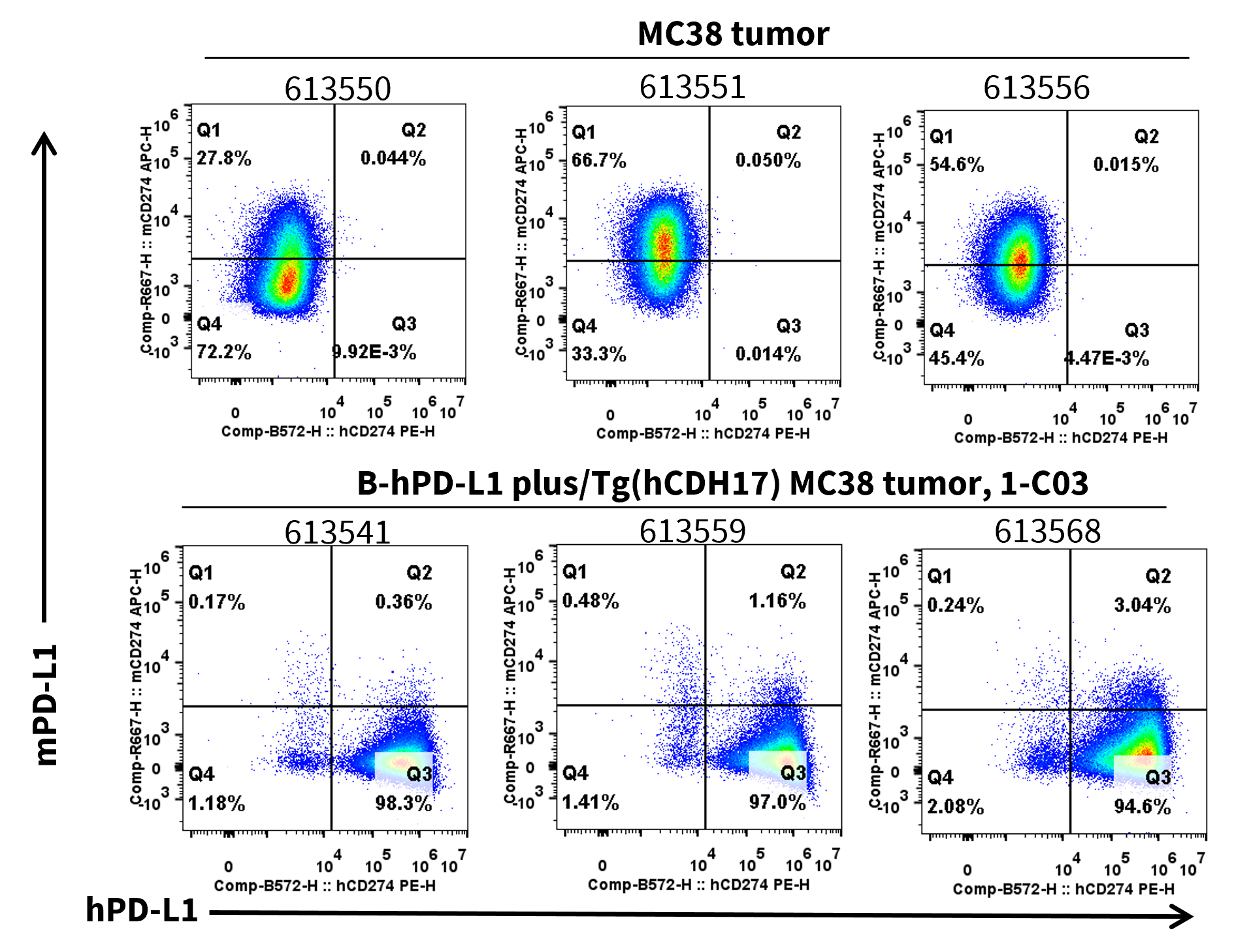
PD-L1 expression was evaluated on B-hPD-L1 plus/Tg(hCDH17) MC38 by flow cytometry. These cells were subcutaneously transplanted into B-hCDH17 mice (n=6). At the end of the experiment, tumor cells were harvested and analyzed for both mouse and human PD-L1 expression by flow cytometry.
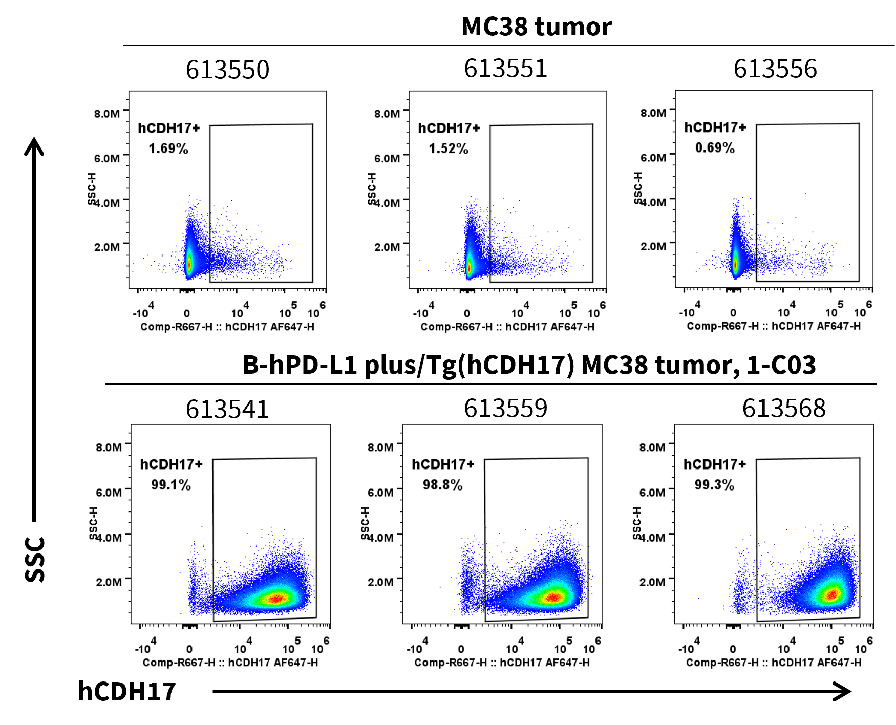
CDH17 expression was evaluated on B-hPD-L1 plus/Tg(hCDH17) MC38 by flow cytometry. These cells were subcutaneously transplanted into B-hCDH17 mice (n=6). At the end of the experiment, tumor cells were harvested and analyzed for human CDH17 expression by flow cytometry.