Soluble TL1A Protein Expression Analysis in BMDC Supernatants
- Soluble human TL1A was exclusively detectable in homozygous B-hTL1A/hIL23A/hIL12B/hα4β7 mice but not wild-type C57BL/6JNifdc mice.
Soluble TL1A expression analysis in B-hTL1A/hIL23A/hIL12B/hα4β7 mice by ELISA. Bone marrow derived dendritic cells (BMDC) were produced by culturing the bone marrow from wild-type C57BL/6JNifdc mice (+/+) and homozygous B-hTL1A/hIL23A/hIL12B/hα4β7 mice (H/H;H/H;H/H;H/H;H/H), which were stimulated with LPS in vitro. After stimulation, the supernatants were collected and the level of soluble TL1A was analyzed by ELISA. Soluble human TL1A was exclusively detectable in homozygous B-hTL1A/hIL23A/hIL12B/hα4β7 mice, but not in wild-type C57BL/6JNifdc mice. Values are expressed as mean ± SEM. ND: not detectable.
IL23 Protein Expression Analysis in BMDC Supernatants
- Human IL23 was exclusively detectable in homozygous B-hTL1A/hIL23A/hIL12B/hα4β7 mice but not wild-type C57BL/6JNifdc mice.
Mouse IL23 and human IL23 expression analysis in B-hTL1A/hIL23A/hIL12B/hα4β7 mice by ELISA. BMDCs were produced by culturing the bone marrow from wild-type C57BL/6JNifdc mice (+/+) and homozygous B-hTL1A/hIL23A/hIL12B/hα4β7 mice (H/H;H/H;H/H;H/H;H/H), which were stimulated with LPS in vitro. After stimulation, the supernatants were collected and the levels of mouse and human IL23 were analyzed by ELISA (R&D, M2300; R&D, D2300B). Mouse IL23 was only detectable in wild-type C57BL/6JNifdc mice. Human IL23 was exclusively detectable in homozygous B-hTL1A/hIL23A/hIL12B/hα4β7 mice. Values are expressed as mean ± SEM. ND: not detectable.
α4β7 Protein Expression Analysis in Spleen
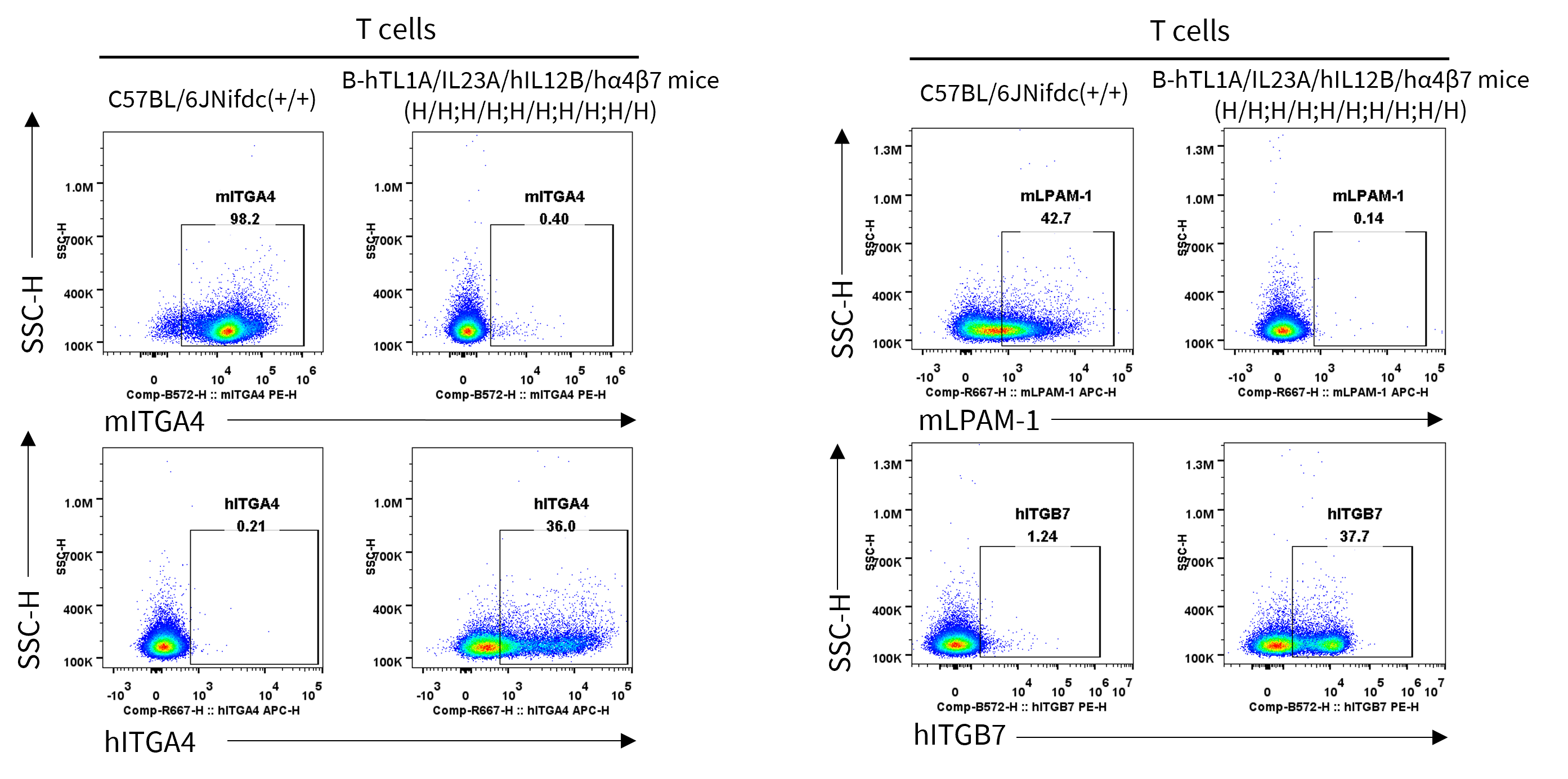
- Human ITGA4 and ITGB7 were exclusively detectable in T cells of homozygous B-hTL1A/hIL23A/hIL12B/hα4β7 mice but not wild-type C57BL/6JNifdc mice.
Strain specific ITGA4 and ITGB7 expression analysis in wild-type C57BL/6JNifdc mice and homozygous humanized B-hTL1A/hIL23A/hIL12B/hα4β7 mice by flow cytometry. Splenocytes were collected from wild-type C57BL/6JNifdc mice (+/+) and homozygous B-hTL1A/hIL23A/hIL12B/hα4β7 mice (H/H;H/H;H/H;H/H;H/H). Protein expression was analyzed with anti-mouse ITGA4 antibody (Biolegend, 103705), anti-mouse ITGB7 antibody (Biolegend, 120607), anti-human ITGA4 antibody (Biolegend, 304307) anti-human ITGB7 antibody (Invitrogen, MA5-23541) by flow cytometry. Mouse ITGA4 and ITGB7 were only detectable in T cells of wild-type C57BL/6JNifdc mice. Human ITGA4 and ITGB7 were exclusively detectable in T cells of homozygous B-hTL1A/hIL23A/hIL12B/hα4β7 mice.
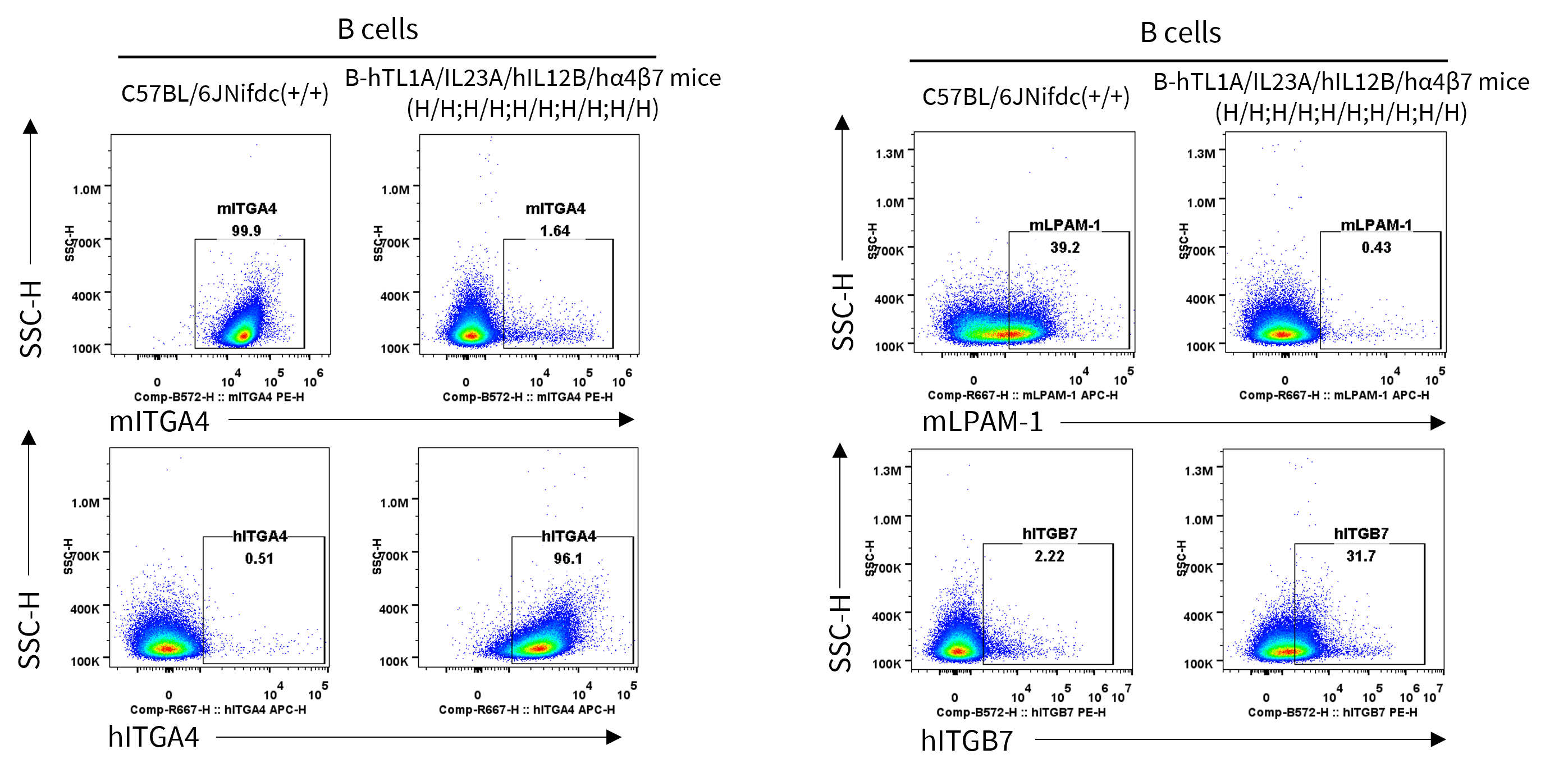
- Human ITGA4 and ITGB7 were exclusively detectable in B cells of homozygous B-hTL1A/hIL23A/hIL12B/hα4β7 mice but not wild-type C57BL/6JNifdc mice.
Strain specific ITGA4 and ITGB7 expression analysis in wild-type C57BL/6JNifdc mice and homozygous humanized B-hTL1A/hIL23A/hIL12B/hα4β7 mice by flow cytometry. Splenocytes were collected from wild-type C57BL/6JNifdc mice (+/+) and homozygous B-hTL1A/hIL23A/hIL12B/hα4β7 mice (H/H;H/H;H/H;H/H;H/H). Protein expression was analyzed with anti-mouse ITGA4 antibody (Biolegend, 103705), anti-mouse ITGB7 antibody (Biolegend, 120607), anti-human ITGA4 antibody (Biolegend, 304307) anti-human ITGB7 antibody (Invitrogen, MA5-23541) by flow cytometry. Mouse ITGA4 and ITGB7 were only detectable in B cells of wild-type C57BL/6JNifdc mice. Human ITGA4 and ITGB7 were exclusively detectable in B cells of homozygous B-hTL1A/hIL23A/hIL12B/hα4β7 mice.
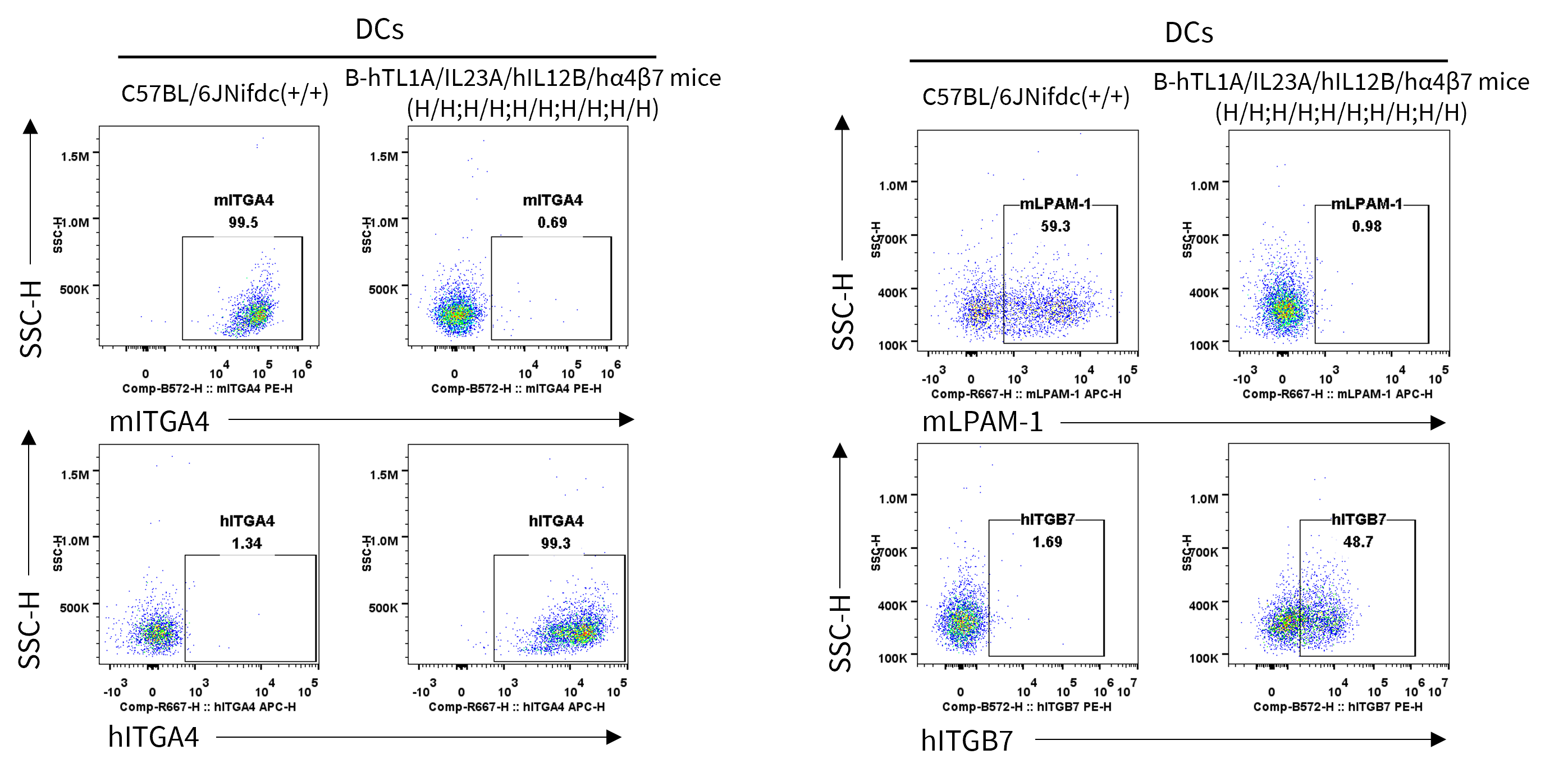
- Human ITGA4 and ITGB7 were exclusively detectable in DCs of homozygous B-hTL1A/hIL23A/hIL12B/hα4β7 mice but not wild-type C57BL/6JNifdc mice.
Strain specific ITGA4 and ITGB7 expression analysis in wild-type C57BL/6JNifdc mice and homozygous humanized B-hTL1A/hIL23A/hIL12B/hα4β7 mice by flow cytometry. Splenocytes were collected from wild-type C57BL/6JNifdc mice (+/+) and homozygous B-hTL1A/hIL23A/hIL12B/hα4β7 mice (H/H;H/H;H/H;H/H;H/H). Protein expression was analyzed with anti-mouse ITGA4 antibody (Biolegend, 103705), anti-mouse ITGB7 antibody (Biolegend, 120607), anti-human ITGA4 antibody (Biolegend, 304307) anti-human ITGB7 antibody (Invitrogen, MA5-23541) by flow cytometry. Mouse ITGA4 and ITGB7 were only detectable in DCs of wild-type C57BL/6JNifdc mice. Human ITGA4 and ITGB7 were exclusively detectable in DCs of homozygous B-hTL1A/hIL23A/hIL12B/hα4β7 mice.
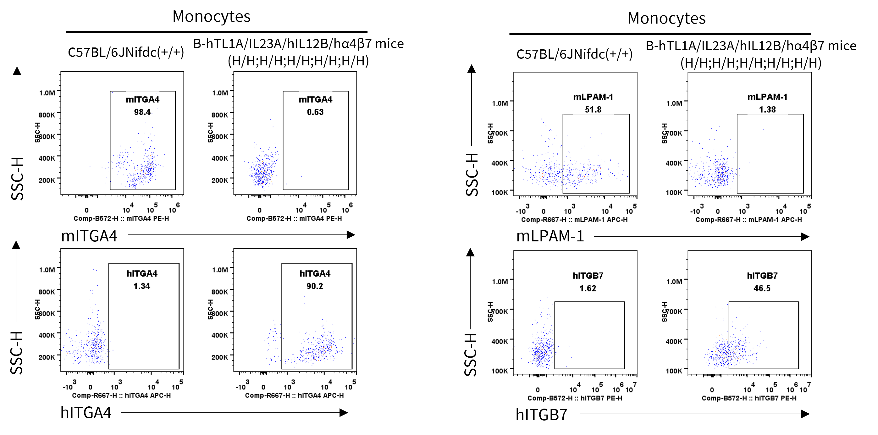
- Human ITGA4 and ITGB7 were exclusively detectable in monocytes of homozygous B-hTL1A/hIL23A/hIL12B/hα4β7 mice but not wild-type C57BL/6JNifdc mice.
Strain specific ITGA4 and ITGB7 expression analysis in wild-type C57BL/6JNifdc mice and homozygous humanized B-hTL1A/hIL23A/hIL12B/hα4β7 mice by flow cytometry. Splenocytes were collected from wild-type C57BL/6JNifdc mice (+/+) and homozygous B-hTL1A/hIL23A/hIL12B/hα4β7 mice (H/H;H/H;H/H;H/H;H/H). Protein expression was analyzed with anti-mouse ITGA4 antibody (Biolegend, 103705), anti-mouse ITGB7 antibody (Biolegend, 120607), anti-human ITGA4 antibody (Biolegend, 304307) anti-human ITGB7 antibody (Invitrogen, MA5-23541) by flow cytometry. Mouse ITGA4 and ITGB7 were only detectable in monocytes of wild-type C57BL/6JNifdc mice. Human ITGA4 and ITGB7 were exclusively detectable in monocytes of homozygous B-hTL1A/hIL23A/hIL12B/hα4β7 mice.
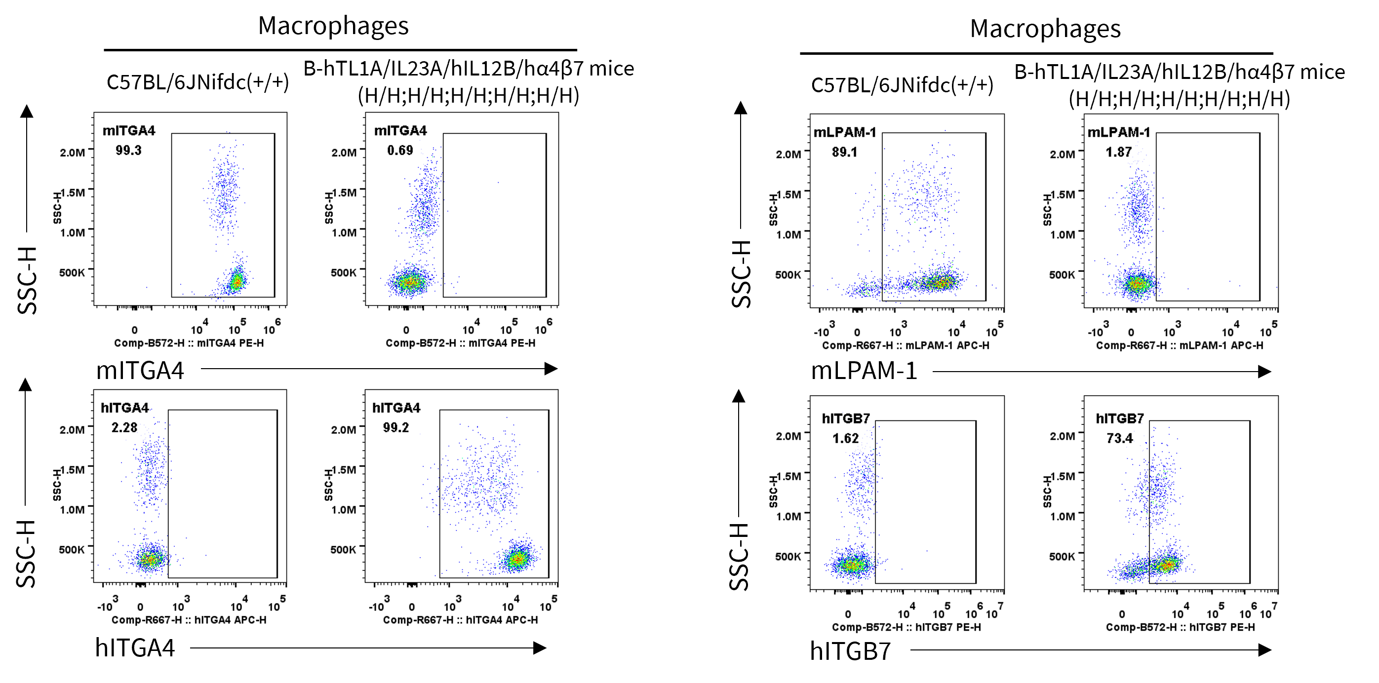
- Human ITGA4 and ITGB7 were exclusively detectable in macrophages of homozygous B-hTL1A/hIL23A/hIL12B/hα4β7 mice but not wild-type C57BL/6JNifdc mice.
Strain specific ITGA4 and ITGB7 expression analysis in wild-type C57BL/6JNifdc mice and homozygous humanized B-hTL1A/hIL23A/hIL12B/hα4β7 mice by flow cytometry. Splenocytes were collected from wild-type C57BL/6JNifdc mice (+/+) and homozygous B-hTL1A/hIL23A/hIL12B/hα4β7 mice (H/H;H/H;H/H;H/H;H/H). Protein expression was analyzed with anti-mouse ITGA4 antibody (Biolegend, 103705), anti-mouse ITGB7 antibody (Biolegend, 120607), anti-human ITGA4 antibody (Biolegend, 304307) anti-human ITGB7 antibody (Invitrogen, MA5-23541) by flow cytometry. Mouse ITGA4 and ITGB7 were only detectable in macrophages of wild-type C57BL/6JNifdc mice. Human ITGA4 and ITGB7 were exclusively detectable in macrophages of homozygous B-hTL1A/hIL23A/hIL12B/hα4β7 mice.
Functional Validation
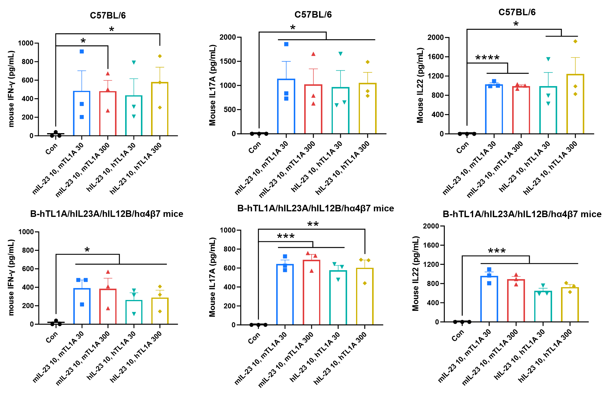
- The synergistic stimulation of TL1A and IL23 could promote the production of downstream cytokines in wild-type C57BL/6 mice and homozygous B-hTL1A/hIL23A/hIL12B/hα4β7 mice.
- Both mTL1A and hTL1A could bind to mouse DR3.
Ex vivo functional analysis in B-hTL1A/hIL23A/hIL12B/hα4β7 mice. Splenocytes were collected from wild-type C57BL/6 mice (+/+) and homozygous B-hTL1A/hIL23A/hIL12B/hα4β7 mice (H/H;H/H;H/H;H/H;H/H), then the production of mouse IFN-γ, mouse IL17A, and mouse IL22 in supernatants were assessed after 72 h of incubation with mIL23 (10 ng/mL), mTL1A (30, 300 ng/mL), hIL23 (10 ng/mL) and hTL1A (30, 300 ng/mL) in vitro.
MAdCAM-1 binding analysis
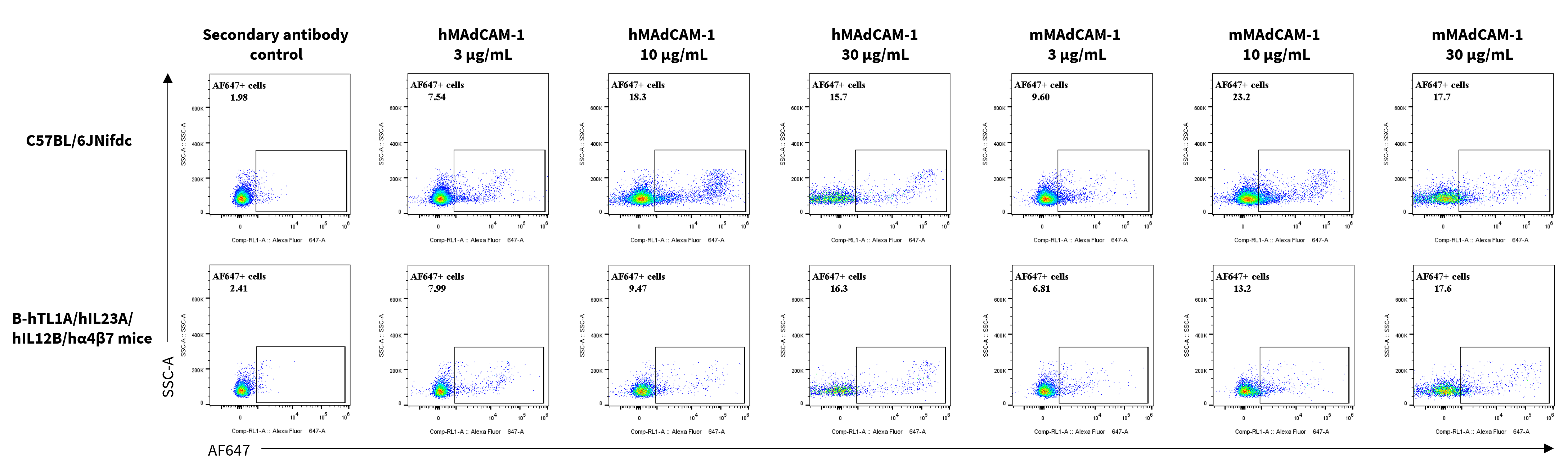
- Both mouse MAdCAM-1 and human MAdCAM-1 could bind to mouse α4β7, and both mouse MAdCAM-1 and human MAdCAM-1 could bind to human α4β7.
Assessment of MAdCAM-1 binding in C57BL/6 and B-hTL1A/hIL23A/hIL12B/hα4β7 mice. Splenocytes were collected from homozygous B-hTL1A/hIL23A/hIL12B/hα4β7 mice (H/H;H/H;H/H;H/H;H/H), and were incubated with human MAdCAM-1 protein (3, 10, 30 μg/mL) or mouse MAdCAM-1 protein (3, 10, 30 μg/mL) in vitro for 1 h. The binding of MAdCAM-1 to α4β7 were detected by flow cytometry.
Analysis of Leukocyte Subpopulations
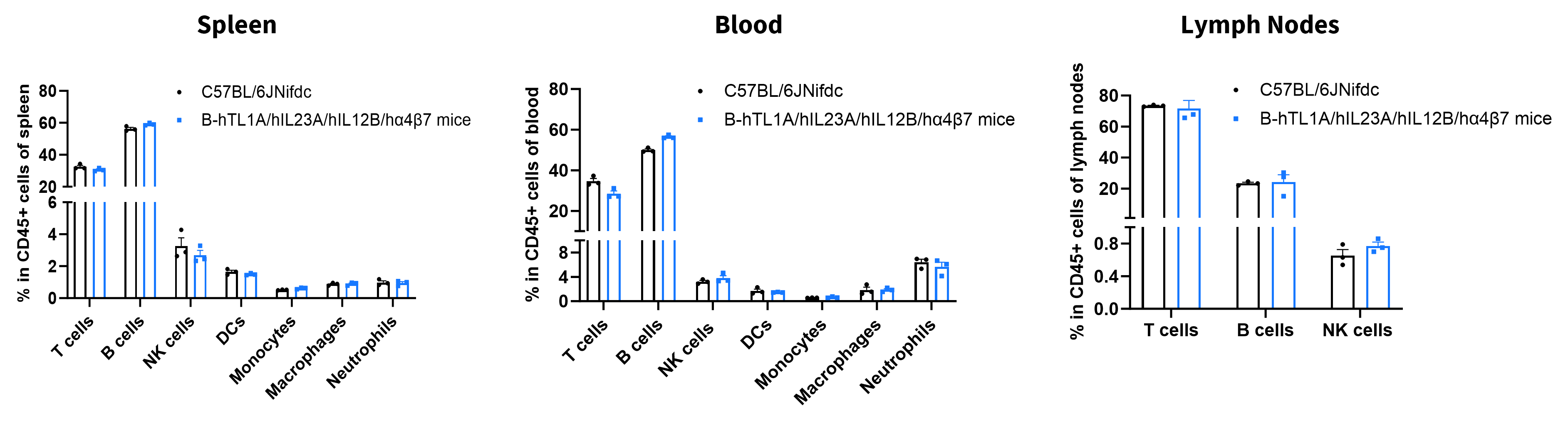
- The percentages of T cells, B cells, NK cells, DCs, monocytes, macrophages, and neutrophils in homozygous B-hTL1A/hIL23A/hIL12B/hα4β7 mice were similar to those in C57BL/6JNifdc mice.
- Humanization of TL1A, IL23A, IL12B, ITGA4, and ITGB7 does not affect normal immune cell development or splenic distribution.
Analysis of leukocyte subpopulations by flow cytometry in immune organs and blood. Splenocytes, peripheral blood, and lymph nodes were isolated from female C57BL/6JNifdc and B-hTL1A/hIL23A/hIL12B/hα4β7 mice (female, 8-week-old, n = 3). Single live cells were gated on the CD45⁺ population and analyzed by flow cytometry as indicated. Values are expressed as mean ± SEM.
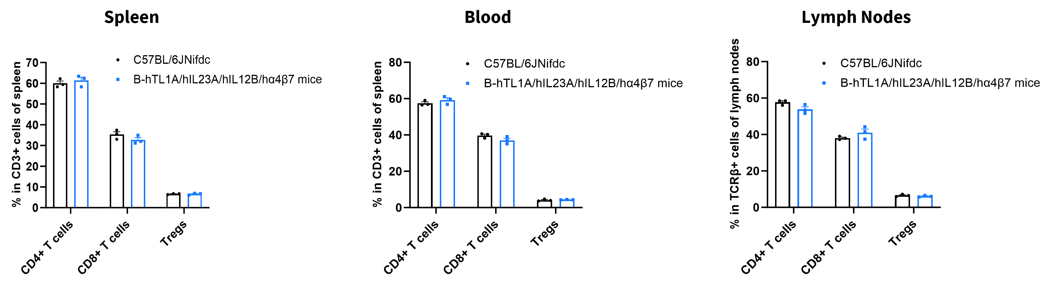
Analysis of T Cell Subpopulations
- The proportions of CD4⁺ T cells, CD8⁺ T cells, and Tregs in homozygous B-hTL1A/hIL23A/hIL12B/hα4β7 mice were comparable to those in C57BL/6JNifdc mice.
- Humanization of TL1A, IL23A, IL12B, ITGA4, and ITGB7 does not affect normal T cell development, differentiation, or splenic distribution.
Analysis of T-cell subpopulations by flow cytometry in immune organs and blood. Splenocytes, peripheral blood, and lymph nodes were isolated from female C57BL/6JNifdc and B-hTL1A/hIL23A/hIL12B/hα4β7 mice (female, 8-week-old, n = 3). Single live cells were gated on the CD3⁺ T-cell population and analyzed by flow cytometry as indicated. Values are expressed as mean ± SEM.
* When publishing results obtained using this animal model, please acknowledge the source as follows: The animal model [B-hTL1A/hIL23A/hIL12B/hα4β7 mice] (Cat# 113826) was purchased from Biocytogen.