
you may also like
Aging is a complex, systemic biological process and the leading risk factor for many chronic diseases, including Alzheimer’s disease, cardiovascular disease, and diabetes. As populations age and the burden of age-related diseases continues to grow, the development of anti-aging therapeutics has become an increasingly active area of biomedical research. Reflecting this momentum, the global anti-aging drug market is projected to grow from $25.1 billion in 2020 to $44.2 billion by 2030.
Pharmaceutical companies are increasingly investing in geroscience to advance the discovery and development of anti-aging therapeutics. Novartis has partnered with BioAge Labs to discover aging-related targets, GSK is leveraging its immunology expertise to investigate inflammaging, and Pfizer has launched a Healthy Aging Initiative focused on metabolic and neurodegenerative diseases. Regulatory momentum is also building. In January 2026, the FDA cleared the first cell reprogramming therapy designed to reverse cellular aging for clinical testing in patients with optic nerve disorders.
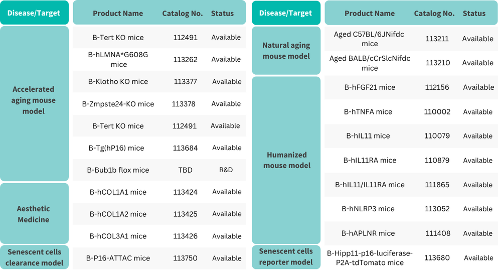
Despite growing commercial momentum, translating anti-aging candidates into the clinic remains challenging. Aging is driven by interconnected biological processes, including cellular senescence, mitochondrial dysfunction, epigenetic alterations, and chronic inflammation, making single-pathway evaluation insufficient. To address this complexity, Biocytogen has established a wide-ranging preclinical model portfolio to systematically validate next-generation interventions:
♦ Naturally aged models: aged C57BL/6J and BALB/c lines - closely recapitulate physiological aging.
♦ Accelerated aging models: Tert KO, Klotho KO, Zmpste24 KO, and hLMNA*G608G - shorten study timelines while reproducing key aging phenotypes.
♦ Senescent cell reporter and clearance models: the P16-ATTAC series - track and eliminate senescent cells in vivo.
♦ Target-humanized mouse models for aging-related targets: the IL11/IL11RA series - support in vivo evaluation of human-targeted therapeutics.
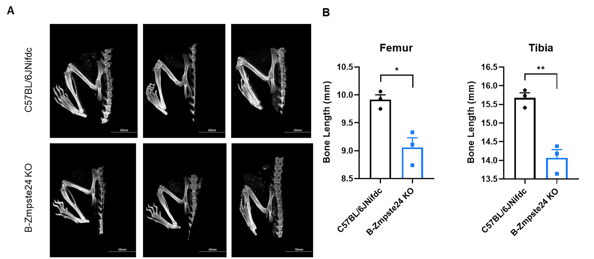
Micro-CT analysis of femur and tibia length in homozygous B-Zmpste24 KO mice. In the homozygous B-Zmpste24 KO mice, the lengths of the femur and tibia were shortened.
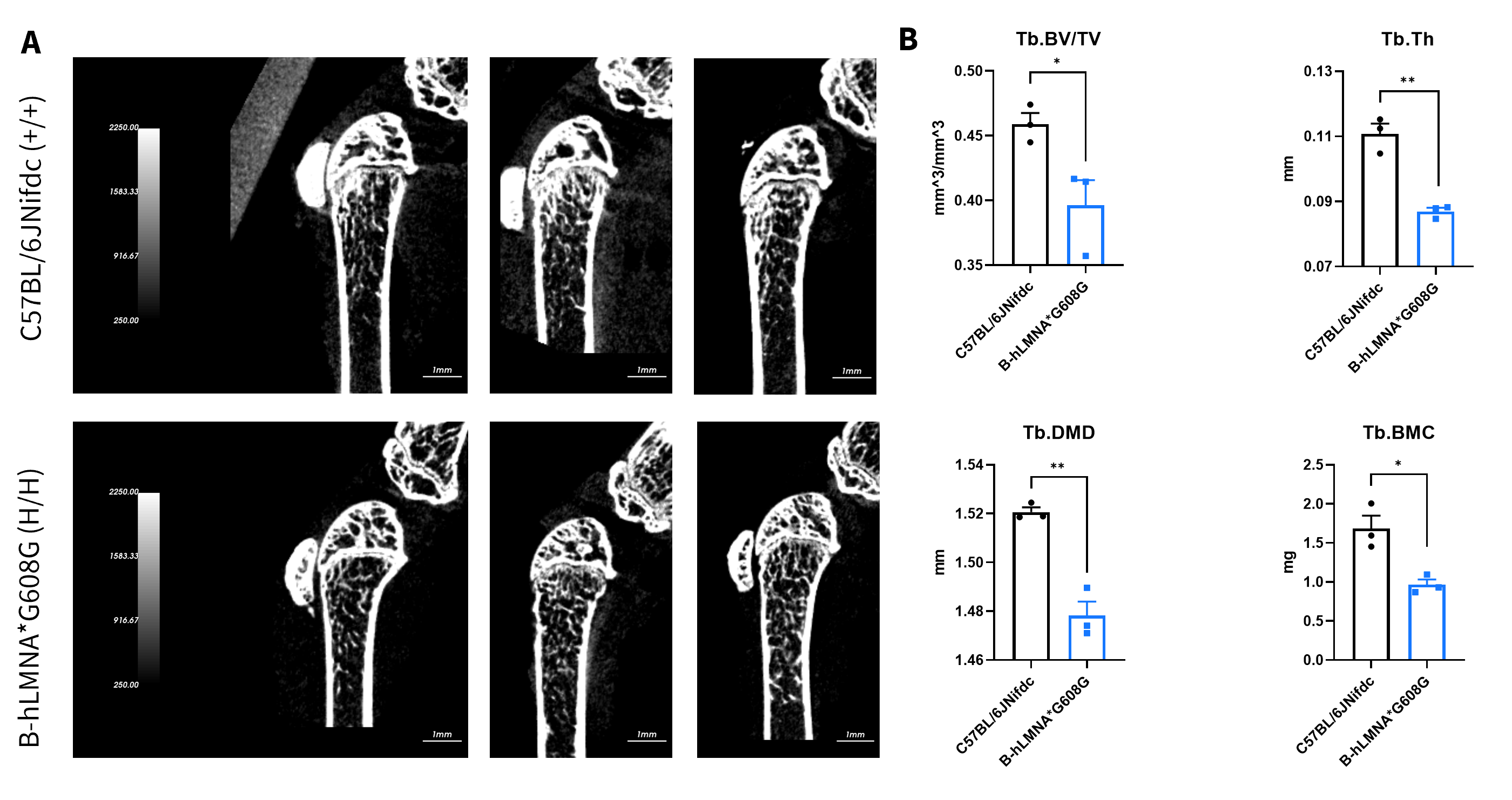
Micro-CT analysis of bone microarchitecture in homozygous B-hLMNA*G608G mice. (A) Representative micro-CT images of trabecular bone from wild-type C57BL/6JNifdc mice (n=3, male, 11-week-old) and homozygous B-hLMNA*G608G mice (n=3, male, 11-week-old). (B) Quantitative analysis of bone volume fraction (Tb.BV/TV), trabecular thickness (Tb.Th), trabecular bone mineral density (Tb.BMD) and trabecular bone mineral content (Tb.BMC). Decreased Tb.BV/TV, Tb.Th, Tb.BMC indicated osteoporotic deterioration or loss of trabecular bone structure.
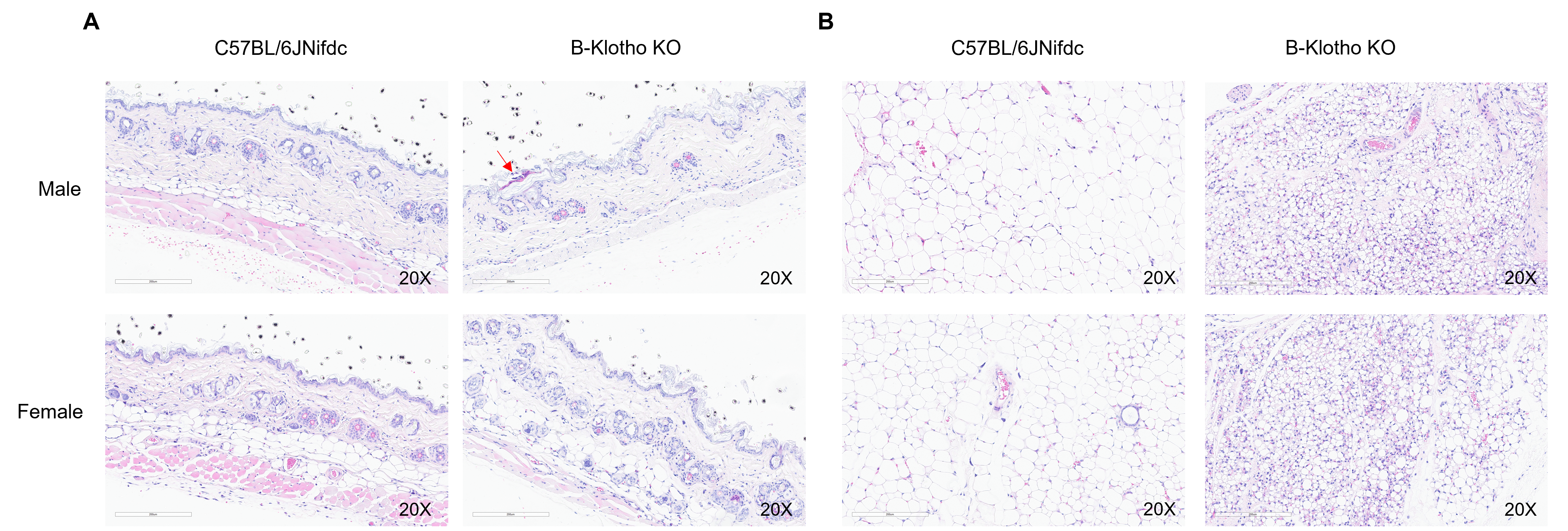
Histopathological analysis of dorsal skin and subcutaneous adipose tissue in homozygous B-Klotho KO mice. (A) H&E-stained sections showed skin pathological changes with punctate crusts, hyperkeratosis and loss of subcutaneous fat layer in homozygous B-Klotho KO mice (-/-, 7-week-old, 1 male and 1 female) compared with wild-type C57BL/6JNifdc mice (+/+, 7-week-old, 1 male and 1 female). (B) H&E-stained subcutaneous adipose tissue showed adipocyte atrophy in homozygous B-Klotho KO mice compared with wild-type C57BL/6JNifdc.
As anti-aging drug discovery advances toward clinical translation, selecting the right preclinical model has become increasingly important for generating meaningful and translatable data. With a comprehensive portfolio spanning naturally aged, accelerated aging, senescent cell, and target-humanized mouse models, Biocytogen provides researchers with flexible solutions to support diverse therapeutic strategies across aging and age-related diseases.
Aging is driven by interconnected changes across multiple biological systems, including cellular senescence, mitochondrial dysfunction, chronic inflammation, and epigenetic alterations. Because no single model can capture this complexity, a diverse model portfolio allows researchers to select the most appropriate biological context and endpoints for each therapeutic strategy.
Natural aging models are generally preferred because they better reflect physiological aging, but they require longer study timelines. Accelerated aging models offer a faster alternative when their underlying mechanism aligns with the therapeutic target.
Accelerated aging models can also be crossed with target-humanized mice, enabling target-specific therapeutic evaluation in an aged background while avoiding the long lead time required to generate naturally aged humanized cohorts. The optimal choice depends on the study objective, target biology, and timeline.
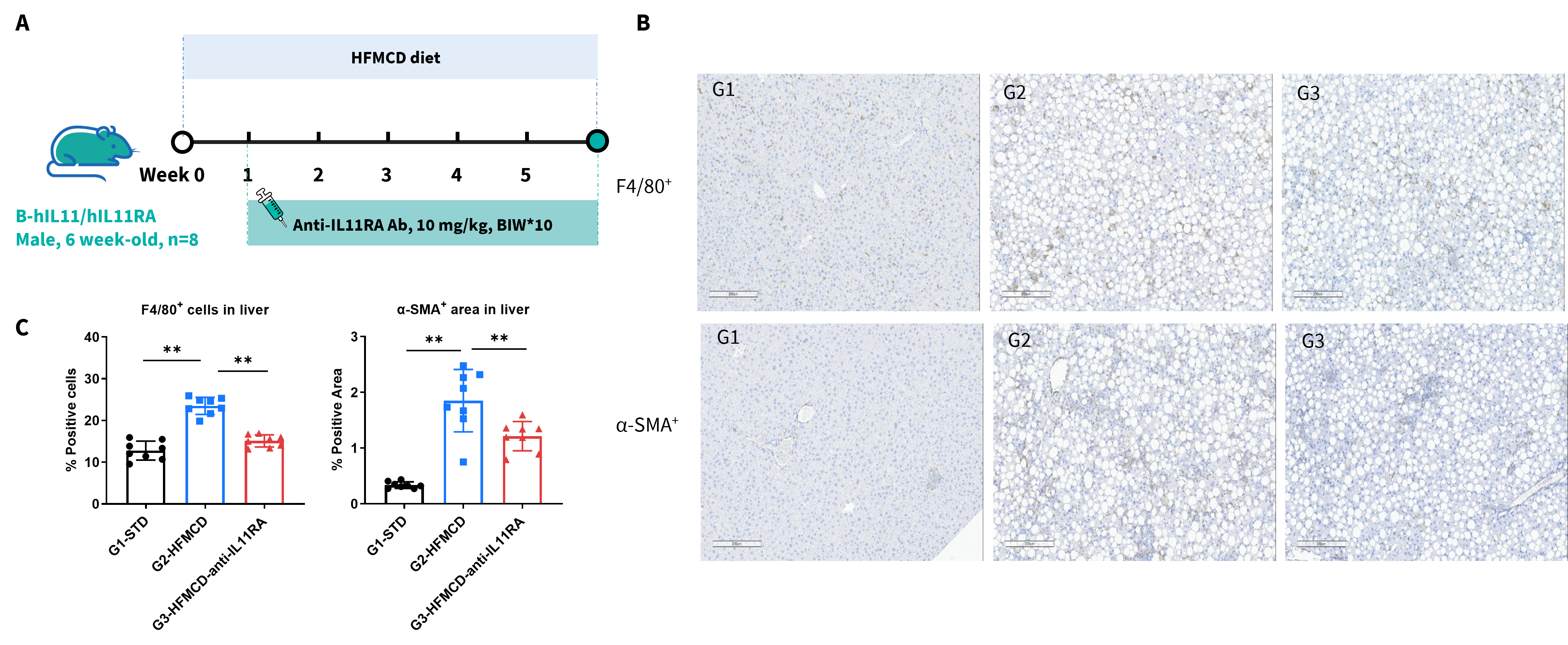
Humanized target models introduce human target biology into a physiologically relevant whole-animal system, enabling direct evaluation of therapeutics designed to recognize human targets. Models such as B-hIL11/hIL11RA mice can support target-specific efficacy, pharmacodynamic, mechanism-of-action, and toxicity studies in aging-related disease settings.
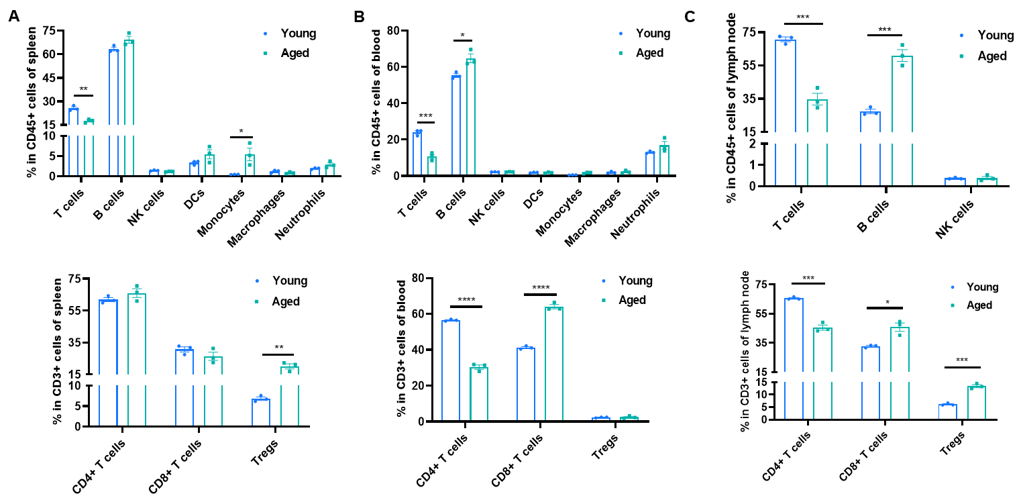
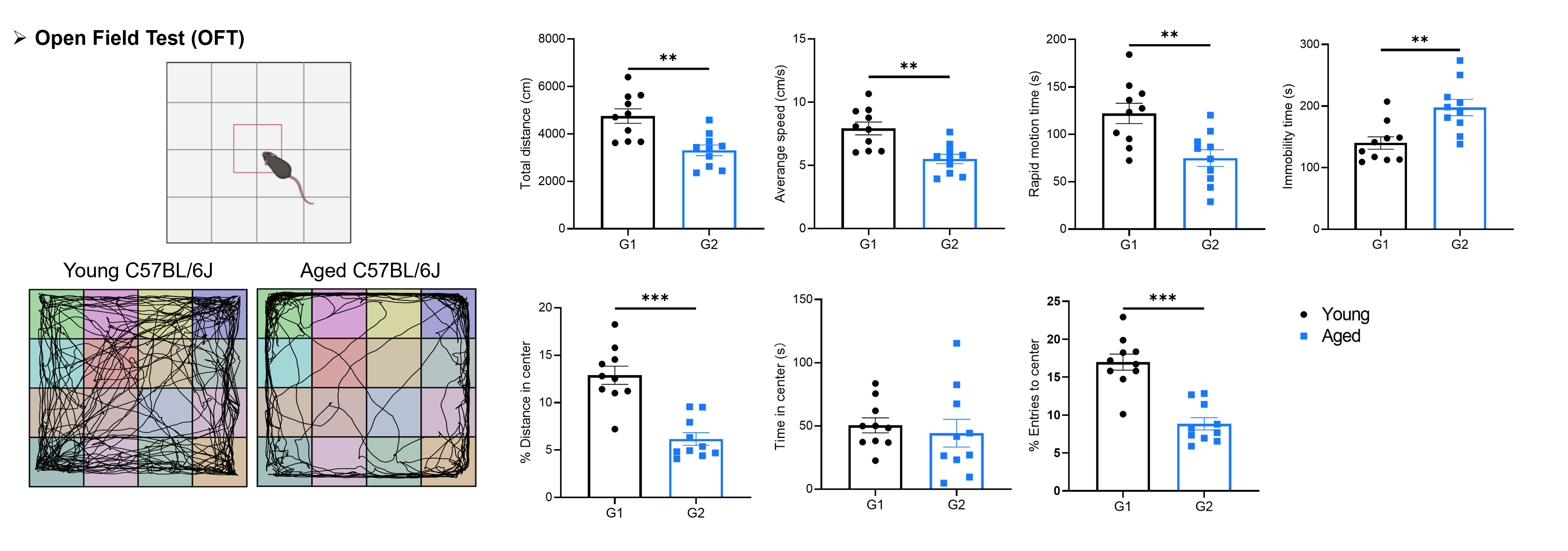
Typical aging-study readouts include senescence biomarkers such as GDF15, P16, P21, mitochondrial DNA damage markers, and SA-β-gal staining. Additional assessments may include hematology, blood biochemistry, metabolic markers, immune-cell profiling, bone parameters measured by micro-CT, and behavioral or motor-function tests. Biocytogen can support these analyses and tailor the readout panel to specific study objectives.
