you may also like
Animal models remain a critical foundation of oncology drug development. However, traditional subcutaneous xenograft models often fail to fully capture the complex biology of human tumors, particularly the influence of the native tumor microenvironment. This limitation can reduce their ability to accurately predict tumor progression, invasion, and metastatic behavior.
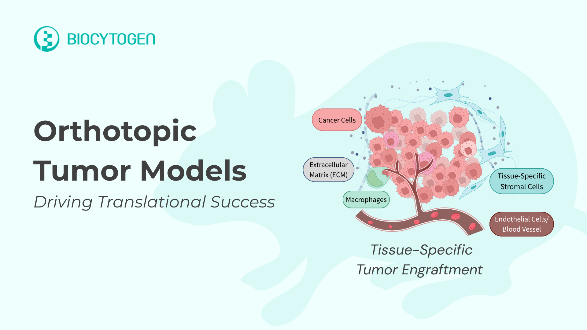
To address these challenges and improve translational relevance, researchers are increasingly turning to orthotopic tumor models, in which tumors are implanted at their tissue of origin. By preserving the native anatomical and microenvironmental context, orthotopic models more accurately reflect physiologically relevant tumor growth, local invasion, and metastatic spread. As a result, they provide a more robust and predictive platform for evaluating therapeutic efficacy and informing clinical decision-making.
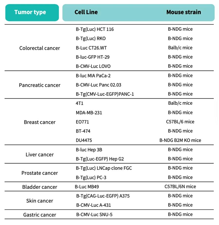
Biocytogen offers a comprehensive portfolio of orthotopic models spanning breast, brain, liver, pancreatic, colorectal, prostate, bladder, and skin cancers.
Powered by cutting-edge gene-editing technologies and robust preclinical platforms, Biocytogen’s orthotopic models are optimized for both monotherapy and combination therapy studies. Key capabilities include:
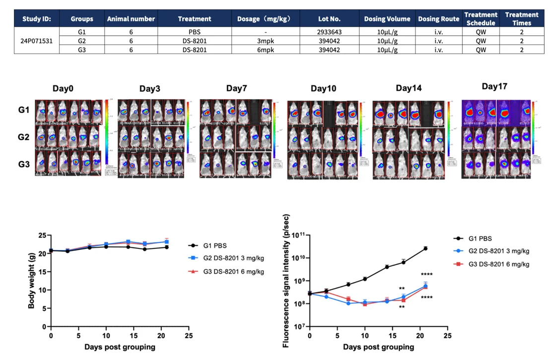
Together, these capabilities generate multidimensional, clinically relevant datasets that accelerate the development of antibody therapeutics, antibody–drug conjugates (ADCs), and immuno-oncology programs.
|
Accelerate Oncology Drug Development with Biocytogen |
By integrating organ-relevant tumor biology with advanced imaging, immune profiling, and metastasis analysis, Biocytogen’s orthotopic tumor model platforms enable more predictive preclinical decision-making. These platforms are designed to help researchers de-risk development programs and advance promising therapeutics toward the clinic with greater confidence.
Partner with Biocytogen to bring clinically meaningful oncology innovations closer to patients!
Orthotopic tumor models implant tumors into their organ of origin, preserving the native tumor microenvironment. This enables more physiologically relevant growth, invasion, and metastasis, making them highly valuable for preclinical oncology research and translational studies.
Unlike subcutaneous xenografts, orthotopic xenograft models grow tumors in the correct anatomical site. This results in more clinically predictive data for cancer drug development, particularly for therapies targeting invasion, metastasis, and immune interactions.
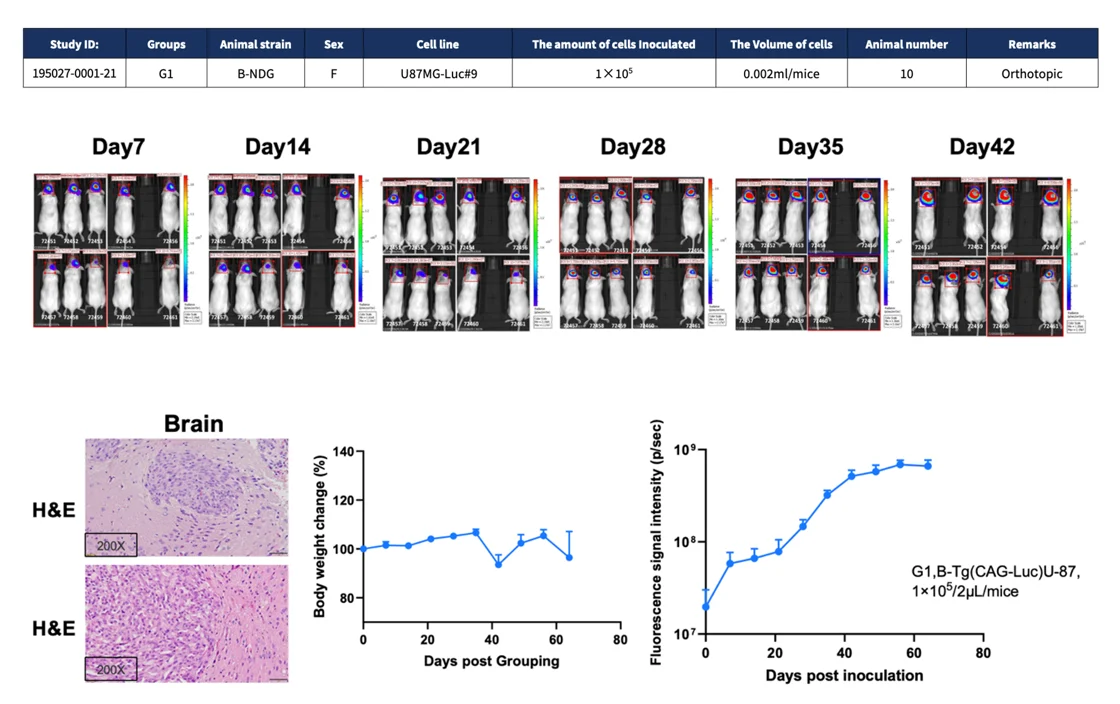
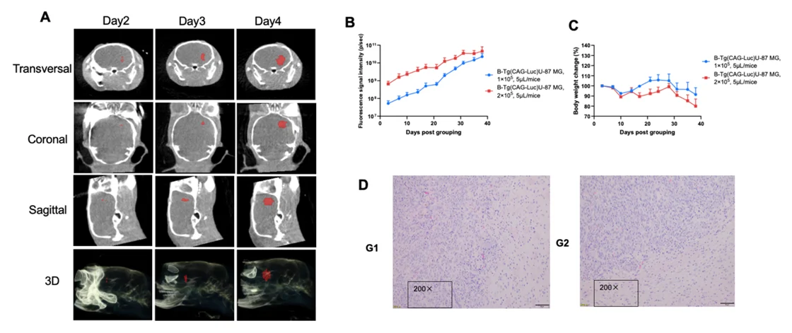
Orthotopic tumor models are used across major solid tumors, including breast, brain, liver, pancreatic, colorectal, prostate, bladder, and skin cancers, supporting diverse in vivo oncology studies.
By integrating bioluminescence imaging, immune profiling, and metastasis monitoring, Biocytogen's orthotopic models deliver multidimensional, clinically relevant data to accelerate antibodies, ADCs, and immuno-oncology programs.