you may also like
The 2026 Alzheimer’s disease drug development pipeline includes 192 clinical trials evaluating 158 therapeutic candidates, yet the field continues to face a low success rate of just 0.4%. Despite rapid advances in antibody engineering and bispecific designs, the gap between preclinical findings and clinical outcomes remains substantial.
Recent progress highlights both the potential and the challenge. Roche’s trontinemab, a TfR1/amyloid bispecific antibody based on Brainshuttle™ technology, has demonstrated that receptor-mediated blood–brain barrier (BBB) transport can achieve rapid and robust target engagement—reaching 91% amyloid PET negativity within 28 weeks at 3.6 mg/kg, while maintaining a low incidence of ARIA-E (<5%). As illustrated in the mechanism schematic, this approach leverages TfR1-mediated transcytosis to shuttle the antibody across the BBB and enable efficient engagement with amyloid plaques in the brain parenchyma. However, most preclinical systems still lack the capability to properly evaluate therapeutics that engage human TfR1.
Two major limitations continue to constrain translational success.
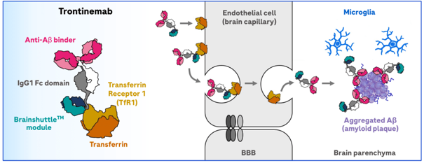
Trontinemab Brainshuttle™ Mechanism via TfR1-Mediated BBB Transport (Source: Medically, Roche Science Hub)
BioMice has developed a suite of TfR1-humanized mice models designed to better align preclinical evaluation with the biological requirements of next-generation CNS therapeutics. By integrating human-relevant BBB transport mechanisms with disease-relevant amyloid pathology, these models enable simultaneous assessment of brain exposure and pharmacodynamic efficacy within a single in vivo system.
The 5xFAD mouse model is widely used in Alzheimer’s disease research for its rapid amyloid pathology and cognitive phenotypes. However, while Tg(5xFAD) captures key disease pathology, species differences in transferrin receptor 1 (TfR1) limit the binding of human TfR1-directed antibodies and prevent reliable assessment of receptor-mediated BBB transport. As a result, this model lacks a functional human TfR1 pathway, creating a key gap for evaluating TfR1-based BBB shuttle strategies for Alzheimer’s disease.
To address this gap, BioMice developed a humanized TfR1 mouse model (B-hTfR1) using targeted gene replacement. The extracellular domain of mouse TfR1 is replaced with the human sequence while preserving endogenous regulatory control, enabling physiologically relevant receptor expression.
The humanized TfR1 retains its native endocytic and transcytotic functions, supporting receptor-mediated transport across the blood–brain barrier (BBB). Consistent with this, pharmacokinetic studies show that TfR1-targeting antibodies achieve measurable brain exposure in B-hTfR1 mice.
This model was further crossed with Tg(5xFAD) mice to generate a dual-feature system, B-hTFR1/B-Tg(5xFAD), combining Alzheimer’s pathology with a functional human TfR1 transport pathway.
Together, these features allow researchers to evaluate both brain penetration and therapeutic efficacy of TfR1-targeting biologics within a single, translationally relevant in vivo system.
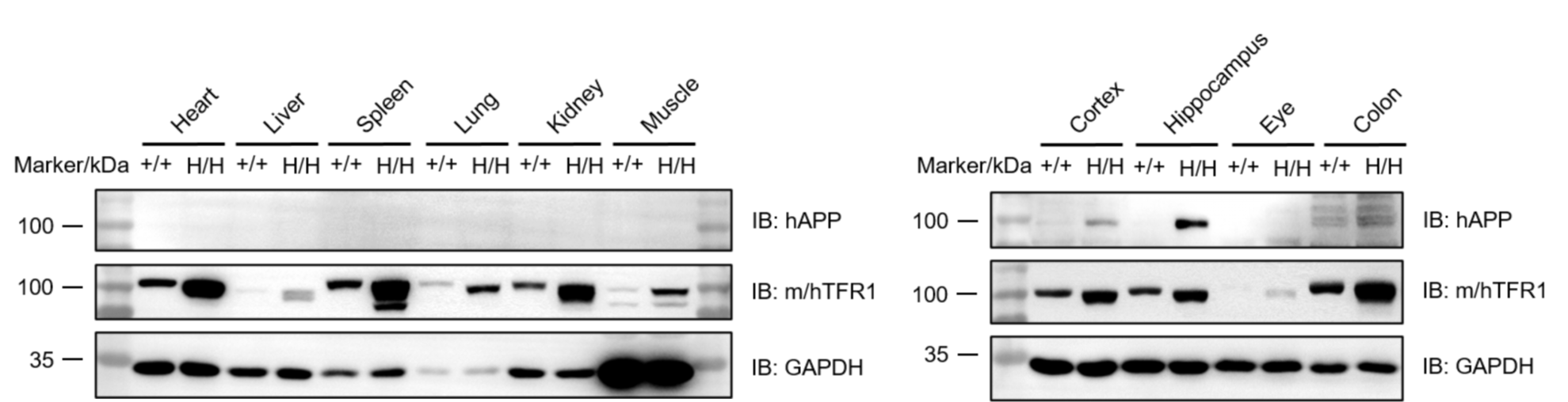
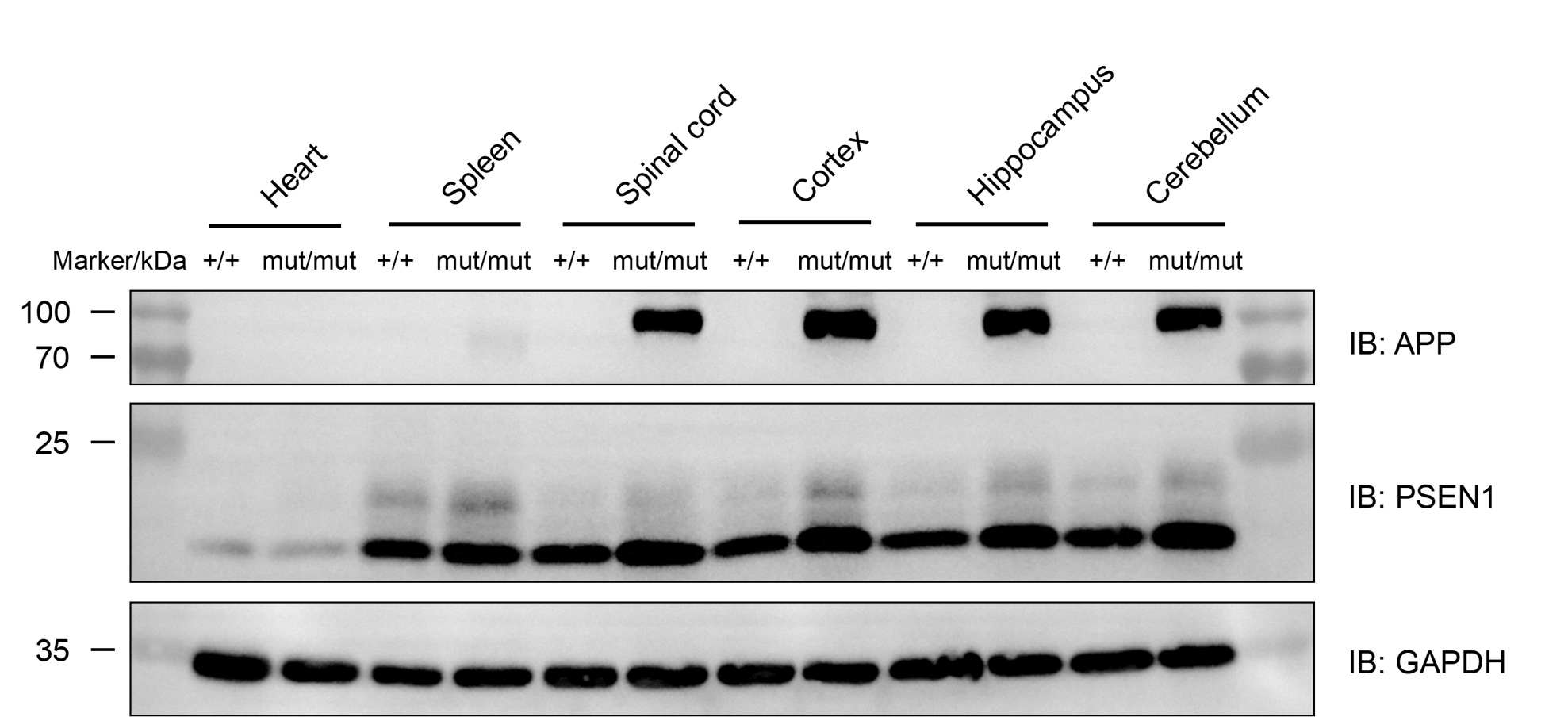
Western blot analysis of APP and TfR1 protein expression in B-hTFR1/B-Tg(5XFAD) mice plus. Various tissue lysates were collected from wild-type C57BL/6 mice (+/+) and B-hTFR1/B-Tg(5XFAD) mice plus (H/H), and then analyzed by western blot with anti-APP antibody and anti-TfR1 antibody (cross-reacts with mouse and human TfR1). Human APP was detected in the cortex and hippocampus from B-hTFR1/B-Tg(5XFAD) mice plus but not in wild-type mice. TfR1 was detected in various tissues from wild-type mice and B-hTFR1/B-Tg(5XFAD) mice plus.
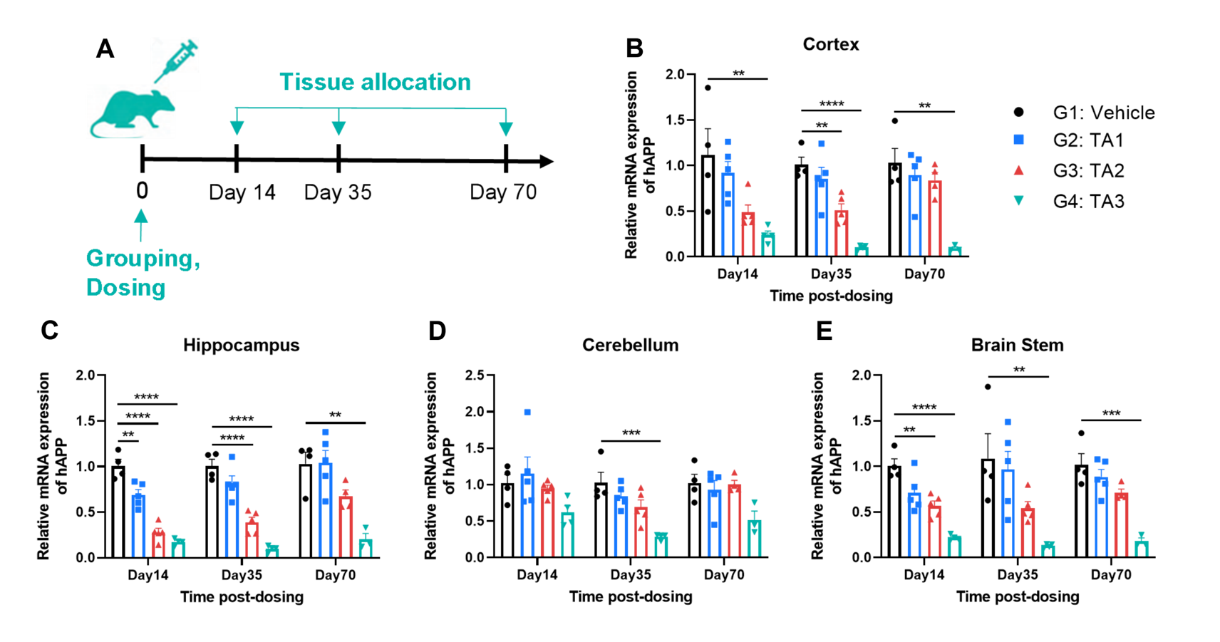
In vivo inhibition of human APP in B-hTFR1/B-Tg(5XFAD) mice plus by oligonucleotide therapeutics. B-hTFR1/B-Tg(5XFAD) mice plus were randomly divided into four groups (n=3-5/group, 10-week-old, male). The oligonucleotide drugs TA1, TA2, TA3 (provided by client) and vehicle were administered to the mice individually. Then nervous tissues were collected to detect the human APP mRNA by RT-qPCR. (A) The experimental schema. (B-E) The expression of human APP mRNA in various nervous tissues. Values are expressed as mean ± SEM. Significance was determined by one-way ANOVA. *P < 0.05, **P < 0.01, ***P < 0.001.
While TfR1-humanized models address the critical need for evaluating BBB transport, Alzheimer’s disease research often requires diverse model systems to capture different aspects of disease biology. To support this, BioMice has developed a broader portfolio of Alzheimer’s disease models. For example, B-App NL-F/Psen1*P117L mouse model incorporates clinically relevant knock-in mutations without relying on APP overexpression, enabling more physiologically relevant amyloid pathology and disease progression. This makes it well suited for studying disease mechanisms and therapeutic responses.
Western blot analysis of APP and PSEN1 protein expression in homozygous B-App NL-F/Psen1*P117L mice. Various tissue lysates were collected from wild-type C57BL/6JNifdc mice (+/+) and homozygous B-App NL-F/Psen1*P117L mice (mut/mut), and then analyzed by western blot with anti-amyloid precursor antibody and anti-PSEN1 antibody. Humanized Aβ was detected in various nerous tissues of homozygous B-App NL-F/Psen1*P117L mice, but not in wild-type mice. PSEN1 was detected in various tissues both in wild-type and homozygous B-App NL-F/Psen1*P117L mice.
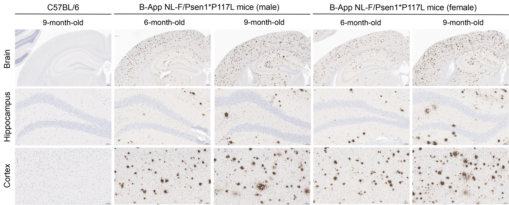
Histopathological analysis of Aβ in homozygous B-App NL-F/Psen1*P117L mice. Brain was collected from wild-type C57BL/6 mice and homozygous B-App NL-F/Psen1*P117L miceand processed into paraffin sections. The Aβ plaque was detected in the cortex and hippocampus of homozygous B-App NL-F/Psen1*P117L mice with anti-human β-Amyloid antibody. The burden of Aβ immunoreactivity was increased in an age-dependent manner. Scale bar: 50 μm.
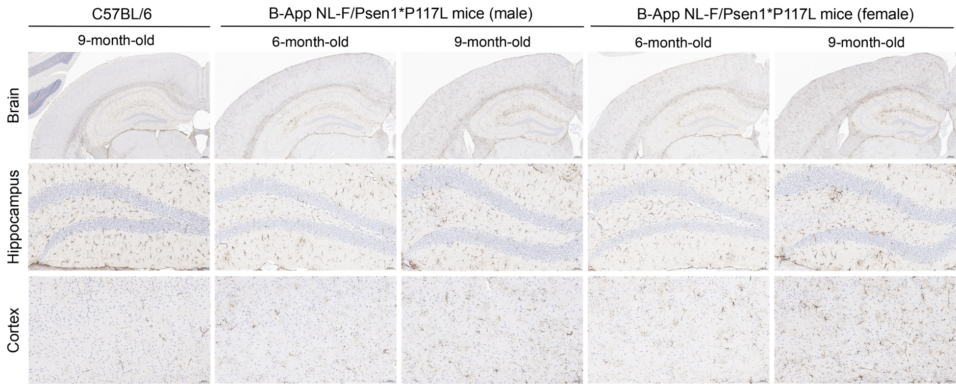
Histopathological analysis of astrocytes in homozygous B-App NL-F/Psen1*P117L mice. Brain was collected from wild-type C57BL/6 mice and homozygous B-App NL-F/Psen1*P117L mice and processed into paraffin sections. The expression of GFAP in the cortex and hippocampus of C57BL/6JNifdc mice and homozygous B-App NL-F/Psen1*P117L mice was detected by IHC with anti-GFAP antibody. Compared to wild-type mice, the number of activated astrocytes in the cortex and hippocampus of homozygous B-App NL-F/Psen1*P117L mice was increased in an age-dependent manner. Scale bar: 50 μm.
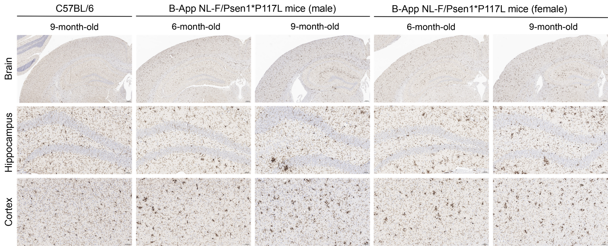
Histopathological analysis of microglia cells in homozygous B-App NL-F/Psen1*P117L mice. Brain was collected from wild-type mice and homozygous B-App NL-F/Psen1*P117L mice and processed into paraffin sections. The expression of Iba1 in the cortex and hippocampus of C57BL/6 mice and B-App NL-F/Psen1*P117L mice was detected by IHC with anti-Iba1 antibody. Compared to wild-type mice, the number of activated microglia cells in the cortex and hippocampus of homozygous B-App NL-F/Psen1*P117L mice was increased in an age-dependent manner. Scale bar: 50 μm.
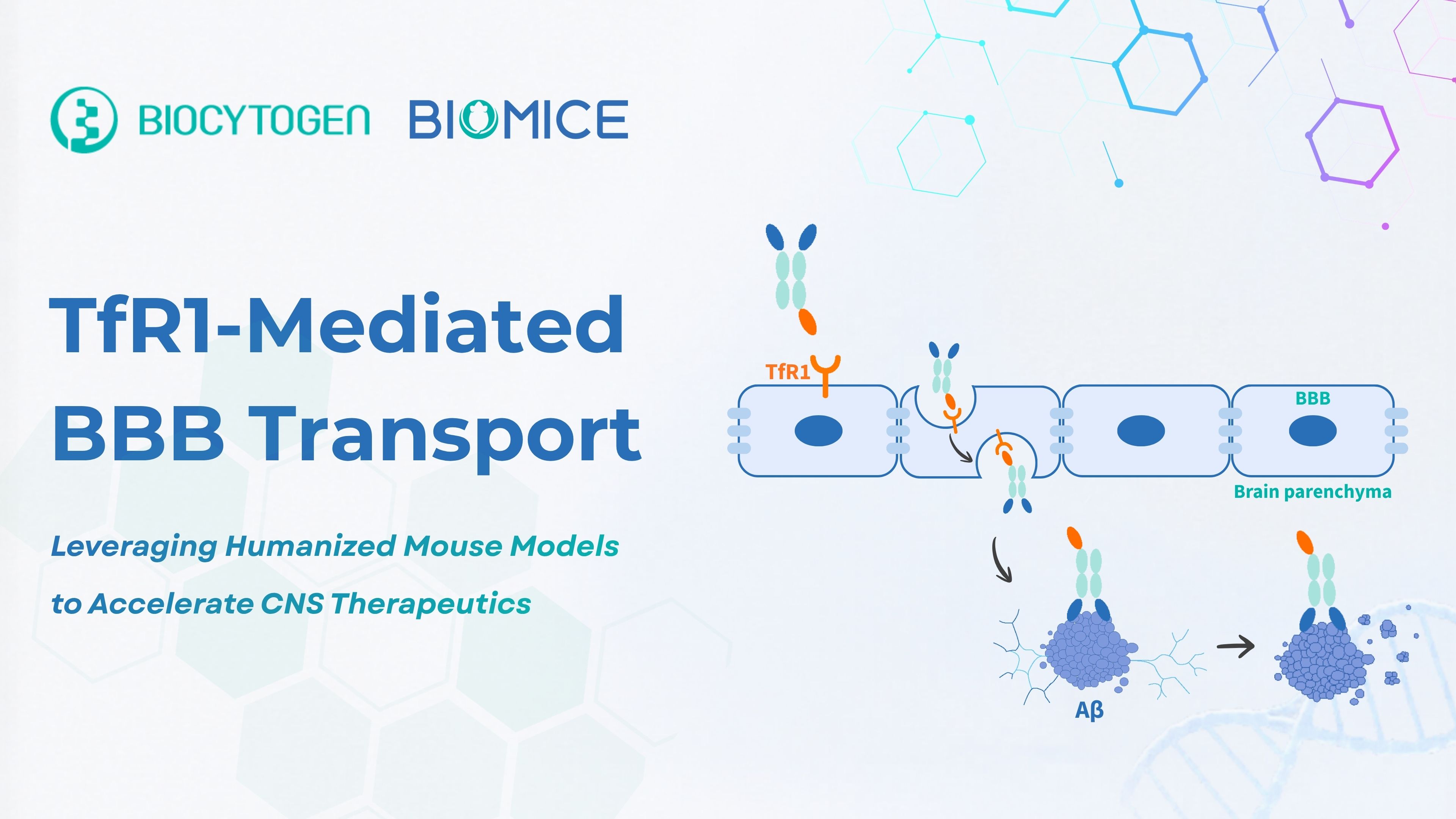
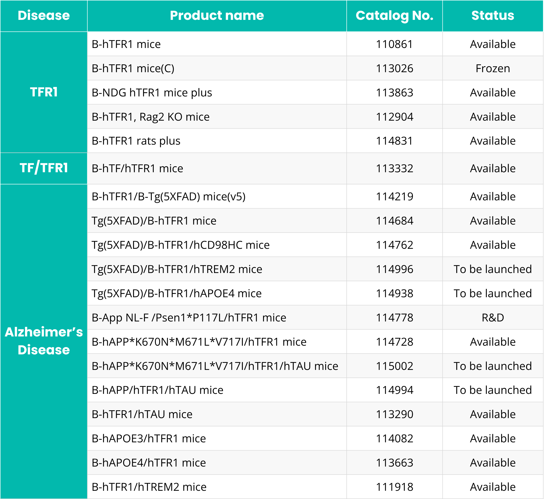
BioMice TfR1-Related Humanized Models for CNS and Alzheimer’s Disease Research
A4: TfR1-targeted antibodies, bispecific antibodies, BBB shuttle biologics, fusion proteins, and other CNS-directed large molecules may benefit from evaluation in a human TfR1-relevant in vivo system.