you may also like

Vitiligo is rapidly emerging as an active area of immunology and dermatology drug development, as advances in disease biology shift therapeutic strategies from managing visible depigmentation toward targeting the immune mechanisms that drive melanocyte loss and disease recurrence. Recent clinical and regulatory progress reflects this momentum:
Together, these programs highlight growing interest in JAK-STAT signaling, T-cell activity, tissue-resident memory T cells, and cytokine pathways as therapeutic opportunities in vitiligo.
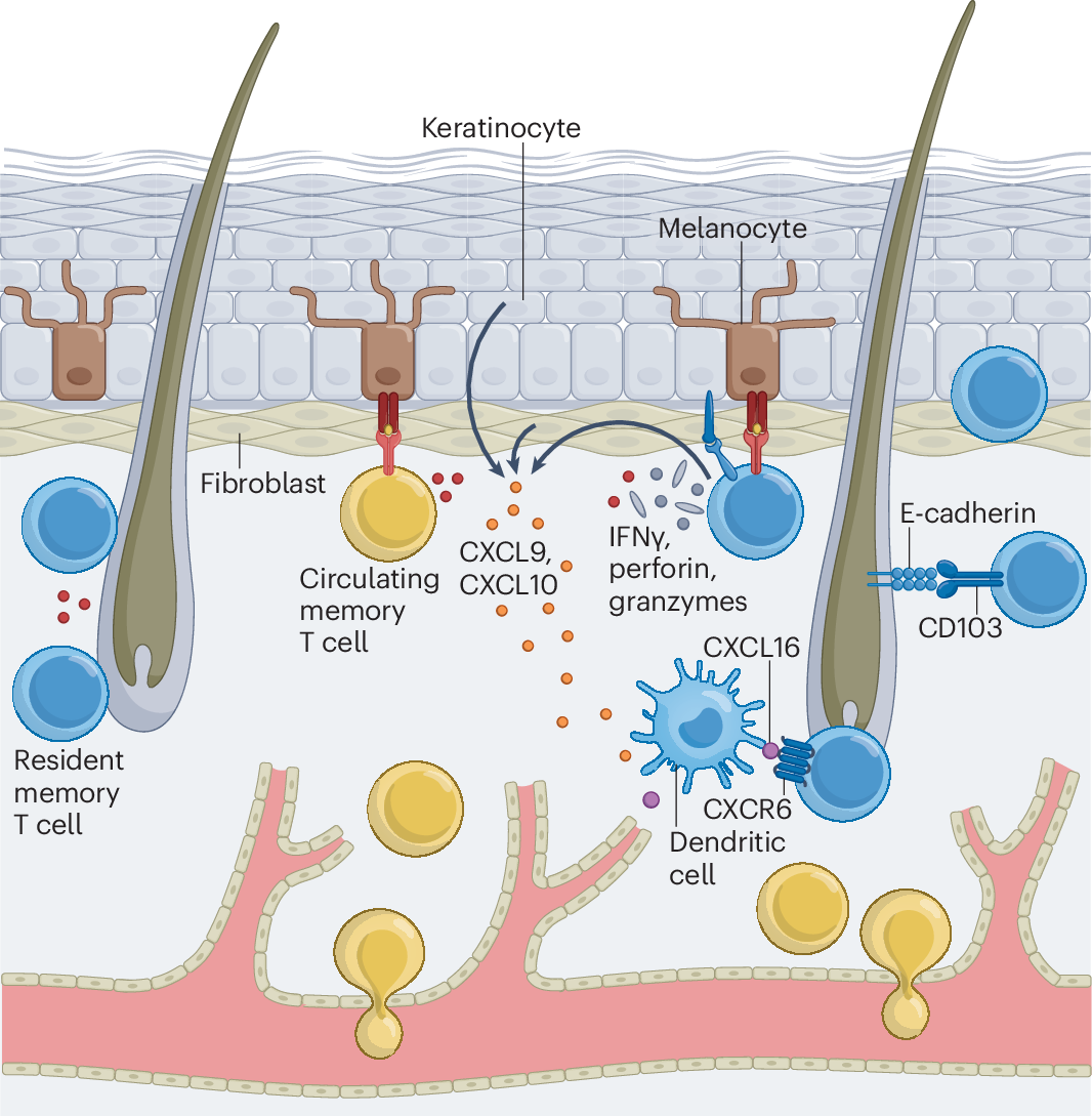
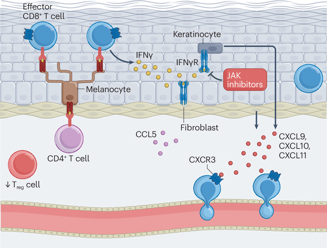
The Immunology of Vitiligo. (Turk, M.J., Huang, Y.H. 2026)
This expanding therapeutic landscape also creates a more demanding preclinical challenge. Vitiligo is not defined by pigmentation changes alone: melanocyte damage, CD8⁺ T-cell infiltration, IFN-γ-associated signaling, and persistent tissue-resident memory T cells (TRM) can all contribute to disease progression and recurrence. As a result, evaluating emerging therapies requires complementary readouts of both the visible disease phenotype and the underlying immune response, such as longitudinal pigmentation, immune-cell profiling, and histopathology.
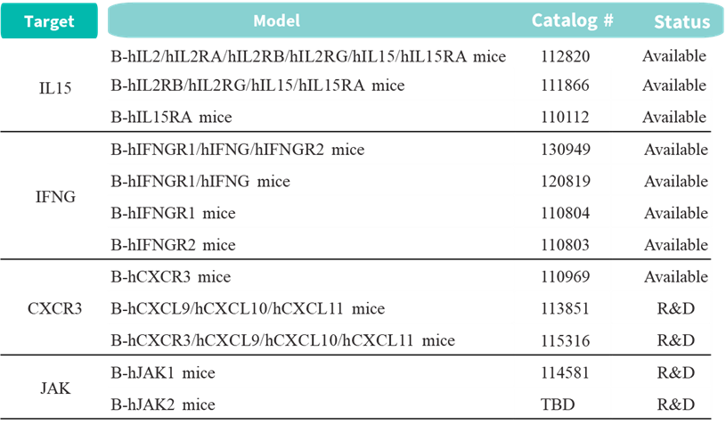
Biocytogen supports vitiligo research with complementary disease-induced models and target-humanized mouse models, providing options for mechanistic studies, efficacy evaluation, and target-specific therapeutic assessment:
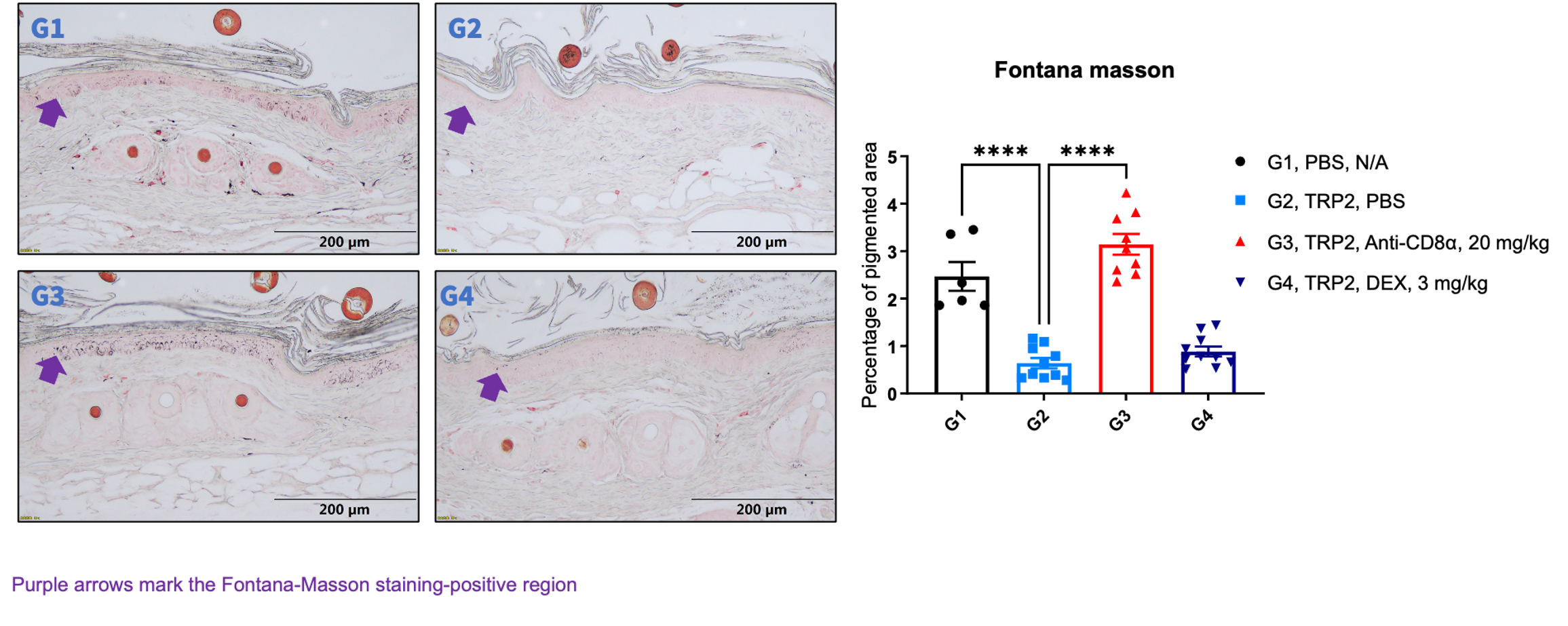
TRP2(180-188)-induced vitiligo model: An antigen-driven model that induces melanocyte-targeted immune responses and progressive depigmentation, enabling longitudinal pigmentation monitoring, CTL/TRM profiling, Fontana–Masson/HALO-based melanin quantification, and in vivo evaluation of immunomodulatory therapies such as anti-CD8α and dexamethasone.
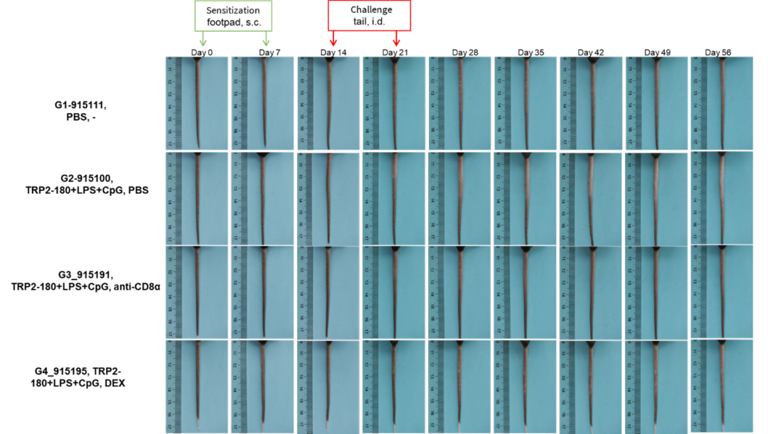
Longitudinal changes in tail skin pigmentation in the TRP2(180-188)-induced vitiligo model. Progressive depigmentation was observed from Day 21, enabling dynamic monitoring of model development and treatment response over time.
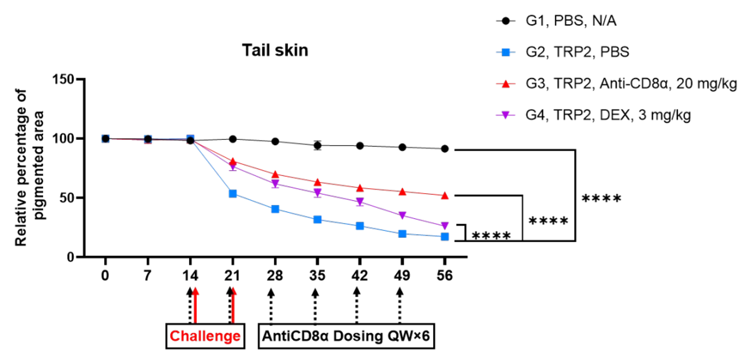
Longitudinal quantification of tail skin pigmentation in the TRP2(180-188)-induced vitiligo model. ImageJ analysis showed a progressive decline in pigmented area beginning at Day 21 in model mice. Treatment with anti-CD8α or dexamethasone significantly attenuated pigment loss over time, with anti-CD8α showing the stronger protective effect.
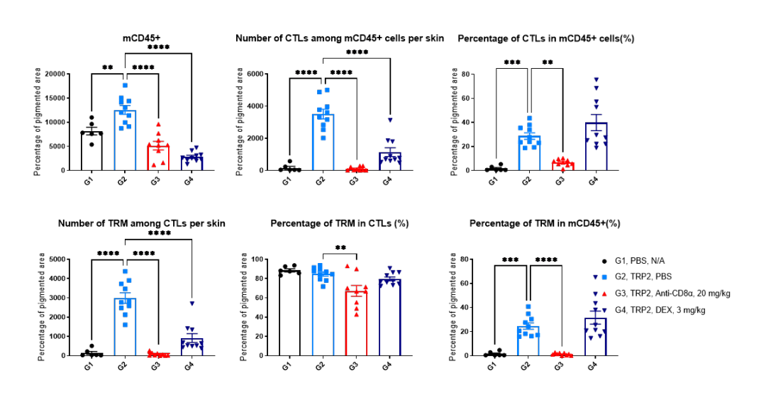
Flow cytometric analysis of tail skin immune cell populations at the study endpoint in the TRP2(180-188)-induced vitiligo model. Compared with vehicle controls, model mice showed significant increases in mCD45⁺ immune cells, CTLs, and TRM cells. Anti-CD8α treatment markedly reduced these immune cell populations, while dexamethasone produced partial reductions in mCD45⁺ cells and CTLs, supporting modulation of the local immune response.
|
As vitiligo research moves toward immune-targeted therapies, preclinical evaluation increasingly requires more than monitoring depigmentation alone. Disease models that enable longitudinal pigmentation analysis, immune-cell profiling, and histological readouts can provide a more comprehensive view of treatment response and underlying immune activity.
Biocytogen offers TRP2(180-188)- and B16F10-induced vitiligo models, together with a portfolio of target-humanized mice covering pathways such as IL-15, IFN-γ, CXCR3, and JAK, supporting vitiligo research from disease modeling and mechanistic studies to in vivo therapeutic evaluation.
Biocytogen has established TRP2(180-188)-induced and B16F10-induced vitiligo mouse models for in vivo efficacy and mechanistic studies. The TRP2(180-188) model provides quantitative pigmentation, immune-cell, and histological readouts, while the B16F10-induced model offers a complementary immune-induction strategy.
Activated CD8⁺ T cells and other immune cells can produce IFN-γ, which activates JAK-STAT signaling and promotes CXCL9 and CXCL10 expression. These chemokines can recruit additional CXCR3⁺ T cells into the skin, helping sustain the local inflammatory response described in vitiligo.
The current Biocytogen vitiligo portfolio includes humanized models targeting different components of the IFN-γ pathway, including IFNGR1 humanized mice, IFNGR2 humanized mice, IFNGR1/IFNG humanized mice, and IFNGR1/IFNG/IFNGR2 humanized mice. These models are positioned for studies requiring interaction with human IFN-γ pathway components.
The article identifies JAK-STAT signaling as a key therapeutic pathway in vitiligo and lists JAK1 humanized mice and JAK2 humanized mice within Biocytogen's target-humanized mice portfolio. These models may provide future options for evaluating therapeutics that require interaction with human JAK targets; the current draft does not present vitiligo efficacy data from these models.
Disease-induced models such as TRP2(180–188)- and B16F10-induced vitiligo models enable evaluation of depigmentation, immune-cell changes, and treatment response in vivo. Target-humanized mouse models further support studies of therapeutics directed against human targets such as IFNGR1, CXCR3, IL-15 pathway components, and JAK proteins. Together, these models provide complementary tools for studying disease mechanisms and evaluating target-specific therapeutic strategies.
Turk MJ, Huang YH. The immunology of vitiligo. Nature Reviews Immunology. 2026;26(4):284–297. doi:10.1038/s41577-025-01249-z
Wang Y, et al. Innate immune activation in vitiligo: mechanisms and pathophysiological implications. Frontiers in Immunology. 2025;16:1631074. doi:10.3389/fimmu.2025.1631074
Howell MD, et al. Targeting the Janus Kinase Family in Autoimmune Skin Diseases. Frontiers in Immunology. 2019;10:2342. doi:10.3389/fimmu.2019.02342
Riding RL, Harris JE. The Role of Memory CD8+ T Cells in Vitiligo. Journal of Immunology. 2019;203(1):11–19. doi:10.4049/jimmunol.1900027
Rosmarin D, et al. Two Phase 3, Randomized, Controlled Trials of Ruxolitinib Cream for Vitiligo. New England Journal of Medicine. 2022;387(16):1445–1455. doi:10.1056/NEJMoa2118828