B-hDLL3 B16-F10
Catalog Number: 321826
---
Licensing option available

• 321826
on this page
The mouse Dll3 gene was replaced by chimeric human DLL3 coding sequence in B-hDLL3 B16-F10 cells. Human DLL3 is highly expressed on the surface of B-hDLL3 B16-F10 cells.
Gene targeting strategy for B-hDLL3 B16-F10 cells. The exogenous promoter and chimeric human DLL3 coding sequence were inserted to replace part of murine exon 2 and all of exons 3-5. The insertion disrupts the endogenous murine Dll3 gene, resulting in a non-functional transcript.
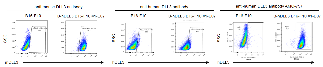
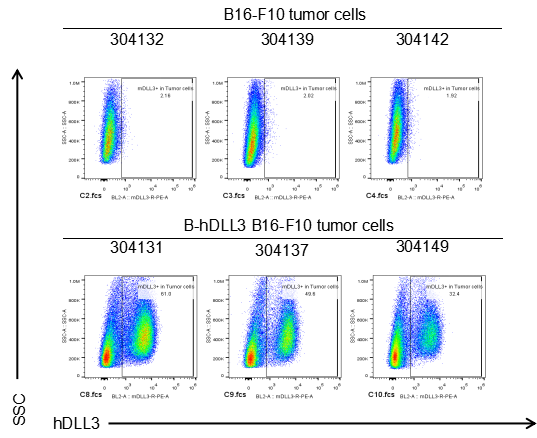
DLL3 expression analysis in B-hDLL3 B16-F10 cells by flow cytometry. Single cell suspensions from wild-type B16-F10 and B-hDLL3 B16-F10 cultures were stained with anti-DLL3 antibody. The mouse antibody is cross-reactive between human and mouse. Human DLL3 was detected on the surface of B-hDLL3 B16-F10 cells, but not on the surface of wild-type B16-F10 cells. The 1-E07 clone of B-hDLL3 B16-F10 cells was used for in vivo tumor growth assays.
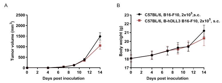
Subcutaneous homograft tumor growth of B-hDLL3 B16-F10 cells. B-hDLL3 B16-F10 cells (2x10⁵) and wild-type B16-F10 cells (2x10⁵) were subcutaneously implanted into C57BL/6 mice (female, 7-week-old, n=8). Tumor volume and body weight were measured three times a week. (A) Average tumor volume ± SEM. (B) Body weight (Mean ± SEM). Volume was expressed in mm3 using the formula: V=0.5 X long diameter X short diameter2. As shown in panel A, B-hDLL3 B16-F10 cells were able to form tumors in vivo and can be used for efficacy studies.
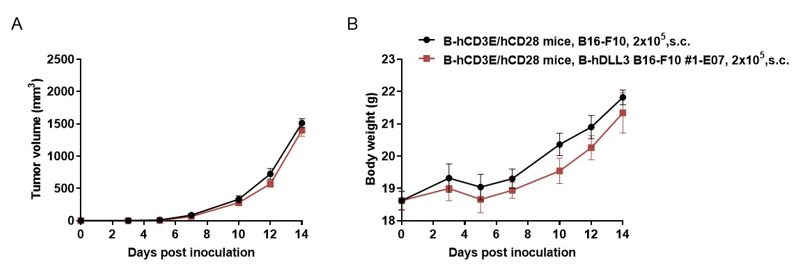
Subcutaneous homograft tumor growth of B-hDLL3 B16-F10 cells. B-hDLL3 B16-F10 cells (2x10⁵) and wild-type B16-F10 cells (2x10⁵) were subcutaneously implanted into B-hCD3E/hCD28 mice (female, 7-week-old, n=5). Tumor volume and body weight were measured three times a week. (A) Average tumor volume ± SEM. (B) Body weight (Mean ± SEM). Volume was expressed in mm3 using the formula: V=0.5 X long diameter X short diameter2. As shown in panel A, B-hDLL3 B16-F10 cells were able to form tumors in vivo and can be used for efficacy studies.
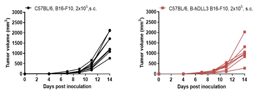
B-hDLL3 B16-F10 tumor growth of individual mice. B-hDLL3 B16-F10 cells (2x10⁵) and wild-type B16-F10 cells (2x10⁵) were subcutaneously implanted into C57BL/6 mice (female, 7-week-old, n=8). As shown in panel, B-hDLL3 B16-F10 cells were able to establish tumors in vivo and can be used for efficacy studies.
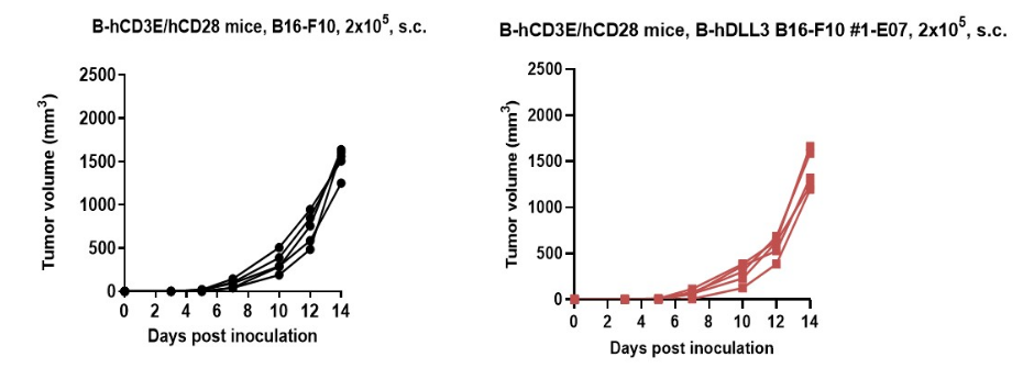
B-hDLL3 B16-F10 tumor growth of individual mice. B-hDLL3 B16-F10 cells (2x10⁵) and wild-type B16-F10 cells (2x10⁵) were subcutaneously implanted into B-hCD3E/hCD28 mice (female, 7-week-old, n=5). As shown in panel, B-hDLL3 B16-F10 cells were able to establish tumors in vivo and can be used for efficacy studies.