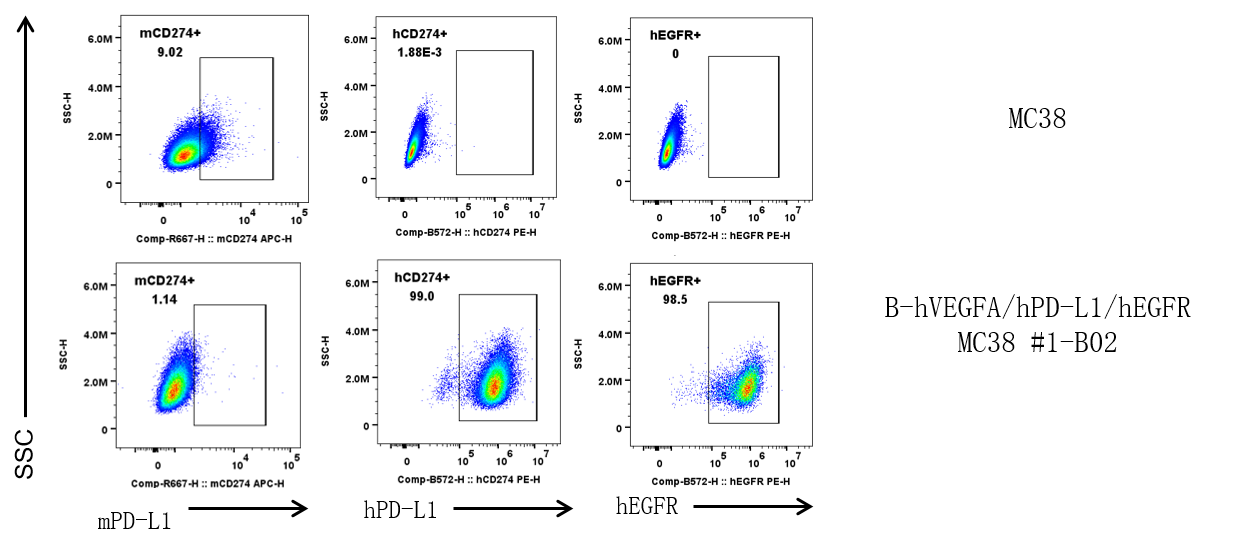
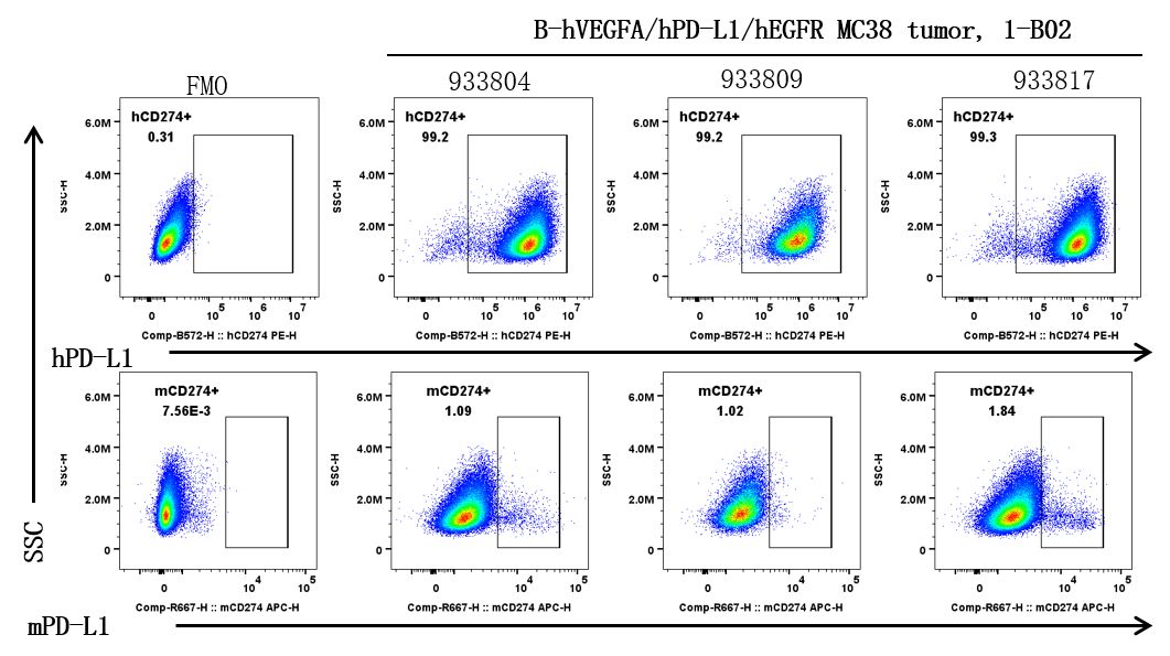
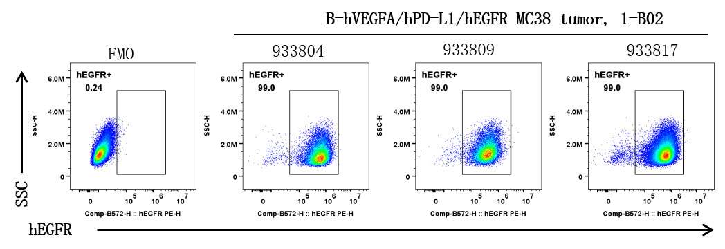
B-hVEGFA/hPD-L1/hEGFR MC38
Catalog Number: 322558
Strain Name: NA
Strain Background: C57BL/6
Aliases: Vpf, Vegf, L-VEGF; B7h1, Pdl1, Pdcd1l1, Pdcd1lg1; Erbb, Erbb1
---
Licensing option available