

C57BL/6-Tnfsf8tm2(TNFSF8)Bcgen/Bcgen • 113224
on this page

CD30L: A crucial co-stimulatory driver that amplifies T-cell activation

CD30L

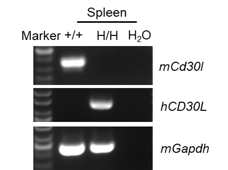
Species specific analysis of CD30L gene expression in wild-type C57BL/6JNifdc mice and homozygous humanized B-hCD30L mice by RT-PCR. Spleen was collected from wild-type C57BL/6JNifdc mice (+/+) and homozygous B-hCD30L mice (H/H). Mouse Cd30l mRNA was only detectable in wild-type C57BL/6JNifdc mice. Human CD30L mRNA was only detectable only in homozygous B-hCD30L mice, but not in wild-type C57BL/6JNidfc mice.
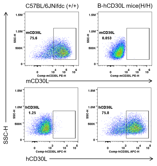
Strain specific CD30L expression analysis in wild-type C57BL/6JNifdc mice and homozygous humanized B-hCD30L mice by flow cytometry. Splenocytes were collected from wild-type C57BL/6JNifdc mice (+/+) and homozygous B-hCD30L mice (H/H). Splenocytes are incubated in a medium containing Cell Activation Cocktail (without Brefeldin A) (Biolegend, 423303) before analysis of CD30L surface expression on gated T cells. Protein expression was analyzed with anti-mouse CD30L antibody (Biolegend, 106405) and anti-human CD30L antibody (RD, FAB1028A) by flow cytometry.
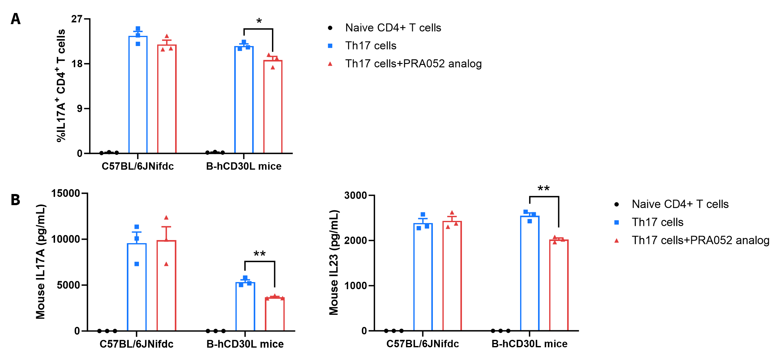
Ex vivo functional analysis in wide-type C57BL/6JNifdc and homozygous B-hCD30L mice. Naïve CD4+ T cells were sorted from the splenocytes of wild-type C57BL/6JNifdc mice (+/+) and homozygous B-hCD30L mice (H/H), and were assessed after 72 h of incubation with rmIL6, rmIL23, rmTGF-β1, anti-mouse IFN-γ antibody and anti-mouse IL4 antibody in combination with bead-associated CD3 and CD28 mAbs. (A) The percentage of mIL17A positive cells were assessed by flow cytometry. The anti-human CD30L antibody PRA052 analog (in house, 50 μg/mL) could reduce the percentage of IL17A+ CD4+ T cells in B-hCD30L mice during Th17 cells differentiation, as PRA052 analog only recognizes human and does not recognize mice. (B) The protein expression of mouse IL17A and mouse IL23 in cell culture supernatant were measured by ELISA. The anti-human CD30L antibody PRA052 analog (in house, 50 μg/mL) could reduce the production of mouse IL17A and mouse IL23 in B-hCD30L mice during Th17 cells differentiation. Values are expressed as mean ± SEM. Significance was determined by unpaired t test. *P < 0.05, **P < 0.01.
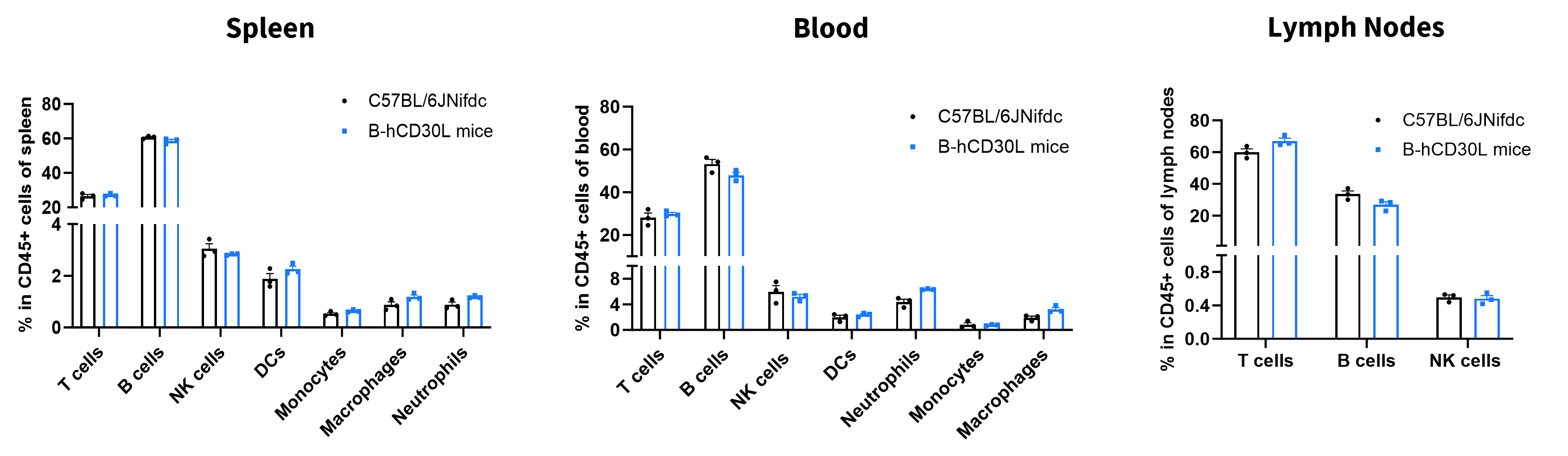
Analysis of leukocyte subpopulations by flow cytometry in immune organs and blood. Splenocytes, peripheral blood, and lymph nodes were isolated from female C57BL/6JNifdc and B-hCD30L mice (female, 6-week-old, n=3). Single live cells were gated on the CD45⁺ population and analyzed by flow cytometry as indicated. Values are expressed as mean ± SEM.
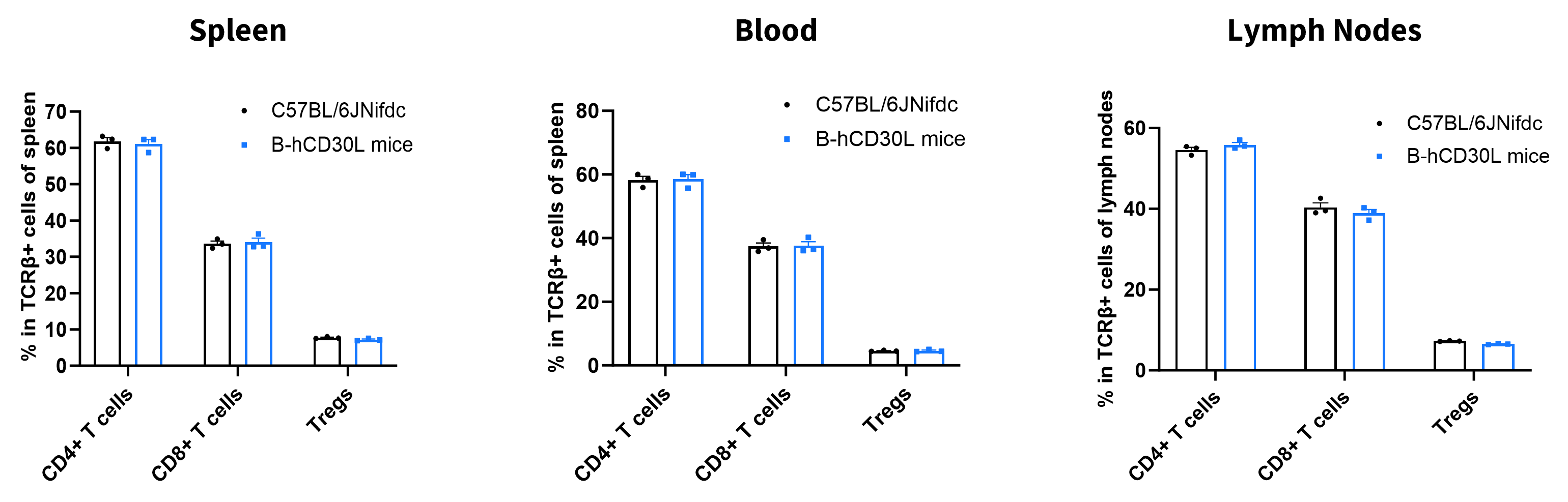
Analysis of T-cell subpopulations by flow cytometry in immune organs and blood. Splenocytes, peripheral blood, and lymph nodes were isolated from female C57BL/6JNifdc and B-hCD30L mice (female, 6-week-old, n = 3). Single live cells were gated on the CD3⁺ T-cell population and analyzed by flow cytometry as indicated. Values are expressed as mean ± SEM.

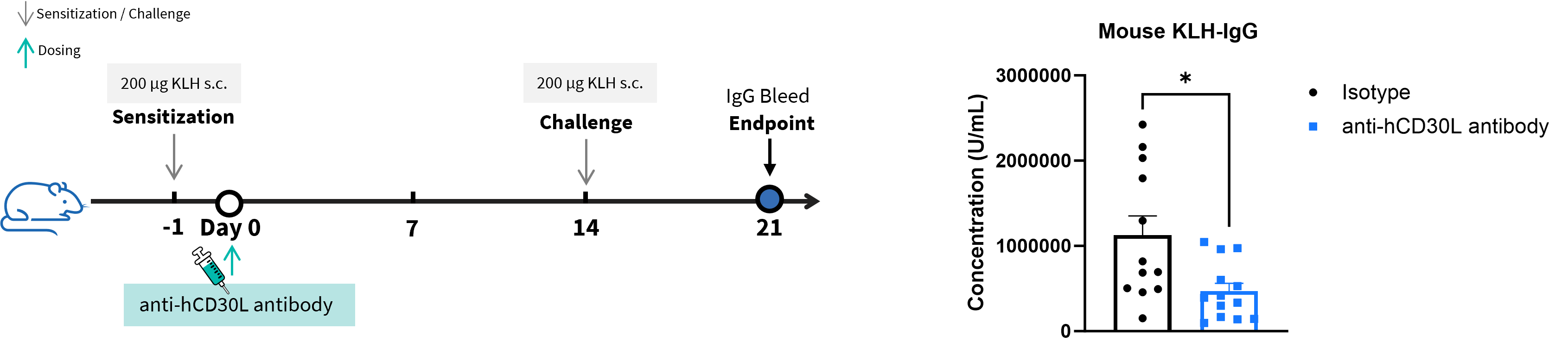
T-cell-dependent antibody response assay in homozygous B-hCD30L mice. B-hCD30L mice were used for the TDAR assay to evaluate the efficacy of the anti-hCD30L antibody (provided by the client). On Day 0, the mice were subcutaneously injected with 200 μg of KLH for immunization, another subcutaneous injection of 200 μg KLH was administered for boosting on Day 14. On Day 21, serum was collected from the mice to assess the levels of KLH-specific IgG. Treatment with the anti-hCD30L antibody (provided by the client) significantly reduced the level of KLH-specific IgG in mouse serum, indicating that the anti-hCD30L antibody could block T cell-dependent B cell activation. This data validated the effectiveness of B-hCD30L mice as a preclinical evaluation model for anti-hCD30L antibodies. Values are expressed as mean ± SEM. Significance was determined by unpaired t test. *P < 0.05.
Note: This experiment was conducted by the client using B-hCD30L mice.
