Description
- CD3 consists of four protein chains (CD3E, CD3D, CD3G and CD3Z), which are important biological markers on the T cell membrane. CD3 can form a TCR/CD3 complex with the T cell receptor, participating in the regulation of T cell antigen recognition, signal transduction and T cell development.
- FOLR1 (Folate Receptor Alpha) and FOLR2 (Folate Receptor Beta) are core members of the folate receptor family. They mediate folate transport to regulate cellular metabolism and signaling, and beyond folate transport, they regulate tumor cell proliferation and metastasis, playing critical roles in the tumor immune microenvironment. FOLR1 exhibits low expression in normal tissues but is highly expressed in solid tumors such as ovarian cancer, triple-negative breast cancer, and non-small cell lung cancer (NSCLC). FOLR1 promotes tumor infiltration, metastasis, and progression, making it an attractive therapeutic target. FOLR2 plays a crucial role in macrophage function and potentially in cancer and inflammatory diseases. Its expression in macrophages, particularly tumor-associated macrophages, its potential as a therapeutic target.
- Chimeric human CD3EDG were expressed, while mouse Cd3edg were knocked out in B-hCD3EDG/hFOLR1/hFOLR2 mice. The exons 3-6 and 3’UTR of mouse Folr1 gene and the exons 2-5 of mouse Folr2 gene that encode the full-length were replaced by the exons 3-6 and 3’UTR of human FOLR1 gene and the exons 2~5 of human FOLR2 gene in B-hCD3EDG/hFOLR1/hFOLR2 mice.
- Mouse CD3E was detectable on the T cells of wild-type mice. Human CD3E was only detectable on the T cells of homozygous B-hCD3EDG/hFOLR1/hFOLR2 mice but not on wild-type mice. Human FOLR2 was only detectable on M2-like macrophages of homozygous mice. FOLR1 was detectable in lung, kidney, brain, salivary gland, liver and choroid plexus of wild-type mice and homozygous B-hCD3EDG/hFOLR1/FOLR2 mice due to the cross-reactivity of antibodies.
- Application: This product is used to evaluate the pharmacodynamics and toxicity of bispecific antibody-drug conjugates (BsADCs) or trispecific antibodies for treating solid tumors such as ovarian cancer, breast cancer, lung cancer, and others in preclinical models.
Targeting Strategy
CD3EDG
- The chimeric human CD3EDG was expressed, while mouse Cd3edg were knocked out in B-hCD3EDG/hFOLR1/hFOLR2 mice.
FOLR1, FOLR2
- The exons 3-6 and 3’UTR of mouse Folr1 gene and the exons 2-5 of mouse Folr2 gene that encode the full-length were replaced by the exons 3-6 and 3’UTR of human FOLR1 gene and the exons 2-5 of human FOLR2 gene in B-hCD3EDG/hFOLR1/hFOLR2 mice.
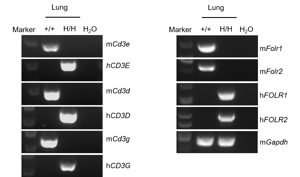
mRNA Expression Analysis
- Mouse Cd3e, Cd3d, Cd3g, Folr1 and Folr2 mRNA was only detectable in wild-type mice, but not in homozygous B-hCD3EDG/hFOLR1/hFOLR2 mice. Human CD3E, CD3D, CD3G, FOLR1 and FOLR2 mRNA was exclusively detectable in homozygous B-hCD3EDG/hFOLR1/hFOLR2 mice.
Strain specific analysis of CD3EDG, FOLR1 and FOLR2 mRNA expression in wild-type C57BL/6JNifdc mice and B-hCD3EDG/hFOLR1/hFOLR2 mice by RT-PCR. Lung RNA were isolated from wild-type C57BL/6JNifdc mice (+/+) and homozygous B-hCD3EDG/hFOLR1/hFOLR2 mice (H/H), then cDNA libraries were synthesized by reverse transcription, followed by PCR with mouse or human CD3E, CD3D, CD3G, FOLR1 and FOLR2 primers. Mouse Cd3e, Cd3d, Cd3g, Folr1 and Folr2 mRNA was only detectable in wild-type mice. Human CD3E, CD3D, CD3G, FOLR1 and FOLR2 mRNA was exclusively detectable in homozygous B-hCD3EDG/hFOLR1/hFOLR2 mice but not in wild-type mice.
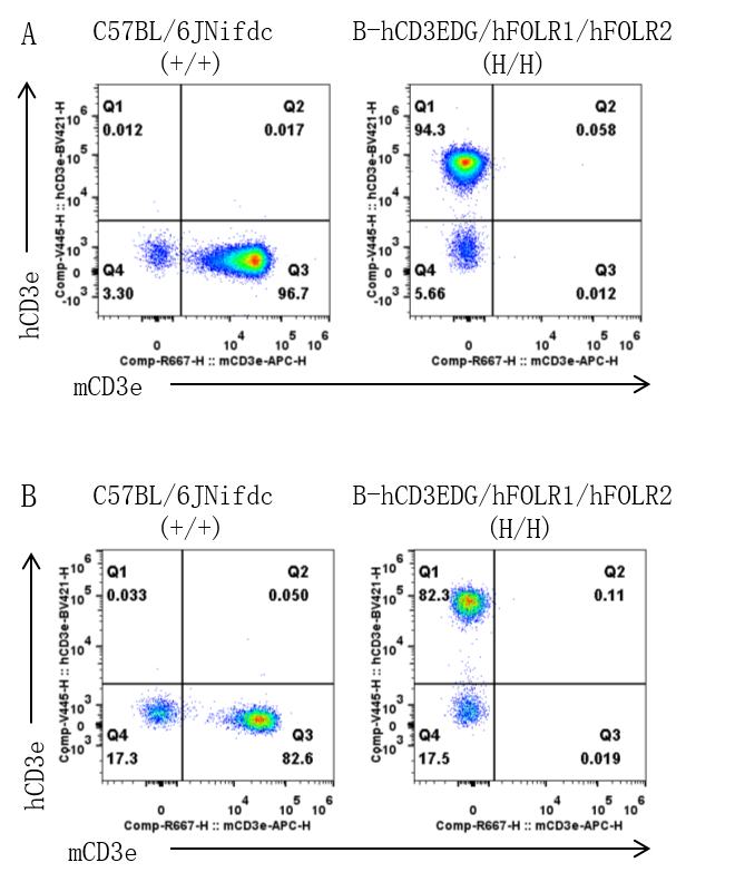
CD3E Protein Expression Analysis
- Mouse CD3E was detectable on the T cells of wild-type mice, but not on homozygous B-hCD3EDG/hFOLR1/hFOLR2 mice. Human CD3E was only detectable on the T cells of homozygous B-hCD3EDG/hFOLR1/hFOLR2 mice.
Strain specific CD3E expression analysis in wild-type mice and homozygous B-hCD3EDG/hFOLR1/hFOLR2 mice by flow cytometry. Spleen(A) and Blood(B) was collected from wild-type C57BL/6JNifdc (+/+) mice and homozygous B-hCD3EDG/hFOLR1/hFOLR2 mice (H/H) and analyzed by flow cytometry with anti-mouse CD3E antibody (Biolegend, 100312) and anti-human CD3E antibody (BD Biosciences, 562426).
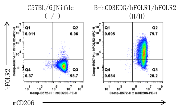
FOLR2 Protein Expression Analysis
- Human FOLR2 was detectable only on M2-like macrophages derived from homozygous B-hCD3EDG/hFOLR1/hFOLR2 mice.
Strain specific FOLR2 expression analysis in wild-type mice and homozygous B-hCD3EDG/hFOLR1/hFOLR2 mice by flow cytometry. Bone marrow cells were collected from wild-type C57BL/6JNifdc (+/+) mice and homozygous B-hCD3EDG/hFOLR1/hFOLR2 mice (H/H), cultured in 6-well plates stimulated with M-CSF (20ng/mL) for 7 days, then treatment with mIL-4 (50 ng/mL) and mIL-10 (75 ng/mL) for 18 h to induce M2-like macrophages. Macrophages were subsequently collected and analyzed by flow cytometry with anti-human FOLR2 antibody(Biolegend, 391705).
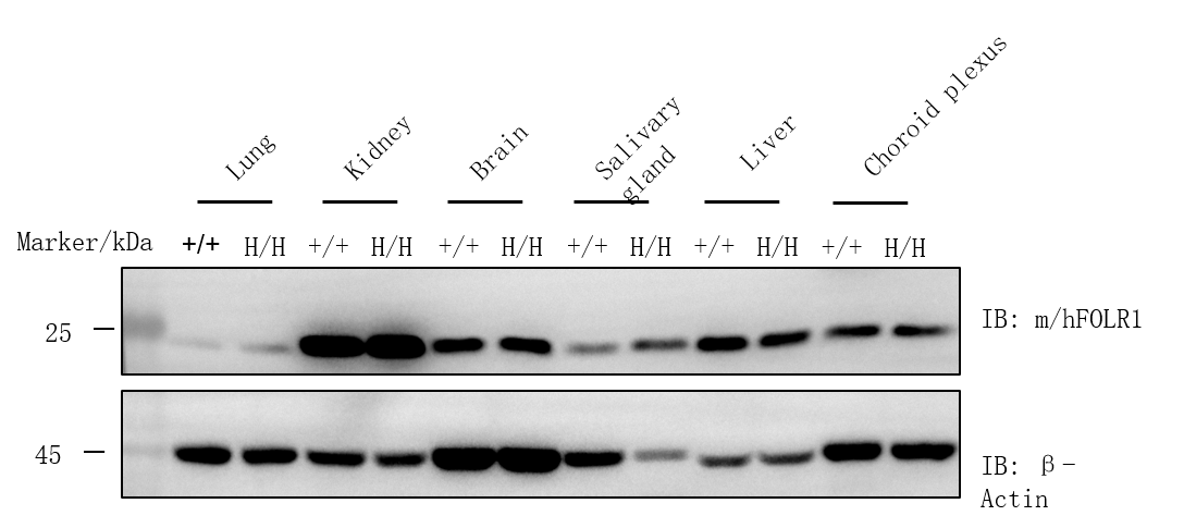
FOLR1 Protein Expression Analysis
- FOLR1 was detectable in lung, kidney, brain, salivary gland, liver and choroid plexus of wild-type mice and homozygous B-hCD3EDG/hFOLR1/FOLR2 mice due to the cross-reactivity of antibody.
Western blot analysis of FOLR1 protein expression in wild-type mice and homozygous B-hCD3EDG/hFOLR1/FOLR2 mice. Various tissue lysates were collected from wild-type C57BL/6JNifdc (+/+) mice and homozygous B-hCD3EDG/hFOLR1/FOLR2 mice (H/H), and then analyzed by western blot with anti-FOLR1 antibody(abcam, ab230469). 40 μg or 30 μg total proteins were loaded for western blotting analysis.
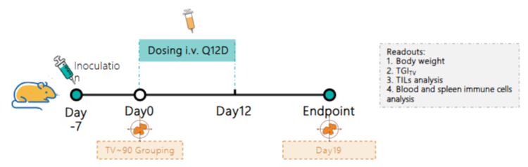
Efficacy Evaluation of CD3/FOLR1 BsAb in the Treatment of the SubcutaneousB-hFOLR1 MC38 Model in B-hCD3EDG/hFOLR1 /hFOLR2 Mice
Establishment of a B-hFOLR1 MC38 model and in vivo efficacy study of an anti-human CD3/FOLR1 bispecific antibody. B-hFOLR1 MC38 colon cancer cells were implanted subcutaneously into homozygous B-hCD3EDG/hFOLR1/hFOLR2 mice (female, 7-weeks-old, n = 6). When the average tumor volume reached approximately 90 mm³, mice were randomized and subsequently administered the anti-human CD3/FOLR1 bispecific antibodies (provided by a client ) via intravenous injection.
Efficacy Evaluation of Treatment of the Subcutaneous B-hFOLR1 MC38 Model in B-hCD3EDG/hFOLR1/hFOLR2 Mice
Efficacy of CD3/FOLR1 bispecific antibody in B-hCD3EDG/hFOLR1/hFOLR2 mice. (A) Tumor growth curves. (B) Body weight changes during treatment. As shown in panel A, CD3/FOLR1 bispecific antibodies (provided by a client ) was efficacious in controlling tumor growth in B-hCD3EDG /hFOLR1 /hFOLR2 mice in a dose-dependent manner, demonstrating that the B-hCD3EDG /hFOLR1 /hFOLR2 mice provide a powerful preclinical model for in vivo evaluation of anti-human CD3/FOLR1 bispecific antibodies. Values are expressed as mean ± SEM.
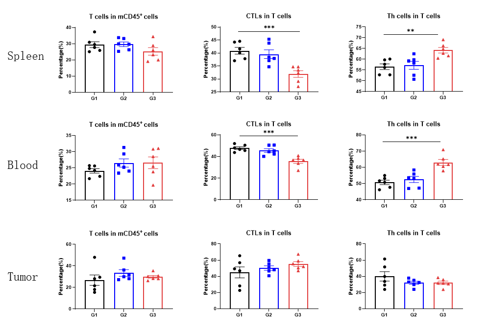
CD3/FOLR1 BsAb Induces T Cell Activation and Proliferation in Spleen and Blood
- CD45+ cells and T cell subgroups in Spleen, Blood and Tumor.
- The proportions of CTL cells in the blood and spleen decreased in the CD3/FOLR1 BsAb treatment group.
- The proportions of Th cells in the blood and spleen increased in the CD3/FOLR1 BsAb treatment group.
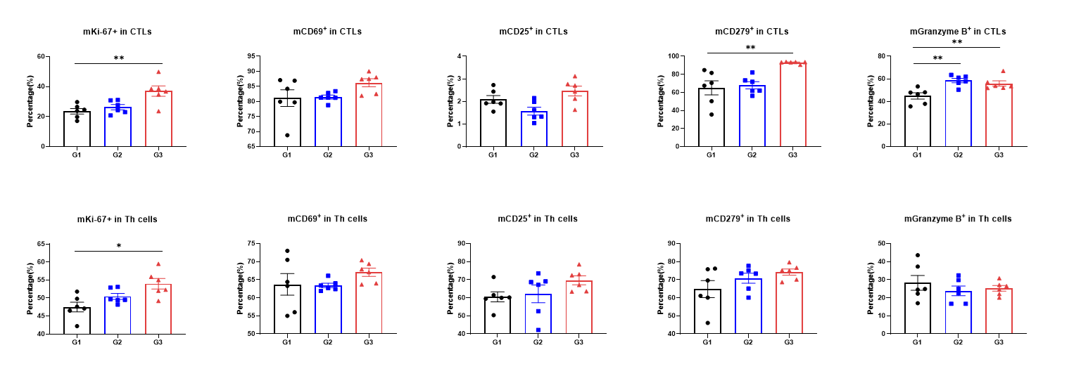
CD3/FOLR1 BsAb Induces Proliferation and Cytotoxic Killing of Intratumoral CTLs
- The proportions of mKi-67+ CTL cells, mCD279+ CTL cells and mGranzyme B+ CTL cells in the tumor were increased in the high dose CD3/FOLR1 BsAb treatment group.
* When publishing results obtained using this animal model, please acknowledge the source as follows: The animal model [B-hCD3EDG/hFOLR1/hFOLR2 mice] (Cat# 113968) was purchased from Biocytogen.