Description
Biological Features and Therapeutic Potential of CD3 and MUC1
- Gene Information: CD3: Encoded by CD3E, D, G; part of the Ig superfamily. It forms the essential signaling backbone of the T-cell receptor (TCR) complex. MUC1: a transmembrane mucin glycoprotein composed of a heavily glycosylated extracellular domain and a conserved cytoplasmic tail. MUC1 plays an important role in epithelial protection and barrier function.
- Protein Expression: CD3: Constitutive and universal marker for all mature T cells (CD4+, CD8+). Always present on the cell surface. MUC1: normally expressed on the apical surface of epithelial cells in the respiratory, gastrointestinal, genitourinary, and mammary tracts. In many solid tumors, MUC1 is overexpressed and aberrantly glycosylated, making it a common tumor-associated antigen.
- Signaling Pathway: CD3: Operates via ITAM phosphorylation and ZAP-70. It triggers the initial "on" switch, Ca2+ flux, and immediate cytotoxicity. MUC1: functions as an oncogenic signaling molecule through its cytoplasmic domain (MUC1-C), activating pathways including PI3K/AKT, MAPK/ERK, NF-κB, and Wnt/β-catenin. These pathways promote tumor cell proliferation, survival, metastasis, and immune evasion.
- Therapeutic Inhibition: CD3×MUC1 bispecific antibodies recruit and activate T cells by simultaneously binding CD3 on T cells and MUC1 on tumor cells, resulting in targeted killing of MUC1-positive cancer cells. Additional therapeutic strategies include monoclonal antibodies, CAR-T cells, antibody–drug conjugates, and cancer vaccines targeting MUC1.
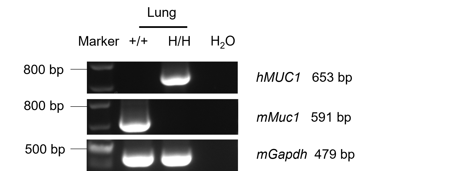
MUC1 mRNA Expression by RT-PCR
- Humanized MUC1 mRNA was detectable only in lung of homozygous B-hCD3EDG/hMUC1 mice, but not in wild-type C57BL/6 mice.
- Mouse Muc1 mRNA was detectable only in lung of wild-type C57BL/6 mice .
Mouse and human MUC1 analysis in lung by RT-PCR and sequencing. Lung RNA was isolated from wild-type C57BL/6 mice (+/+) and homozygous B-hCD3EDG/hMUC1 mice (H/H), and then cDNA libraries were synthesized by reverse transcription, followed by PCR with mouse or human MUC1 primers.
CD3D,CD3G mRNA Expression by RT-PCR
- Humanized CD3D and CD3G mRNA were detectable only in spleen of homozygous B-hCD3EDG/hMUC1 mice, but not in wild-type C57BL/6 mice.
- Mouse Cd3d and Cd3g mRNA were detectable only in spleen of wild-type C57BL/6 mice.
Mouse or human CD3D and CD3G analysis in spleen by RT-PCR and sequencing. Spleen RNA were isolated from wild-type C57BL/6 mice (+/+) and homozygous B-hCD3EDG/hMUC1 mice (H/H), and then cDNA libraries were synthesized by reverse transcription, followed by PCR with mouse or human CD3D and CD3G primers.
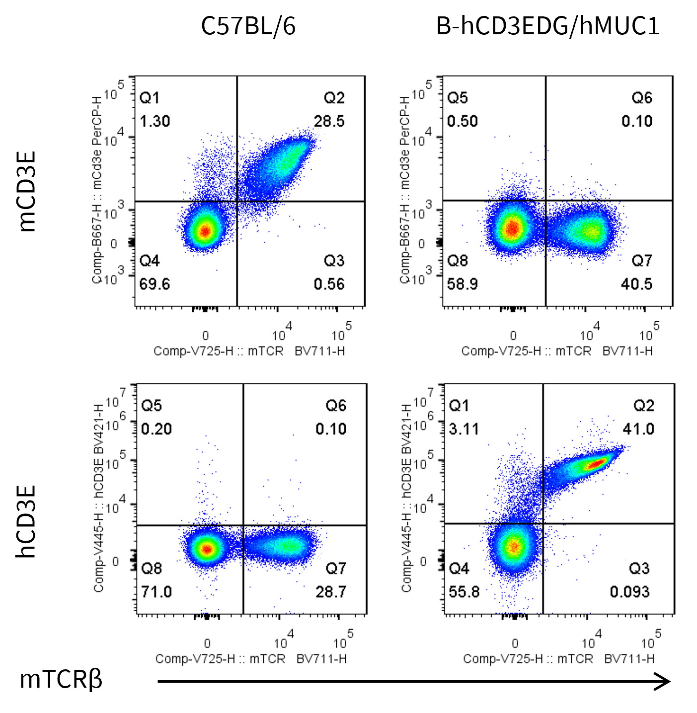
CD3E Protein Expression in Spleen
- Mouse CD3E was detected on T cells populations in wild-type C57BL/6 mice, but not in B-hCD3EDG/hMUC1 mice.
- Human CD3E was detected on T cells populations in B-hCD3EDG/hMUC1 mice, but not in wild-type C57BL/6 mice.
Mouse and human CD3E expression analysis in splenocytes. Splenocytes were collected from wild-type C57BL/6 mice and homozygous B-hCD3EDG/hMUC1 mice (male, 6-week-old, n = 1). CD3E expression on T cells was analyzed by flow cytometry using species-specific anti-CD3E antibodies (anti-human CD3E antibody, BD Horizon, 562426; anti-mouse CD3E antibody, Biolegend, 100312 ).
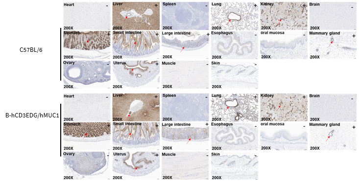
Immunohistochemical Analysis
- Human MUC1 expression was detected in the liver, lung, kidney, stomach, small intestine, large intestine, mammary glad, and uterus of homozygous B-hCD3EDG/hMUC1 mice, but not in the heart, spleen, brain, esophagus, oral mucosa, ovary, muscle and skin.
Immunohistochemical analysis of organs in B-hCD3EDG/hMUC1 mice. Major organs were collected from wild-type C57BL/6 mice and homozygous B-hCD3EDG/hMUC1 mice (female, 8 weeks old), and analyzed by IHC using anti-MUC1 antibody (Abcam, ab245693). The antibody can recognize both human and mouse MUC1. The arrow indicates tissue cells with positive MUC1 staining (brown). "+" and "-" indicate positive and negative tissues, respectively.
* When publishing results obtained using this animal model, please acknowledge the source as follows: The animal model [B-hCD3EDG/hMUC1 mice] (Cat# 114213) was purchased from Biocytogen.