Targeting strategy
Gene targeting strategy for B-hIL1RACP mice. The chimeric IL1RACP coding sequence (human signal peptide, human extracellular domain and mouse transmembrane and intracellular domain) was inserted after the 5’UTR of mouse Il1racp gene in B-hIL1RACP mice. The insertion disrupts the endogenous murine Il1racp gene, resulting in the absence of mouse transcripts.
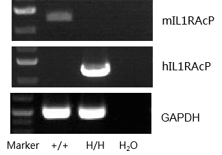
mRNA expression analysis in humanized B-hIL1RACP mice
Species specific analysis of IL1RACP gene expression in wild-type C57BL/6 mice and homozygous humanized B-hIL1RACP mice by RT-PCR. Liver was collected from wild-type C57BL/6 mice (+/+) and homozygous B-hIL1RACPmice (H/H). Mouse Il1racp mRNA was detectable only in wild-type C57BL/6 mice. Human IL1RACP mRNA was detectable only in homozygous B-hIL1RACP mice, but not in wild-type C57BL/6 mice.
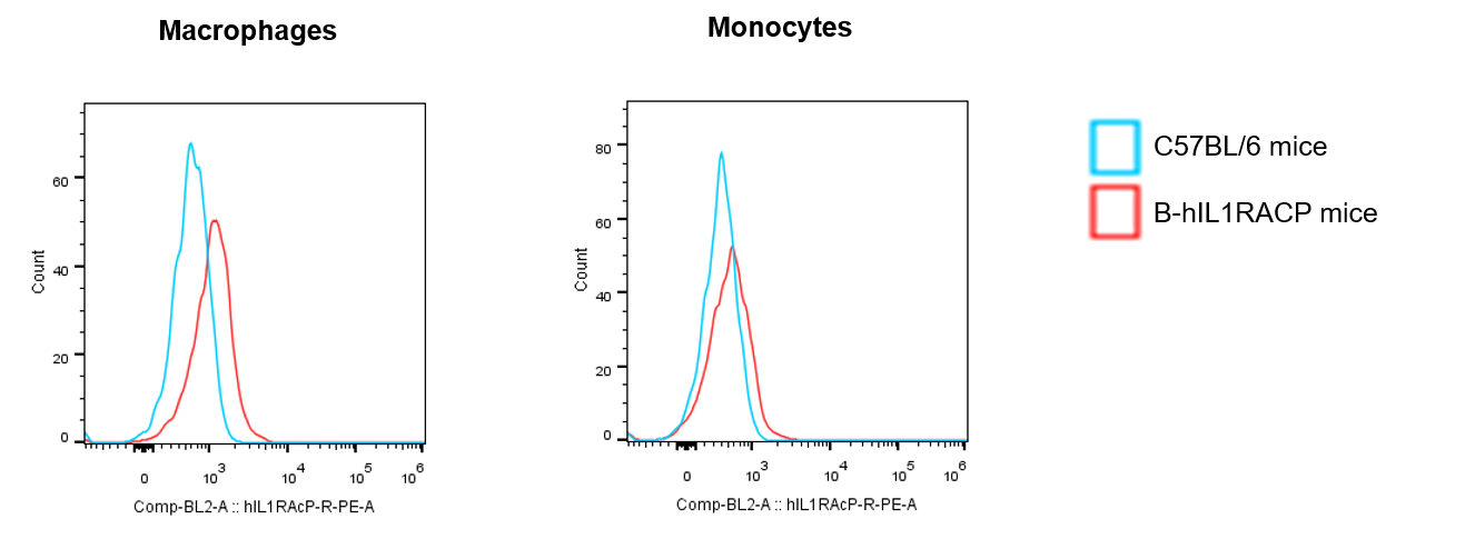
Protein expression analysis in spleen
Strain specific IL1RACP expression analysis in wild-type C57BL/6 mice and homozygous humanized B-hIL1RACP mice by flow cytometry. Splenocytes were collected from wild-type C57BL/6 mice and homozygous B-hIL1RACP mice. Protein expression was analyzed with anti-human IL1RACP antibody (R&D, FAB676P) by flow cytometry. Human IL1RACP was exclusively detectable in homozygous B-hIL1RACP mice, but not in wild-type C57BL/6 mice.
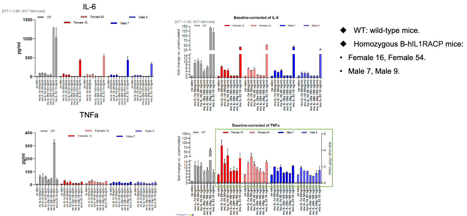
Functional verification of B-hIL1RAcP mice
Splenocytes were collected from wild-type mice and homozygous B-hIL1RAcP mice and stimulated with IL1/IL33/IL36 protein. The expression of cytokines in splenocytes culture medium was detected after 48 hours. The results showed that murine IL1/IL33/IL36 protein effectively activated splenocytes in wild-type mice and triggered the secretion of related cytokines. Murine IL1B and murine IL33 proteins could effectively activate splenocytes in B-hIL1RACP mice and trigger the secretion of related cytokines, while murine IL36 protein could not effectively activate B-hIL1RACP mice splenocytes.
Note: This experiment was performed by the client using B-hIL1RACP mice. All the other materials were provided by the client.
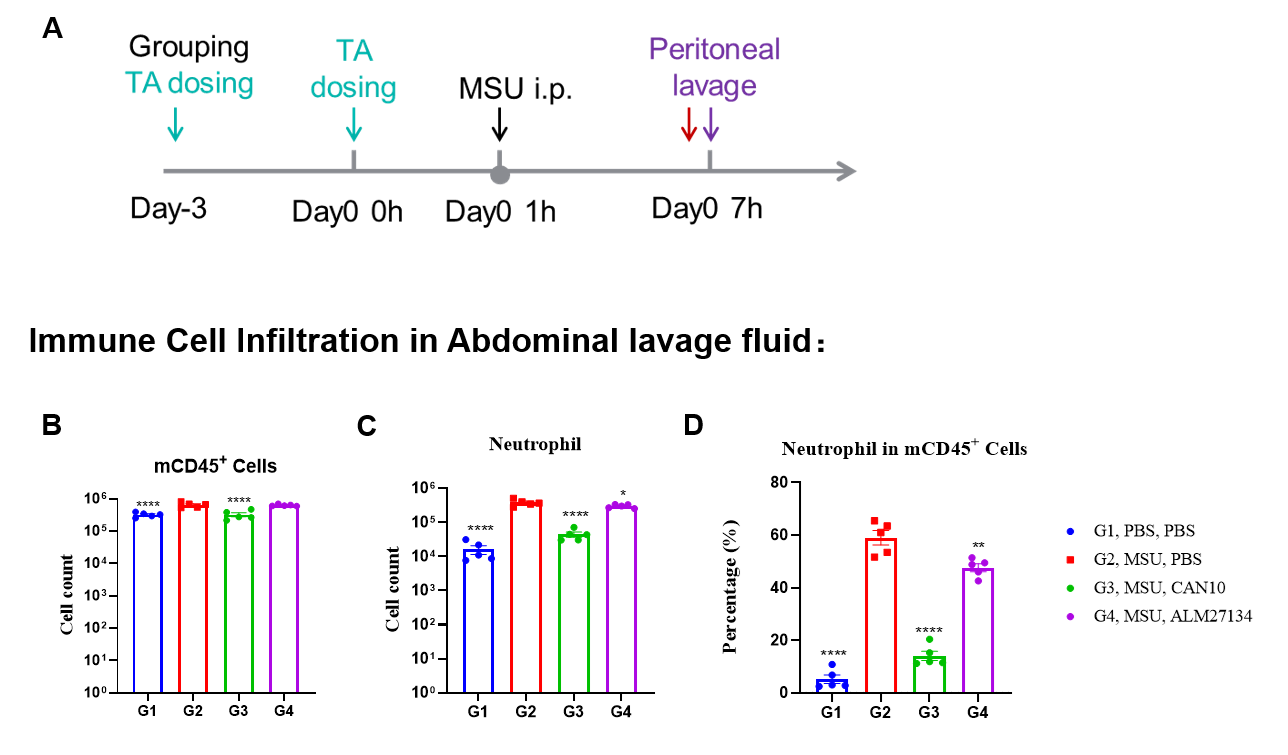
Efficacy Evaluation of Anti-IL1RAcP antibody in peritonitis Model of B-hIL1RAcP mice
Effects of Anti-IL1RAcP antibody on immune cell infiltration in peritoneal lavage fluid in a MSU-induced peritonitis model using B-hIL1RAcP mice. (A) Modeling paradigm of MSU-induced peritonitis model in B-hIL1RAcP mice. (B) Quantification of mCD45⁺ cells in peritoneal lavage fluid across groups. (C) Total neutrophil counts in peritoneal lavage fluid. (D) Percentage of neutrophils within the mCD45⁺ population, demonstrating that the number of neutrophils was significantly inhibited by CAN10 analog (10 mpk, in house) and ALM27134 analog (10 mpk, in house) and almost returned to the level of the control group. Values are expressed as mean ± SEM. Significance was determined by two-way ANOVA test. *P < 0.05, **P < 0.01, ****P < 0.0001. Groups G1, G3, and G4 showed significant differences compared to G2.
Efficacy Evaluation of Anti-IL1RAcP antibody in psoriasis Model of B-hIL1RAcP mice
IMQ-induced skin inflammation in B-hIL1RAcP mice phenotypically resembles psoriasis. Mice (female, 7 week-old, n=5) were scored daily for up to 7 days for body weight and clinical signs of skin inflammation following treatment with imiquimod (IMQ) cream. Mice in each group were treated with CAN10 analog (10 mpk, in-house). (A) Experimental schedule for induction of psoriasis-like skin lesions (B) Phenotypical presentation of mouse back skin at day 6. (C) Body weight during treatment. (D-E) Erythema and scaling score of the back was scored daily. Additionally, the cumulative score (erythema plus scaling) is depicted. Values are expressed as mean ± SEM. Significance was determined by two-way ANOVA test. ***P < 0.001.

Dose dependent effects of antibodies on historical score in IMQ induced psoriasis-like skin lesions in B-hIL1RAcP mice. Back skin was collected at the endpoint and stained with Hematoxylin and eosin (H&E). (A) H&E staining of the back skin. (B) Histological changes were scored. Results indicated that the CAN10 analog (10 mpk, in-house) significantly improved psoriasis-like skin parameters, including hyperkeratosis, crusting, and acanthosis, confirming that B-hIL1RAcP mice provide a powerful model for in vivo evaluation. Values are expressed as mean ± SEM. ***P < 0.001.
* When publishing results obtained using this animal model, please acknowledge the source as follows: The animal model [B-hIL1RACP mice] (Cat# 110072) was purchased from Biocytogen.