

C57BL/6-Il1raptm1(IL1RAP)Bcgen/Bcgen • 110072

Key Advantages
Validation
Application

In B-hIL1RACP mice, a chimeric CDS encoding the human IL1RAcP signal peptide and extracellular domain together with the mouse IL1RAcP transmembrane and cytoplasmic domains was inserted after the 5’ UTR of the mouse Il1racp gene. The chimeric IL1RAcP protein is driven by the endogenous mouse Il1racp promoter, while mouse Il1racp transcription and translation are disrupted.

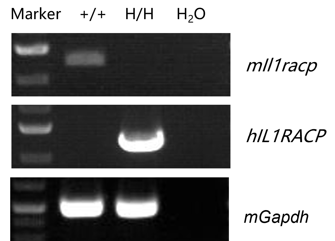
Strain-specific analysis of IL1RAcP gene expression was performed in wild-type C57BL/6 mice and homozygous B-hIL1RACP mice by RT-PCR. Liver was collected from wild-type C57BL/6 mice (+/+) and homozygous B-hIL1RACP mice (H/H), then cDNA libraries were synthesized by reverse transcription and amplified with mouse or human IL1RAcP primers. Mouse Il1racp mRNA was detectable in wild-type C57BL/6 mice, while human IL1RAcP mRNA was detectable in homozygous B-hIL1RACP mice but not in wild-type mice.
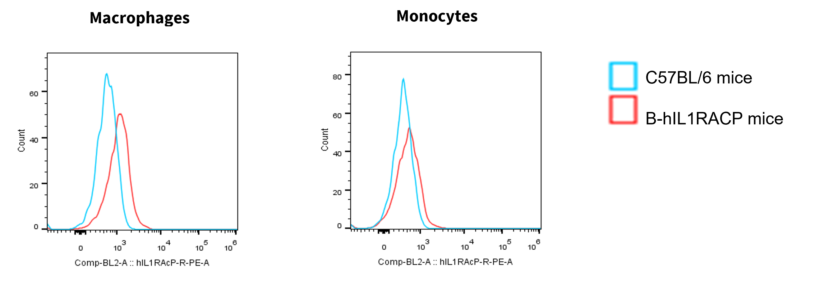
Strain-specific IL1RACP expression was analyzed in wild-type C57BL/6 mice and homozygous B-hIL1RACP mice by flow cytometry. Splenocytes were collected from wild-type C57BL/6 mice and homozygous B-hIL1RACP mice. Protein expression was analyzed with anti-human IL1RACP antibody (R&D, FAB676P). Human IL1RACP was detectable in homozygous B-hIL1RACP mice but not in wild-type C57BL/6 mice.
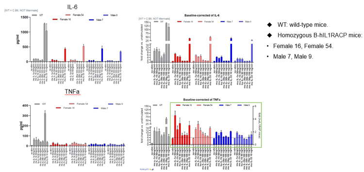
Splenocytes were collected from wild-type mice and homozygous B-hIL1RAcP mice and stimulated with IL1, IL33, or IL36 protein. Cytokines in splenocyte culture medium were detected after 48 hours. Murine IL1/IL33/IL36 proteins effectively activated wild-type splenocytes and triggered cytokine secretion. Murine IL1B and murine IL33 proteins also activated splenocytes in B-hIL1RACP mice, while murine IL36 protein did not effectively activate the splenocytes.
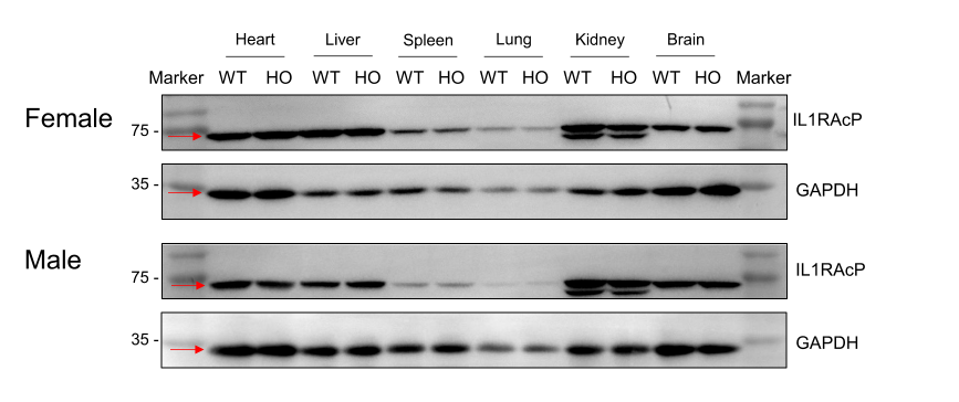
Heart, liver, spleen, lung, kidney, and brain were collected from wild-type C57BL/6JNifdc mice (+/+) and homozygous B-hIL1RAcP mice (H/H) and analyzed by western blot with an IL-1RAP polyclonal antibody (Proteintech, 30966-1-AP). This antibody shows cross-reactivity between human and mouse. IL1RAcP was detected in the heart, liver, spleen, lung, kidney, and brain from wild-type C57BL/6JNifdc mice (+/+) and homozygous B-hIL1RAcP mice (H/H). WT: wild-type C57BL/6JNifdc mice; HO: homozygous B-hIL1RAcP mice.
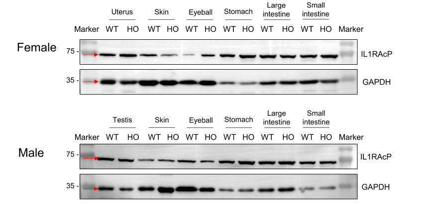
Uterus, testis, skin, eyeball, stomach, large intestine, and small intestine were collected from wild-type C57BL/6JNifdc mice (+/+) and homozygous B-hIL1RAcP mice (H/H) and analyzed by western blot with an IL-1RAP polyclonal antibody (Proteintech, 30966-1-AP). This antibody shows cross-reactivity between human and mouse. IL1RAcP was detected in the uterus, testis, skin, eyeball, stomach, large intestine, and small intestine from wild-type C57BL/6JNifdc mice (+/+) and homozygous B-hIL1RAcP mice (H/H). WT: wild-type C57BL/6JNifdc mice; HO: homozygous B-hIL1RAcP mice.
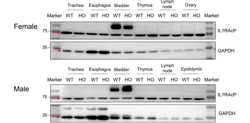
Trachea, esophagus, bladder, thymus, lymph node, ovary, and epididymis were collected from wild-type C57BL/6JNifdc mice (+/+) and homozygous B-hIL1RAcP mice (H/H) and analyzed by western blot with an IL-1RAP polyclonal antibody (Proteintech, 30966-1-AP). This antibody shows cross-reactivity between human and mouse. IL1RAcP was detected in the trachea, esophagus, bladder, thymus, lymph node, ovary, and epididymis from wild-type C57BL/6JNifdc mice (+/+) and homozygous B-hIL1RAcP mice (H/H). WT: wild-type C57BL/6JNifdc mice; HO: homozygous B-hIL1RAcP mice.
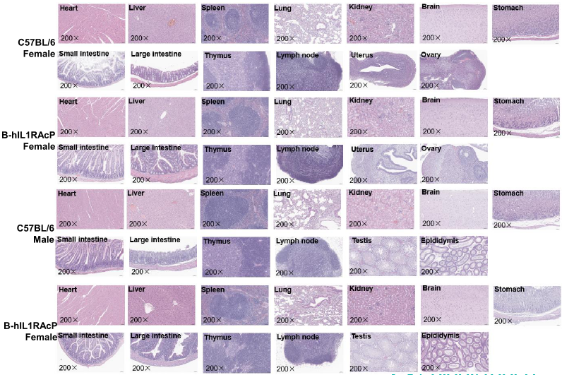
Macroscopic anatomical data of wild-type mice and B-hIL1RAcP mice. 8-week-old wild-type C57BL/6 mice and B-hIL1RAcP mice (10 males and 10 females) were monitored for 8 weeks to assess the overall health of the animals. (A) Absolute body weight and percent weight gain of wild-type mice and B-hIL1RAcP mice over time. (B) Organ weights of wild-type mice and B-hIL1RAcP mice. (C) Organ histology (H&E) of wild-type mice and B-hIL1RAcP mice. Organ morphology and pathological examination of B-hIL1RAcP mice revealed no abnormalities compared to wild-type controls.
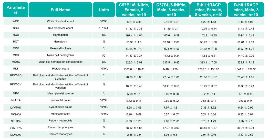
Complete blood count (CBC) of B-hIL1RAcP mice. Values are expressed as mean ± SD.
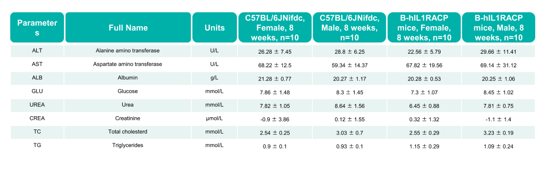
Values are expressed as mean ± SD.

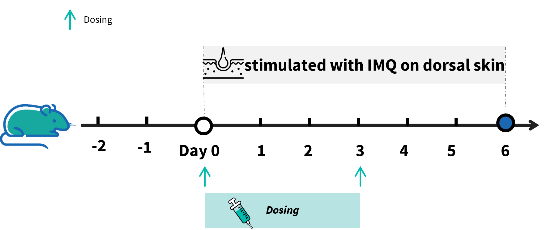
Experimental schedule of the MSU-induced peritonitis model in B-hIL1RAcP mice. Mice(7 week-old) received a daily topical of commercially available IMQ cream on the shaved back for 6 consecutive days to induce IMQ-induced psoriasis model. Control mice were treated similarly with Vaseline cream. Severity of skin inflammation was daily scored, and back skin was collected at the endpoint. IMQ: imiquimod.
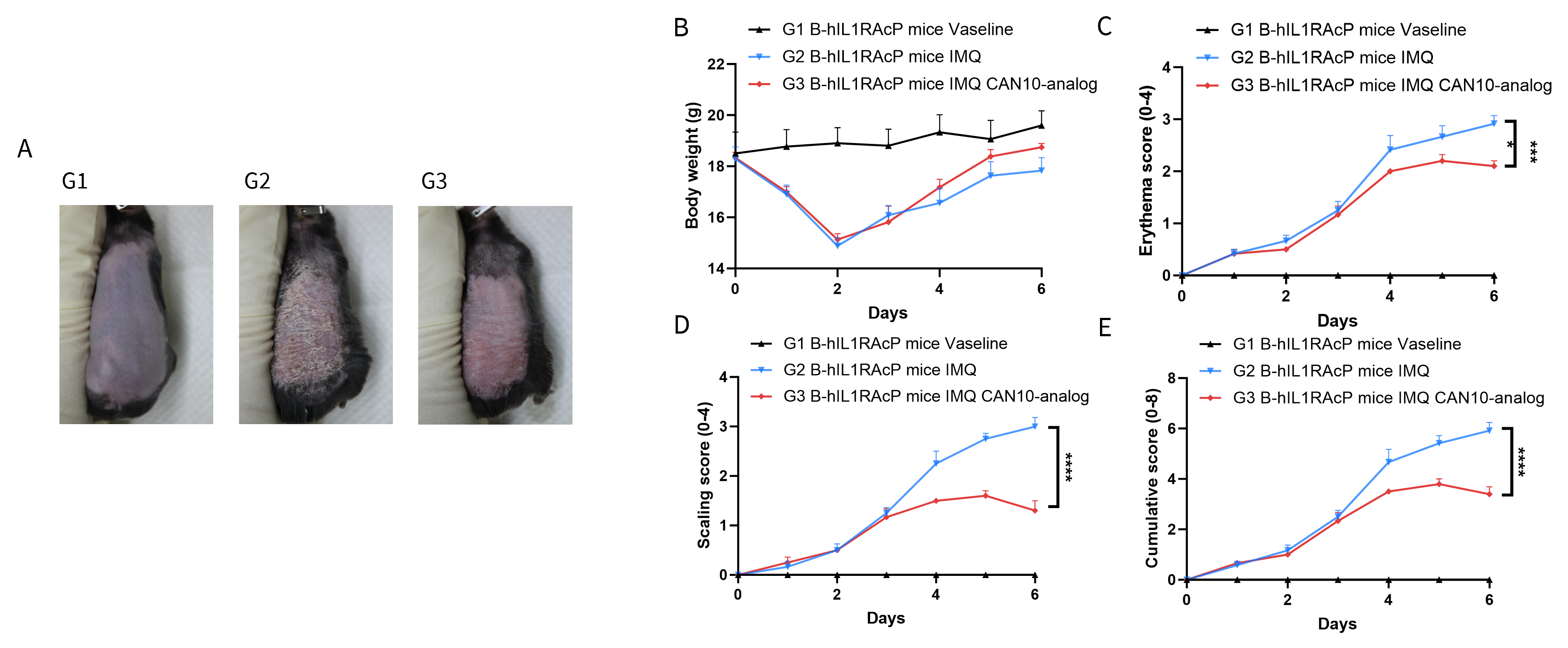
IMQ-induced skin inflammation in B-hIL1RAcP mice phenotypically resembles psoriasis. Mice (female, 7 week-old, n=5) were scored daily for up to 7 days for body weight and clinical signs of skin inflammation following treatment with imiquimod (IMQ) cream. Mice in each group were treated with CAN10-analog (10 mpk, in-house). (A) Experimental schedule for induction of psoriasis-like skin lesions (B) Phenotypical presentation of mouse back skin at day 6. (C) Body weight during treatment. (D-E) Erythema and scaling score of the back was scored daily. Additionally, the cumulative score (erythema plus scaling) is depicted. Significance was determined by two-way ANOVA test. ***P < 0.001.

Dose dependent effects of antibodies on historical score in IMQ induced psoriasis-like skin lesions in B-hIL1RAcP mice. Back skin was collected at the endpoint and stained with Hematoxylin and eosin (H&E). (A) H&E staining of the back skin. (B) Histological changes were scored. Results indicated that the CAN10-analog (10 mpk, in-house) significantly improved psoriasis-like skin parameters, including hyperkeratosis, crusting, and acanthosis, confirming that B-hIL1RAcP mice provide a powerful model for in vivo evaluation. ***P < 0.001.
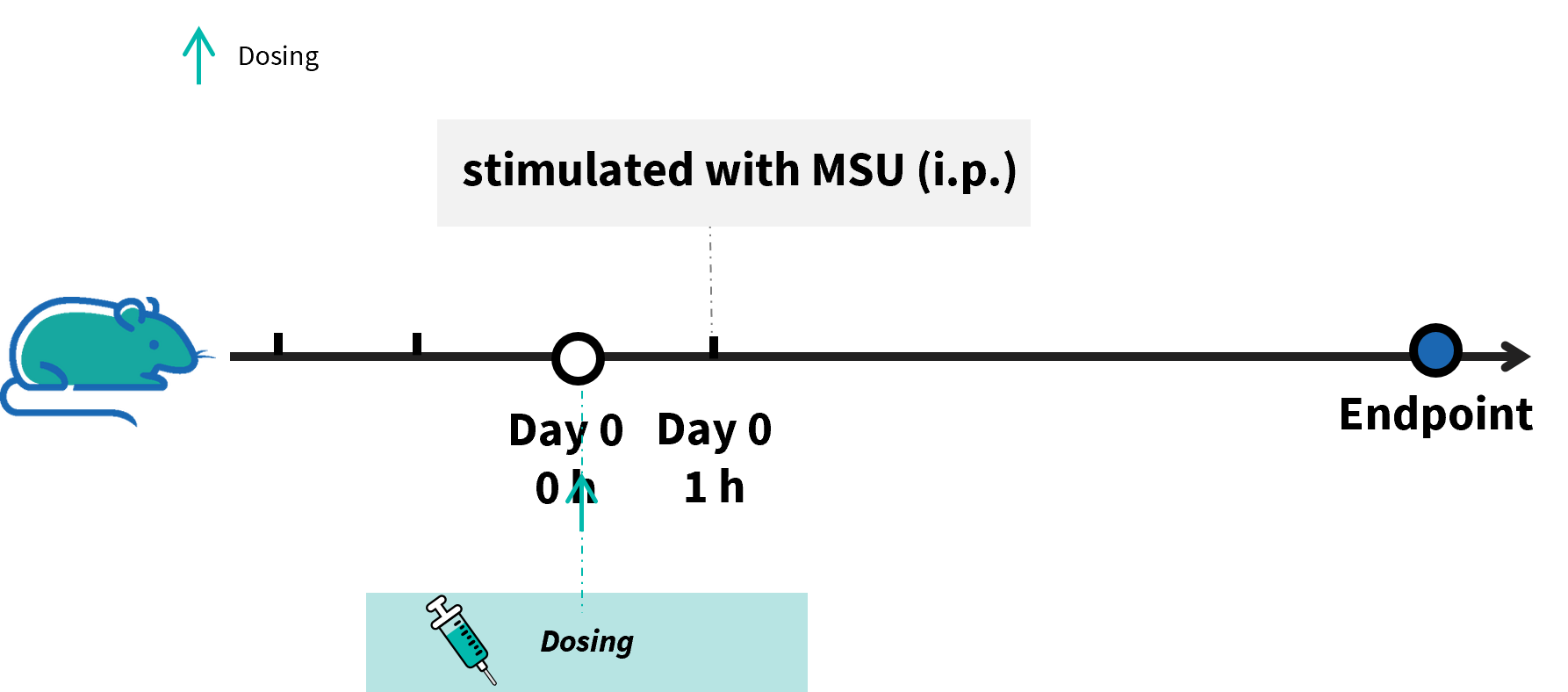
Experimental schedule of MSU-induced peritonitis model in B-hIL1RAcP mice. Male B-hIL1RAcP mice received intraperitoneal (i.p.) administration of Vehicle, CAN10-analog (10 mg/kg), or ALM27134-analog (10 mg/kg) on Day 0 and Day 3. One hour after the initial dosing on Day 0, mice were stimulated with an i.p. injection of MSU to induce the peritonitis model. Samples were collected at the endpoint for efficacy evaluation. MSU: monosodium urate.

Effects of Anti-IL1RAcP antibody on immune cell infiltration in peritoneal lavage fluid in MSU-induced peritonitis model using B-hIL1RAcP mice. (A) Quantification of mCD45⁺ cells in peritoneal lavage fluid across groups. (B) Total neutrophil counts in peritoneal lavage fluid. (C) Percentage of neutrophils within the mCD45⁺ population, demonstrating that the number of neutrophils was significantly inhibited by CAN10-analog (10 mpk, in house) and ALM27134-analog (10 mpk, in house) and almost returned to the level of the control group. Significance was determined by two-way ANOVA test. *P < 0.05, **P < 0.01, ****P < 0.0001.

Q1: What are B-hIL1RACP mice?
B-hIL1RACP mice are human IL1RACP gene-humanized mice on a C57BL/6 background, developed for IL1RACP-targeted inflammation and antibody efficacy research.
Q2: Why is IL1RACP important?
IL1RACP, also known as IL1R3 or IL-1RAcP, is a shared accessory receptor for IL1 family cytokine signaling, including IL1, IL33, and IL36 pathway responses.
Q3: How was human IL1RACP expression validated in B-hIL1RACP mice?
Human IL1RACP mRNA was validated by RT-PCR, and human IL1RACP protein was validated in splenocytes by flow cytometry.
Q4: Can B-hIL1RACP mice be used for antibody efficacy studies?
Yes. B-hIL1RACP mice have efficacy data in IMQ-induced psoriasis-like skin inflammation and MSU-induced peritonitis models using anti-IL1RACP antibody analogs.
Q5: What are the main applications of B-hIL1RACP mice?
Applications include IL1RACP-targeted antibody evaluation, IL1 family signaling research, autoimmune and inflammatory disease studies, psoriasis-like inflammation models, peritonitis models, and preclinical biologics development.
